
Abstract
Type XV is a large collagen-proteoglycan found in all human tissues examined. By light microscopy it was localized to most epithelial and all nerve, muscle, fat and endothelial basement membrane zones except for the glomerular capillaries or hepatic/splenic sinusoids. This widespread distribution suggested that type XV may be a discrete structural component that acts to adhere basement membrane to the underlying connective tissue. To address these issues, immunogold ultrastructural analysis of type XV collagen in human kidney, placenta, and colon was conducted. Surprisingly, type XV was found almost exclusively associated with the fibrillar collagen network in very close proximity to the basement membrane. Type XV exhibited a focal appearance directly on the surface of, or extending from, the fibers in a linear or clustered array. The most common single arrangement was a bridge of type XV gold particles linking thick-banded fibers. The function of type XV in this restricted microenvironment is expected to have an intrinsic dependence upon its modification with glycosaminoglycan chains. Present biochemical characterization showed that the type XV core protein in vivo carries chains of chondroitin/dermatan sulfate alone, or chondroitin/dermatan sulfate together with heparan sulfate in a differential ratio. Thus, type XV collagen may serve as a structural organizer to maintain a porous meshwork subjacent to the basement membrane, and in this domain may play a key role in signal transduction pathways.
Keywords
T
The type XV collagen chain is comprised of 1363 residues (Myers et al. 1992; Kivirikko et al. 1994, and Figure 1). Most of the sequence is non-collagenous and represented in the 530 and 256 residue amino and carboxy domains, respectively. The 577 residue discontinuous collagenous region is highly interrupted by many different-size non-collagenous segments, totaling one-third of the domain sequences. RNA analysis by in situ hybridization and northern blotting showed that type XV is expressed by fibroblast, muscle, endothelial and some epithelial cells (Kivirikko et al. 1995) and is detected in most tissues except for liver and brain (Myers et al. 1996). Consistent with these observations, light microscopy immunohistochemistry illustrated a nearly ubiquitous type XV distribution in human tissues, but a restricted localization in most epithelial, and all nerve, muscle, fat and endothelial BM zones, except for those of the glomerular capillaries or hepatic/splenic sinusoids (Myers et al. 1996,1997; Hagg et al. 1997; Tomono et al. 2002). There was minimal type XV immunoreactivity in the interstitium. Type XV collagen in vivo is a full-time proteoglycan (PG) with a mass of glycosaminoglycan (GAG) chains attached to the N-terminal domain (Li et al. 2000). Digestion with chondroitinase, but not heparitinase, revealed type XV core protein chains of 250/225 kD, which are subject to some degree of carboxy-terminal cleavage. Partial purification of type XV from placenta showed that in the richest source of this collagen, it is a very scarce and very large protein (greater than 1 million molecular weight).
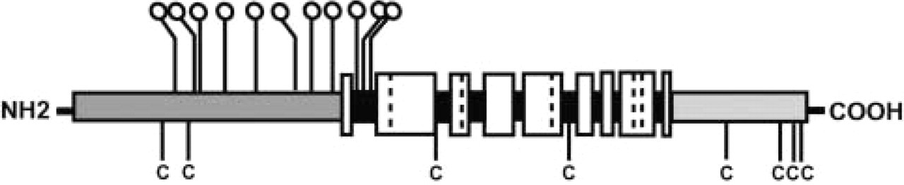
Schematic diagram showing the domain structure in the type XV collagen chain and the location of consensus sites for GAG chain attachment. Sequence information was reported by Myers et al. (1992) and Kivirikko et al. (1994). The amino-terminal non-collagenous region (530 residues) is shown in dark gray and the carboxy domain (256 residues) is in a medium gray shade. The 577 residue discontinuous collagenous region is displayed in white. Large interruptions (45, 31, 24, 11, 34, 14, 7, 10 residues, amino to carboxy) are black, whereas those of 2 or 3 residues are designated by vertical dashed lines. Ball and stick symbols depict the location of 11 consensus sequences for GAG chain attachment (Li et al. 2000); 8 are in the amino-terminal noncollagenous region and 3 are in the first interruption. C, cysteines.
Although no naturally occurring type XV mutations have yet been identified, type XV null mice under imposed mechanical stress have collapsed capillaries and endothelial cell degeneration, manifested in cardiovascular defects and skeletal myopathy (Eklund et al. 2001). The surprisingly mild phenotype created by the absence of type XV in the mouse left ambiguous the importance of this collagen in BM-stromal adhesion. We chose to concentrate on the presence of type XV in normal human tissues by establishing its ultrastructural distribution and its possible association with a morphologically distinct entity. Immunogold localization unexpectedly showed that type XV is an integral and unique part of the fibrillar collagenous network immediately subjacent to the BM. New biochemical data demonstrated that type XV can carry chondroitin/dermatan sulfate (CS/DS) and also heparan sulfate (HS) chains, implying signaling as well as structural roles for this collagen in cell-matrix interactions.
Materials and Methods
Type XV Collagen Antibodies
Development and characterization of the polyclonal type XV carboxy and amino antibodies have been described previously (Myers et al. 1996,1997; Li et al. 2000; Amenta et al. 2000,2003).
Electron Microscopy Immunohistochemistry
Normal human tissues were acquired from the Department of Pathology and Laboratory Medicine at Robert Wood Johnson University Hospital. Three or four experiments were conducted using each tissue. Immunogold localization was carried out with modifications of the procedure reported by Birk et al. (1988). Tissues were minced into small pieces and fixed in 4% paraformaldehyde in 0.2 M sodium phosphate buffer, pH 7.4, for 10-12 min. They were then washed at 4C in 4% sucrose in PBS for 1 hr, followed by 7% glycerol/4% sucrose in PBS for 1 hr. The tissues were snap-frozen in OCT compound (Miles Inc.; Elkhart, IN) in methylbutane at liquid nitrogen temperature and stored at −80C until sectioning. Five-μm sections were prepared on a cryostat at −20C onto poly-L-lysine-coated slides, dried at room temperature, and rehydrated with cold PBS at 4C. Tissue sections were treated with 0.1 mg/ml testicular hyaluronidase (Sigma-Aldrich; St Louis, MO) for 1 hr at 37C, washed in cold PBS, reduced with 0.25 mg/ml sodium borohydride for 15 min and rinsed again in PBS. Chondroitinase ABC (Seikagako Corp.; Rockville, MD) pretreatment (0.2 units/ml at 37C overnight) was included in some initial experiments but later discontinued since it did not change the final results. Sections were pretreated with 5% bovine serum albumin (BSA) in PBS for 1 hr at room temperature and then incubated with purified type XV polyclonal carboxy or amino antibodies (14 μg/ml and 44 μg/ml, respectively) overnight in a humidified chamber at 4C. In the control samples included with each experiment, kidney, placenta and colon tissue sections were incubated with normal rabbit serum or buffer in place of the primary antibody. Slides were rinsed in cold PBS and 1% Tween 20 and incubated as above with 6 nm gold-labeled goat anti-rabbit IgG (Electron Microscopy Sciences; Fort Washington, PA) at a 1:2.5 dilution in 1% BSA. The tissues were fixed with 2.5% glutaraldehyde in 0.1 M sodium cacodylate, pH 7.4, for 15 min, rinsed in the same buffer and treated with 1% osmium tetroxide for 1 hr. The slides were washed with 50% ethanol and dehydrated through an increasing concentration gradient of alcohol and acetone to a final concentration of 100% acetone. The sections were subsequently embedded in epon resin, sectioned and viewed on a Jeol 1200S electron microscope.
Tissue Extracts, Enzyme Digestions and Immunoblotting
Normal human tissues were obtained from the Hospital of the University of Pennsylvania and Robert Wood Johnson University Hospital. Samples were stored at −80C and thawed at 4C or in an ice slurry. One to three grams of tissue were minced and added to 10 vol of extraction buffer (50 mM Tris-HCl, 1.0 M NaCl, 10 mM EDTA, pH 7.5) with protease inhibitors (10 mM N-ethylmaleimide, 0.5 mM phenylmethylsulfonyl fluoride, 5 μg/ml leupeptin, 1 μg/ml aprotinin, obtained from Sigma). The tissues were homogenized at speed 5 (speed 7 = 27,000 rpm) for 8 × 1 min (Polytron; Brinkman Instruments, Westbury, NY) while maintained in an ice slurry. The tissue suspensions were rocked overnight at 4C and centrifuged at 32,500 × g for 30 min at 4C. The supernatant was dialyzed against 50 mM Tris-HCl, 1 mM EDTA, 100 mM NaCl, pH 7.5, plus protease inhibitors. Samples were aliquoted and stored at −80C. Protein concentrations were determined using the BCA reagent (Pierce Biotechnology; Rockford, IL).
The amount of protein extract used for the reactions was as follows: kidney, 44 μg; colon, 28 μg; and placenta, 15 μg. The control samples for the enzyme reactions were incubated exactly as described below but without any glycosidase. Three types of enzyme reactions were carried out: chondroitinase alone [designated R1ch]; chondroitinase and then heparitinase [designated R2ch+hep]; and heparitinase alone [designated R3hep]. All reactions contained the same volume and buffer concentrations and were incubated for the same time and temperature. The first incubation (chondroitinase reaction conditions) was carried out in 15 μl at 37C for 90 min, and the second incubation (heparitinase reaction conditions) was carried out in 24 μl at 43C for 90 min. In the first reaction, 3 μl of 5X buffer was added to give a final concentration of 100 mM Tris-HCl, 30 mM sodium acetate buffer, pH 7.4. [R1ch] and [R2ch+hep] reactions contained 3 μl (30 milliunits) of chondroitinase ABC (Sigma-Aldrich); [R3hep] reactions contained no chondroitinase. After the first incubation, 5 μl of 5X buffer (0.5 M sodium acetate, 50 mM calcium acetate, pH 7.0) was added to each reaction for the second incubation. To [R2ch+hep] and [R3hep] reactions, 4 μl (4 milliunits) of heparitinase (mixture of form I and form II) (Seikagako Corp.; Rockville, MD) was added. To [R1ch] reactions lacking heparitinase, 4 μl of water was added. (In other experiments, a second 90 min incubation was carried out for [R1ch] reactions in which another 30 milliunits of chondroitinase was added. There was no difference in the signal intensity for the type XV bands compared with reactions incubated for the single 90 min duration.)
The samples were boiled for 2 min in 60 mM Tris-HCl, pH 6.8, 4% SDS, 10% glycerol, 50 mM EDTA, 0.025% bromophenol blue, 100 mM DTT and electrophoresed in a 8% SDS-polyacrylamide gel containing 0.05% N, N'-methylene-bis-acrylamide. Immunoblotting has been described previously (Myers et al. 1996). Membranes were incubated for 90 min with the type XV polyclonal carboxy antibody at a concentration of 0.22 μg/ml, washed and incubated with a 1:5000 dilution of secondary antibody (anti-rabbit IgG, peroxidase-linked F(ab')2 fragment from donkey (Amersham Bioscience; Piscataway, NJ). Membranes were developed using ECL reagents (Amersham).
Line densitometry quantitation of bands was determined on several films for each tissue set using the FluorChem 8800 Imaging System (Alpha Innotech Corp.; San Leandro, CA).
Results
Tissues Chosen for Type XV Collagen Analysis
Three tissues were studied to ascertain whether type XV immunogold localization showed a consistent pattern. This choice also favored analysis of a broad spectrum of BM regions. The placenta is characterized by a rapidly developing fetal trophoblast responsible for hormone synthesis, and by epithelial and endothelial BM, which allow for vital maternal-fetal exchange. Colon and kidney have continuous capillaries and BM, and epithelial cells, which function in absorption, secretion and/or filtration. Specialized structures in kidney include fenestrated glomerular capillaries and a trilaminar BM.
By light microscopy (Myers et al. 1996,1997), type XV collagen had been found in the renal capsular, vascular, smooth muscle, neural and lipocyte BM zones, but was virtually absent from the glomerular capillaries, mesangium, and the tubular BM zone, except when adjacent to interstitial capillaries or the glomerular capsule. In placenta, all BM zones, i.e., trophoblastic, vascular, and smooth muscle BM zones of the chorionic villi were reactive for type XV. Similarly, all BM zones in colon stained positive for type XV including the colonic surface epithelial and crypt BM zones.
Immunogold Localization of Type XV Collagen in Human Tissues
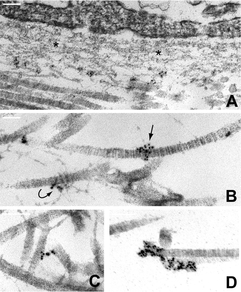
Kidney. The gold particles marking the ultrastructural localization of type XV collagen were concentrated beneath the endothelial BM (Figure 2A) and the epithelial BM of the glomerular capsule (Figure 2B). The BM proper of the glomerulus, vascular endothelium and epithelium was essentially devoid of particles except for an occasional few at the interface with the underlying stroma. Most type XV was found in association with thick-banded collagen fibers of ∼35+ nm in diameter, immediately adjacent and/or inserting into the BM. The type XV particles occurred in clusters or linear arrays; they spanned the width of a fiber or fibers, extended laterally, and formed a bridge between two or more fibers.
Figure 3 is a higher magnification showing typical type XV-fiber arrangements where the gold particles directly overlay the surface of banded fibers. In Figure 3C, type XV particles follow the branched outline of fine fibrils, in contact with one thick fiber and terminating near another. In Figure 3D, one group of gold particles was present at the intersection of a banded fiber and thin fibril; another group was situated between a second banded fiber and the BM. But the most prominent and defined structure was an alignment of type XV grains linking banded fibers. Representative images chosen from numerous examples are portrayed in Figures 3A and 3B, 3E-3J. In Figure 3G, the bridge of type XV gold particles spans at least five fibers. In control sections incubated with normal rabbit serum or buffer in place of the primary antibody, only a few scattered gold particles were observed with no specific distribution.
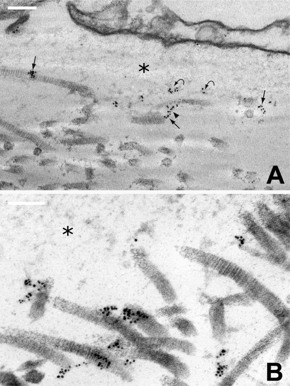
Ultrastructural localization of type XV collagen in human kidney.
Placenta. Ultrastructural localization of type XV collagen in placenta was consistent with the kidney results, and the same distribution pattern was observed using type XV antibodies raised against the carboxy-or amino-terminal domains (Myers et al. 1996,1997; Amenta et al. 2000; Li et al. 2000). Type XV was most prevalent within the stroma immediately adjacent to either the trophoblastic (Figure 4A) or endothelial BM (data not shown). The BM was, for the most part, negative except for some signal at the fibrous interface where typical banded fibers insert. Gold particles were associated with thick-banded fibers (Figures 4A-4D). In Figure 4B, type XV is seen at the junction between two banded fibers, and additional gold particles are located on the surface of adjacent fibers. In Figure 4D, a dense array of type XV particles bridge two thick fibers; others appeared directly on the fiber surface where a fine fibril intersects.
Colon. Type XV collagen was found in the outermost part of the colonic epithelial cell BM and in association with neighboring banded fibers (Figures 5A-5D). In Figure 5B, type XV decorates a fine fibrillar network adjacent to a collagen fiber. A type XV bridge extending from the midpoint of one fiber to the end of another (Figure 5C) is similar to those depicted above for kidney and placenta, and in Figure 5D gold particles overlay two contiguous fibrils.
Glycosaminoglycan Chain Modification of Type XV Collagen In Vivo
Type XV collagen has been characterized as a proteoglycan in several human tissues (Li et al 2000; Amenta et al 2003). By immunoblotting, the 250/225 kD core protein chain was identified by chondroitinase but not heparitinase digestion of extracted proteins. For the first time, kidney was analyzed to determine if type XV in this tissue was also conjugated to GAG chains. Chondroitinase digestion of extracted proteins followed by immunoblotting revealed the type XV core protein (Figure 6A, lane 2). But extended autoradiography of the kidney blot showed a significant amount of diffuse reactivity above the bands (data not shown), prompting us to ask if type XV could also be glycanated with HS chains. Successive digestions with chondroitinase and then heparitinase eliminated the undigested material and resulted in a greatly intensified type XV signal (Figure 6A, lane 3). Heparitinase digestion alone did not reveal the sharp 250/225 kD bands but did convert most of the high molecular weight smear to fuzzy bands above the doublet (Figure 6A, lane 4). These observations caused reassessment of type XV placenta and colon forms, since double digestions had not previously been carried out (Li et al. 2000). Successive incubations (Methods) with both glycosidases (Figures 6B and 6C, lanes 3), compared with chondroitinase alone (Figures 6B and 6C, lanes 2), revealed an increase in the signal of the respective type XV core protein, but much less than the differences seen in kidney. Heparitinase samples (lanes 4 of Figures 6A-6C) compared with untreated samples (lanes 1 of Figures 6A-6C) showed a stronger signal for the type XV smear, due to removal of the HS chains and better transfer of the lower molecular weight collagen XV-CSPG from the gel to the membrane. The ratio of the bands, generated by double digestion (Figures 6A-6C, lanes 3) compared with chondroitinase alone (Figures 6A-6C, lanes 2) was 8.8, 1.6 and 1.5 for kidney, placenta and colon, respectively.
Discussion
The data reported here establish the subjacent BM localization of type XV collagen and provide new insight into its biochemical properties. While invaluable information was obtained from former light microscopy analysis (Myers et al. 1996,1997; Hagg et al. 1997; Tomono et al. 2002), the limitations of this method underscored the need for definitive resolution provided by immunoelectron microscopy. In retrospect, some prior information—discussed below—hinted that type XV was unlikely to be an integral BM component, but its close association with banded fibers was no less than surprising. Type XV has been categorized as a “multiplexin” together with its homolog type XVIII collagen (Oh et al. 1994), rather than a member of the “FACIT” (fibril-associated collagen with interrupted triple-helices) subclass, which shares some common features in primary structure and in a few types have been established as collagen fiber/fibril-associated (Ricard-Blum et al. 2000).
The predominant localization of type XV in kidney, placenta and colon was consistently in the collagen network of fibers just distal to epithelial and endothelial BMs. The occasional gold particles within the outermost aspect of the BM may reflect a real type XV presence or original sites of contact between the BM and focally inserted fibers coated with type XV. In all three tissues, gold particles were identified in linear arrays and in clusters along the surface of banded ∼35+ nm diameter collagen fibers and in some cases, also thinner 10-15 nm fibrils. Type XV particles projecting laterally from one fiber were usually in close apposition to another. Organized type XV bridges linking fibers were striking in appearance and a common finding. These ∼30- to 90-nm-length formations may be a direct consequence of type XV assembly or another matrix protein(s) and visualized here because of the type XV association. Future knowledge of the type XV supramolecular conformation may help to clarify these questions. Use of both type XV amino- and carboxy-domain antibodies showed no difference in orientation of the gold particles. Notably, partial purification of type XV under non-denaturing vs denaturing conditions revealed approximately three times less protein was extracted in the neutral salt buffer, supporting an intricate association of this molecule in the fibrillar collagen matrix (Li et al. 2000 and unpublished data).
Type XV Has a Distinctly Different Distribution from Its Homolog, Type XVIII Collagen
This report helps to resolve the long-standing confusion of the relationship between types XV and XVIII collagen. The homology in primary structure, and their localization to BM zones—albeit different in some tissues especially liver—caused much speculation that these two collagens may have similar roles (Myers et al. 1996,1997; Hagg et al. 1997; Saarela et al. 1998). An additional parallel was drawn when they were discovered to be full time PGs (Halfter et al 1998; Li et al. 2000), a unique feature in the collagen family. But an important distinction was the nature of the GAG side chains—HS for type XVIII, vs CS for type XV or a mixture of both chains. Genetic data first provided evidence that the two collagens evolved to assume different functions. Null mice for the respective genes exhibit a very different phenotype and double deficient mice demonstrated the absence of biological compensation (Eklund et al. 2001; Fukai et al. 2002; Ylikarppa et al. 2003). The type XVIII −/- mice have ocular abnormalities (consistent with human mutations, Sertie et al. 2000) due to abnormal retinal pigment epithelia, whereas type XV −/- mice have stress-induced muscle and vascular defects and morphologically normal BM.
The ultrastructural analysis puts much of the data into perspective. Type XVIII collagen localizes in epidermal and vascular BM (Marneros et al. 2004), in accord with other HSPGs that are found in BM and in close proximity to the cell surface (Iozzo 2000). The presence of type XV peripheral to BM and its association with collagen fibers/fibrils reflects primary glycanation with CS chains, since CSPGs are usually located in the stroma and have in a number of instances been shown to bind to fibrillar collagens (Iozzo 1999; Ezura et al. 2000). The distinct distribution also suggests that a putative type XV carboxy cleavage fragment, analogous to the type XVIII BM endostatin (O'Reilly et al. 1997; Sasaki et al. 2000) may have its own special function and a non-BM distribution like its parent molecule.
Type XV May Help to Protect Collagen Fibers from Proteolysis
The localization of type XV collagen clarifies former light microscopy studies, which showed its distribution in colon and breast carcinomas to be significantly different from integral BM components (Amenta et al. 2000,2003). Type XV was lost from the BM zone early in the invasive process, before fragmentation of the BM as evidenced by linear immunoreactivity for type IV collagen and laminin. This result suggested type XV was more sensitive to proteolysis, and/or was peripheral to the BM and exposed to a stromal environment where matrix metalloproteinases are upregulated in response to soluble factors released by tumor cells (Shapiro 1998). One can now hypothesize that type XV may represent the first line of defense against matrix proteolysis and initially serve to protect type I/III/V collagen fibers from degradation, analogous to the aggregan-type II collagen relationship (Pratta et al. 2003). Once the type XV structure is compromised, it may signal disruption first of the fibrillar collagen network and then of the collagenous-BM interface to facilitate tumor growth and cell migration. Additionally, type XV protein core fragments may possess biological activity, as well as the liberated GAG chain fragments, depending on the CS or HS content. Better understanding of the type XV function in normal tissue could well emerge from ultrastructural study of the type XV-fiber linkages at the in situ stage of tumor progression.
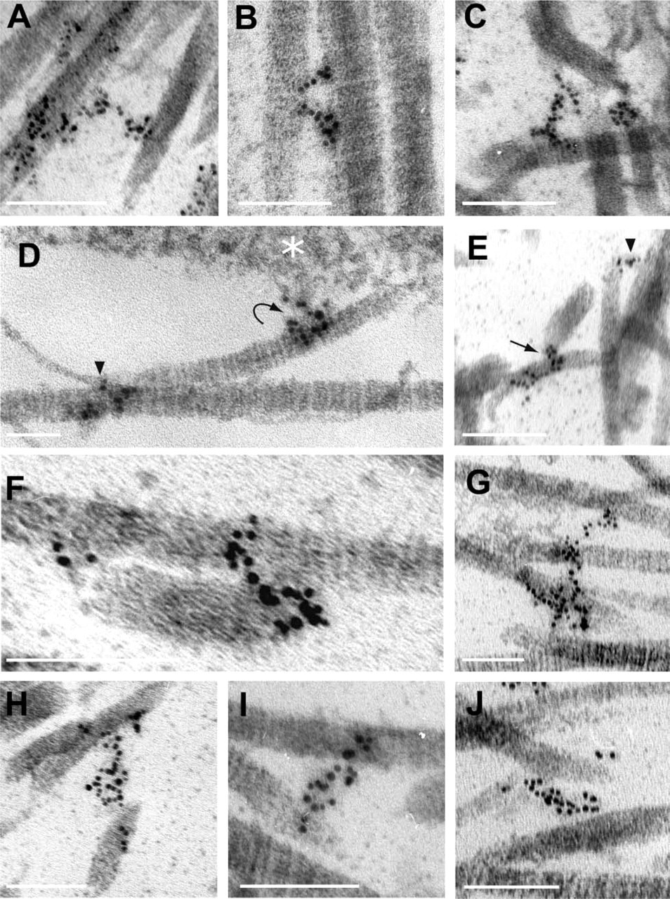
Representative illustrations of type XV association with banded fibers in kidney.
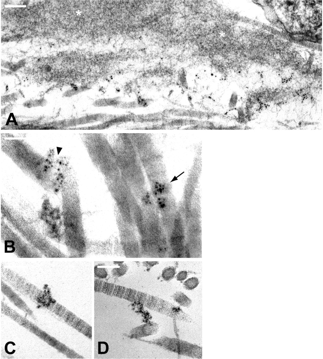
Type XV ultrastructural localization in placenta. (
Ultrastructural localization of type XV collagen in colon. (
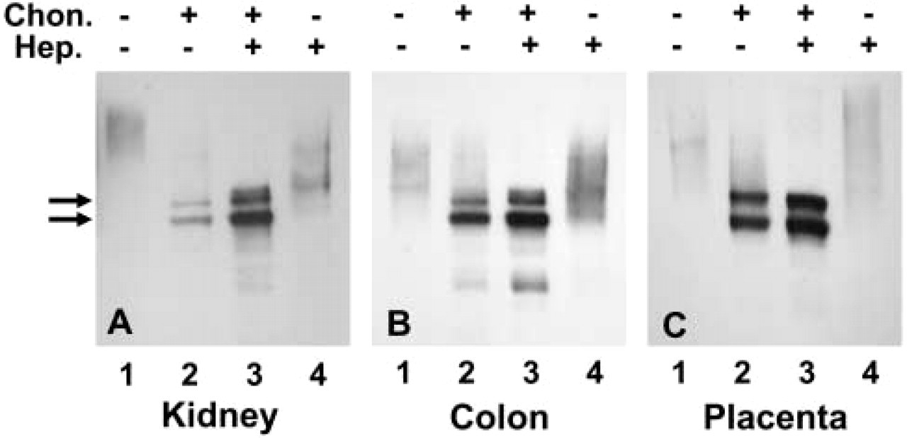
Type XV collagen in vivo is a CSPG and a CS/HSPG hybrid. Extracted proteins from human kidney (44 μg), colon (28 μg), and placenta (15 μg) were incubated in the presence (+) or absence (−) of chondroitinase and/or heparitinase as indicated in the figure and detailed in Methods. Protein extracts in lanes 1 of each panel were incubated in the respective enzyme buffers but without (−) any enzyme. After incubations, all samples were electrophoresed on an 8% SDS-polyacrylamide gel, immunoblotted, and reacted with the type XV carboxy antibody (Methods). Bands were visualized using ECL immunodetection reagents. The exposure times were as follows: (
Type XV Displays Features Intermediate between Interstitial and BM Collagen Proteoglycans
Five collagens, IX, XII, XIV, XV and XVIII, are recognized as PGs. The first three are part-time CSPGs (Ricard-Blum et al. 2000); type XV is a CSPG or CS/HSPG hybrid (Li et al. 2000, and Figure 6) and type XVIII in vivo is a HSPG (Halfter et al. 1998) although in vitro it is decorated with both HS and CS chains (Dong et al. 2003). Type IX is covalently linked to type II collagen fibers in cartilage (van der Rest and Mayne 1988), types XII and XIV are associated with type I collagen bundles (van der Rest and Dublet 1996; Keene et al. 1991), and type XVIII is found in BM (Marneros et al. 2004). Type XV exhibits properties of both groups, seeming to reflect its “intermediate” localization. Like types IX, XII and XIV, type XV is primarily associated with large banded collagen fibers, exists as a CSPG, and is present in the stroma. Like type XVIII, type XV is a full-time PG, can harbor HS side chains, and is present in the BM zone, but probably not BM per se. These features further emphasize the importance of CS chains in the interaction with collagen fibers and the importance of HS chains in BM/zone structure and biological activity.
Type XV Collagen Is Extensively Glycanated with Heparan Sulfate as well as Chondroitin Sulfate Chains
Whereas most HSPGs are known to carry CS chains (Iozzo 2000), to our knowledge the converse is not typical. Type XV is a CSPG that carries HS chains (Li et al. 2000, Figure 6, and unpublished data); moreover, the ratio varies depending on the tissue. There are eight potential sites for GAG chain attachment in the type XV amino-terminus and three in the first interruption (Li et al. 2000 and Figure 1), but in vivo identification of occupancy and GAG chain characterization will likely be precluded by the paucity of type XV in tissues. We had estimated that glycosylation added a minimum of 200-kD mass to each individual 225/250 kD type XV chain (Li et al. 2000). Compared with placenta and colon, the glycosylated kidney type XV chain is even larger than 400 kD and contains considerably more HS than CS chains. As with other PGs, the significance of type XV GAG forms, qualitatively and quantitatively, will be difficult to grasp until at least some model system is established. One would expect that the different forms are synthesized by different cell types recognized by different ligands and modulate different activities, as shown for two transmembrane syndecan-HSPGs that are also modified by CS chains (Carey 1997; Yoneda and Couchman 2003). It has recently been reported in fact that CS chains in syndecans-1 and −4 can “cooperate” with HS chains to bind growth factors for “delivery to the cell surface receptors” (Deepa et al. 2004).
Concluding Remarks
Type XV joins the three well-known non-fibrillar collagen-CSPGs closely associated with collagen fibers. Type IX is thought to mediate interactions between collagen fibrils and the surrounding matrix, type XII has a presumed role in development of stromal architecture and maintenance of fibril organization, and type XIV is believed to help regulate fibrillogenesis (Bateman 2001; Young et al. 2002). These still general statements could extend to type XV and it may be meaningful that the type XV immunogold profile looks similar to that of type XIV in human tendon (Keene et al. 1991). However, only type XV is associated with a restricted collagenous network subjacent to BM and a loose rather than dense array of large collagen fibrils (likely types I, III). There were occasions when smaller fibrils (possibly collagen VI, fibrillin, etc., Keene et al. 1997,1998) were also seen in close proximity to type XV, especially in the portal area of the liver (data not shown). Future determination of their composition will help define interactions between the BM and the heterogeneous underlying stroma (Adachi et al. 1997). Furthermore, the shift in CS to CS/HS modification of the type XV core protein may fundamentally change the properties of the molecule and of the matrix, not only among different tissues but also in epithelial/vascular/muscle BM zones of the same tissue. For example, type XV is strikingly absent from the glomerular capillaries, renal tubules and hepatic/splenic sinusoids where a reduced anionic charge in the environment enhances the filtering exchange (Kanwar et al. 1980; Barnes et al. 1984). This suggests type XV may also be important in maintaining a hydrated matrix that facilitates cell migration and in selectively modulating the transfer of soluble factors.
Type XV is clearly a multifunctional collagen-PG with different characteristics than originally believed. Our focus here has been on specific aspects of this novel molecule, which point already to its involvement in many biological processes and give a validated direction for future studies.
Footnotes
Acknowledgements
These studies were supported by NIH Grants AR-44549 and GM-64777.
We would like to thank Kelly Walton for excellent technical assistance, Rajesh Patel for extensive effort in the electron microscopy photography, and Mary Leonard for valuable help with the illustrations. We are very grateful to Dr David Birk at Thomas Jefferson University for advice on the immunogold procedures.