
Abstract
Kidneys from lambs derived by nuclear transfer are frequently abnormal and are characterized by an enlarged pelvis and narrow medulla, consistent with lower urinary tract obstruction and development of variable hydronephrosis. The precise pathogenesis of this entity is unknown. Immunohistochemical staining for intermediate filaments was used to further characterize the lesions seen in this condition and was compared with age-matched control tissue. Major findings were upregulation of cytokeratin on damaged tubules, desmin and vimentin in undifferentiated mesenchyme, and smooth muscle actin in mesenchyme and on smooth muscle “collars” around dilated tubules. In addition, some cases showed reexpression of vimentin and desmin on proximal tubular epithelial cells. Taken together, these findings provide a valuable database for tracking the expression of intermediate filaments throughout renal development in sheep and have further characterized the nature of the response to injury by the developing kidney, a response that is characterized by proliferation of mesenchyme and both reexpression and upregulation of intermediate filaments within renal cells. In addition, the study has confirmed that the changes in cloned lamb nephropathy are established by day 85 of development.
I
During nephrogenesis, there is a tightly regulated developmental process involving reciprocal induction of epithelial and mesenchymal elements in the developing kidney. Studies on developing human nephrons have identified site-specific expression of vimentin and cytokeratin (CK) intermediate filaments, with recognizable alterations occurring as the nephrons mature into functional adult tissue (Moll et al. 1991). In lambs, ligation of the ureter at day 70 of gestation has been shown to cause marked parenchymal and architectural disorganization (Matsell et al. 1996). Smooth muscle actin (SMA) expression was recorded in the abnormally distributed smooth muscle and fibromuscular collars of cysts and dilated collecting ducts. In addition, CK expression was identified in the epithelia of tubular epithelial structures and most cysts.
In this study we sought to further characterize the nephropathy that occurs in cloned lambs by investigating any alterations in the mesenchymal-to-epithelial transition associated with the entity. Further, the study included as a prerequisite a detailed immunohistochemical (IHC) characterization of normal developing fetal sheep kidneys. Tissue microarrays were constructed to optimize the limited amount of tissue available in some cases, to provide direct comparisons with control tissue, and to preserve reagents. The expression pattern of small and intermediate filaments (SMA, vimentin, desmin, and CK) in a series of control sheep fetuses was compared with a series of kidneys from cloned lambs with nephropathy. Together, these findings provide a valuable database for tracking the expression of intermediate filaments throughout the developmental process and further define the histological and IHC features of cloned lamb nephropathy (CLN).
Materials and Methods
Animals and Sampling
Control material was obtained from fetuses at days 70, 84, 98, 112, 126, 140, and 146 of gestation. Kidneys were removed and fixed in 10% neutral buffered formalin. Material from cloned lambs was obtained as soon as possible post mortem and fixed in 10% neutral buffered formalin as described previously (Denning et al. 2001; Rhind et al. 2003). This study was conducted following approval by Roslin Institute's Animal Ethics Committee and within a project license issued under the Animal (Scientific Procedures) Act 1986.
Tissues and Processing
Tissue samples were processed routinely, dehydrated through graded ethanol, cleared in xylene, and impregnated with Histoplast paraffin (Thermo Shandon; Pittsburgh, PA). Tissue was embedded in fresh Histoplast paraffin before cutting. For initial histopathology and IHC, tissue samples were cut at 4 μm and stained with hematoxylin and eosin (HE). Sections were picked up onto either standard double-frosted microscope slides for HE staining or onto Biobond-coated microscope slides (British Biocell; Cardiff, UK) for IHC studies. Sections were allowed to dry overnight at 37C before IHC staining.
Tissue Microarrays
To aid direct comparison between control samples and clones and to optimize the use of small tissue samples and reagents, tissue microarrays were constructed using the initial HE-stained slide as a guide to select the regions for sampling. The tissue microarrays were prepared on a manual tissue arrayer (Beecher Instruments; Sun Prairie, TX) using 2.0-mm punches.
Constructing the Array
Recipient blocks were molded using paraffin, and holes were created in the empty recipient block using a punch with the depth adjusted to 1 mm above the plastic block holder. The first hole initiated the arraying process with the micrometer set to zero at this point. The punch was pushed downward by hand until the depth stop blocked the downward motion. At this point, the handle in the punch was rotated ~45° to free the paraffin core from the block. The HE slide was aligned over the corresponding tissue block, and the punch was pushed down to retrieve the sample, which was then placed in the hole created by the smaller punch.
The micrometers were adjusted to move the precision guide to the next X-Y position and repeat this cycle to construct the whole array.
Sectioning the Array Block
Before sectioning, the array block surface was smoothed and leveled. The array was removed from the recipient block holder and placed in a warm incubator (37C) for 30 min to promote adherence of the tissue biopsies to the walls of the holes in the array block. A glass microscope slide was then used to level the wax surface, and even pressure was applied to push all tissue cores on the array to the same level. Blocks were then sectioned using standard microtome sectioning techniques.
Immunohistochemistry
The following antibodies and antigen unmasking were used for IHC. SMA: mouse anti-SMA (Sigma Aldrich; Poole, UK) diluted 1:1000 in 0.1 M phosphate-buffered saline (PBS), pH 7.4, for 30 min at room temperature; desmin: mouse anti-desmin (Dakocytomation; Ely, UK) diluted 1:50 in 0.1 M PBS (pH 7.4) for 30 min at room temperature following antigen retrieval using 0.01 M citrate buffer (pH 6.0) in a pressure cooker, using a microwave oven as the heat source for 15 min at 800 W followed by 15 min at 300 W; vimentin: mouse anti-vimentin (Novocastra Laboratories; Newcastle Upon Tyne, UK) diluted 1:50 in 0.1 M PBS (pH 7.4) for 30 min at room temperature following antigen retrieval using 0.01 M citrate buffer (pH 6.0) in a pressure cooker in a microwave oven for 15 min at 800 W followed by 15 min at 300 W; pan-CK: mouse anti-pan CK (Sigma Aldrich) diluted 1:100 in 0.1 M PBS (pH 7.4) for 30 min at room temperature following antigen retrieval using 0.01 M citrate buffer (pH 6.0) in a pressure cooker in a microwave oven for 15 min at 800 W followed by 15 min at 300 W; CK 7: mouse anti-CK 7 (Dakocytomation) prediluted for 30 min at room temperature following antigen retrieval using 0.01 M citrate buffer (pH 6.0) in a pressure cooker in a microwave oven for 15 min at 800 W followed by 15 min at 300 W; CK 18: mouse anti-CK 18 (Abcam; Cambridge, UK) diluted 1:50 in 0.1 M PBS (pH 7.4) for 30 min at room temperature following antigen retrieval using 0.01 M citrate buffer (pH 6.0) in a pressure cooker in a microwave oven for 15 min at 800 W followed by 15 min at 300 W.
Assessment of Histopathology and IHC Staining
Initial examination of HE sections prior to construction of the tissue microarray was used for histopathological assessment of lesions and for identification of affected areas prior to selection of affected areas for production of the tissue microarray.
The histopathological features were recorded on a semiquantitative scale based on the presence of tubular dilations and the presence of mesenchymal tissue. For tubular dilations, the average number of dilations per high-power (×400) field in the cortex was recorded as 0 (average <1), 1 (average 1-3 inclusive), or 2 (average >3). Abnormal mesenchyme was recorded as present (+) or absent (−).
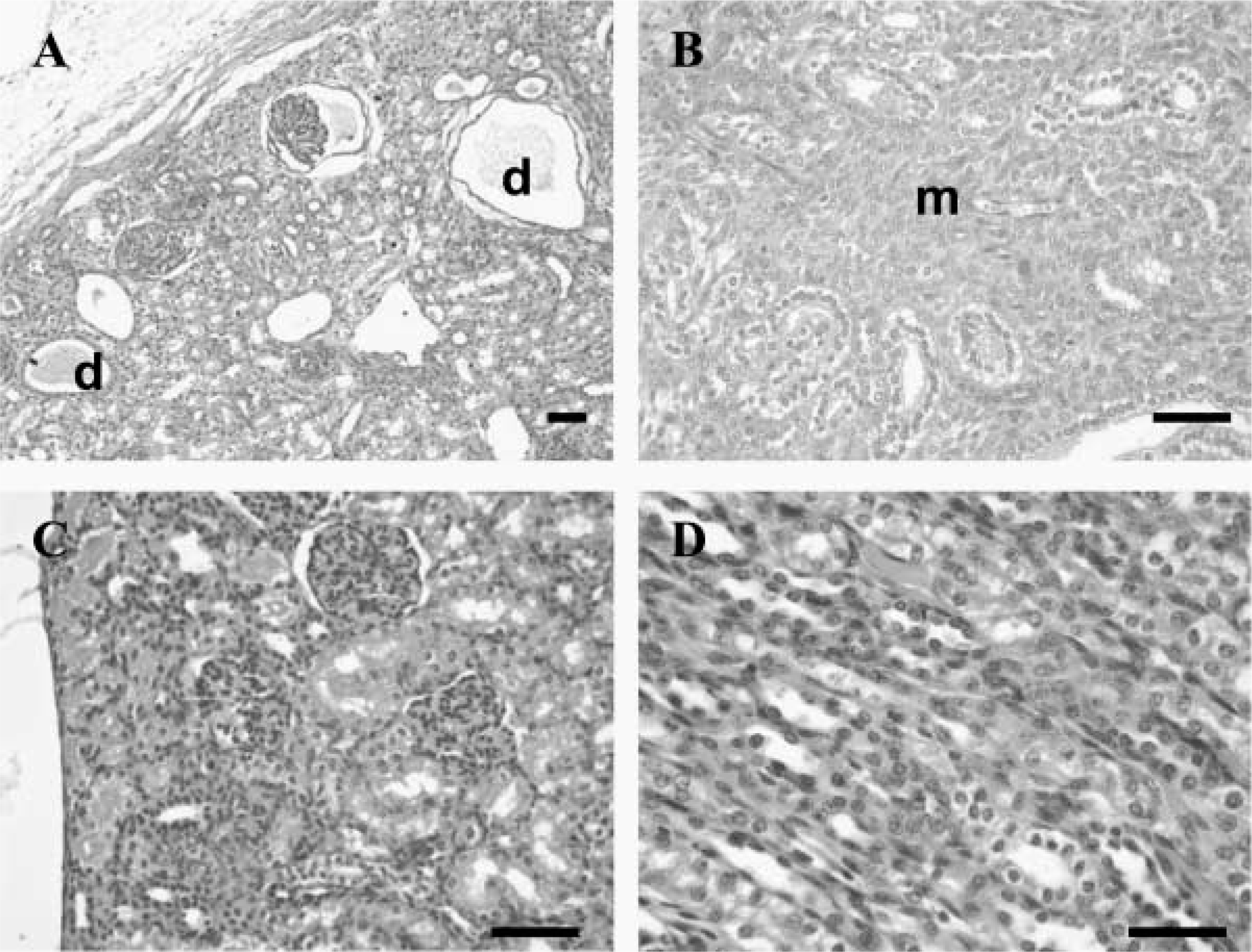
Histological features of cloned lamb nephropathy (CLN) (
Patterns of IHC staining in association with lesions were defined anatomically and assessed as being either diffuse cytoplasmic or punctate.
Results
The typical gross appearance of affected kidneys has been described previously (Rhind et al. 2003). In addition, one case showed macroscopically visible cysts in association with the architectural disruption. With the exception of this case, the macroscopic changes in the kidneys were uniform, with no evidence of inflammatory changes, obvious scarring, or fibrosis.
The typical histological lesions seen in CLN are shown in Figures 1A and 1B in comparison to age-matched control tissue from the same anatomical location (Figures 1C and 1D). There is narrowing of the medulla with dilation of collecting ducts and tubules (Figure 1A). More-severe lesions are characterized by an increase in the amount of mesenchyme surrounding tubular elements in the medulla (Figure 1B).
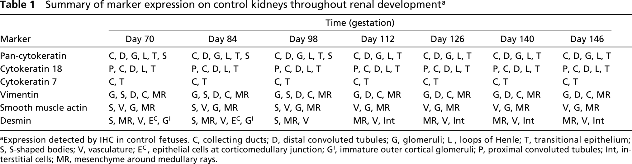
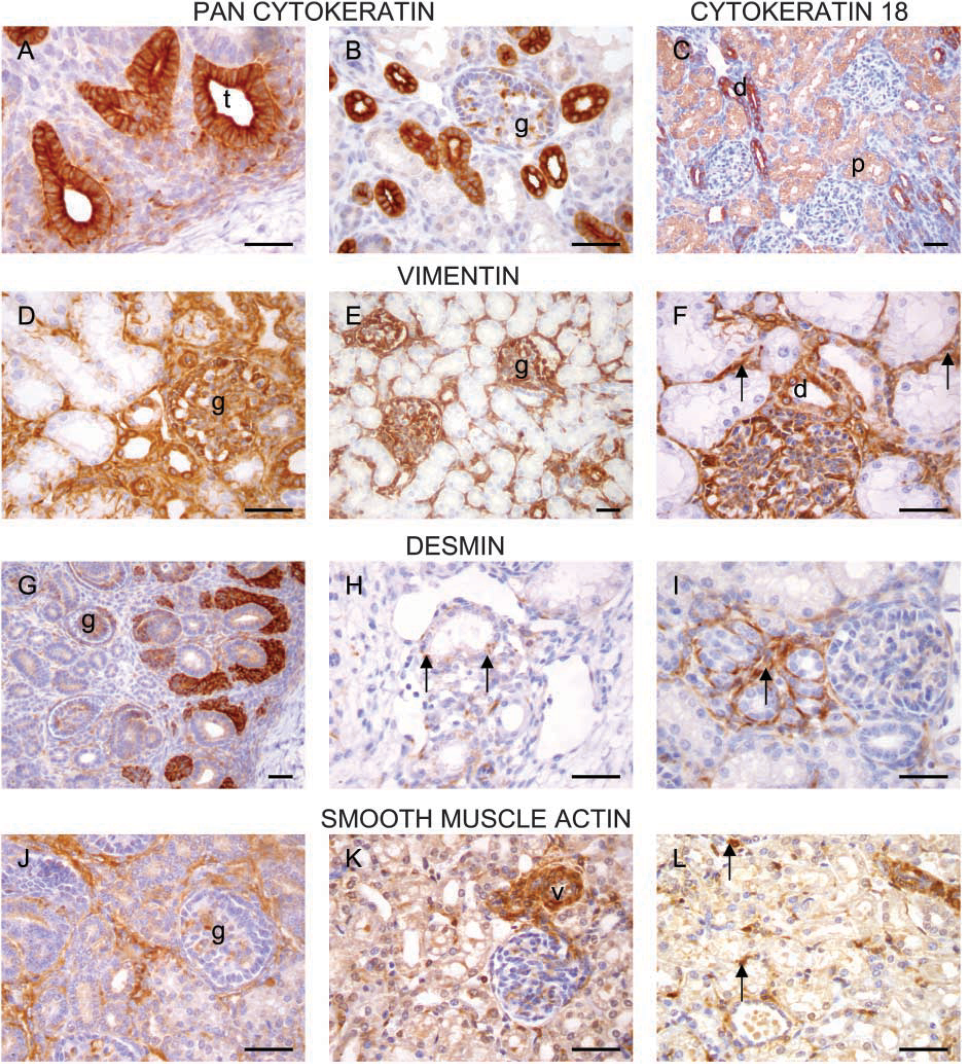
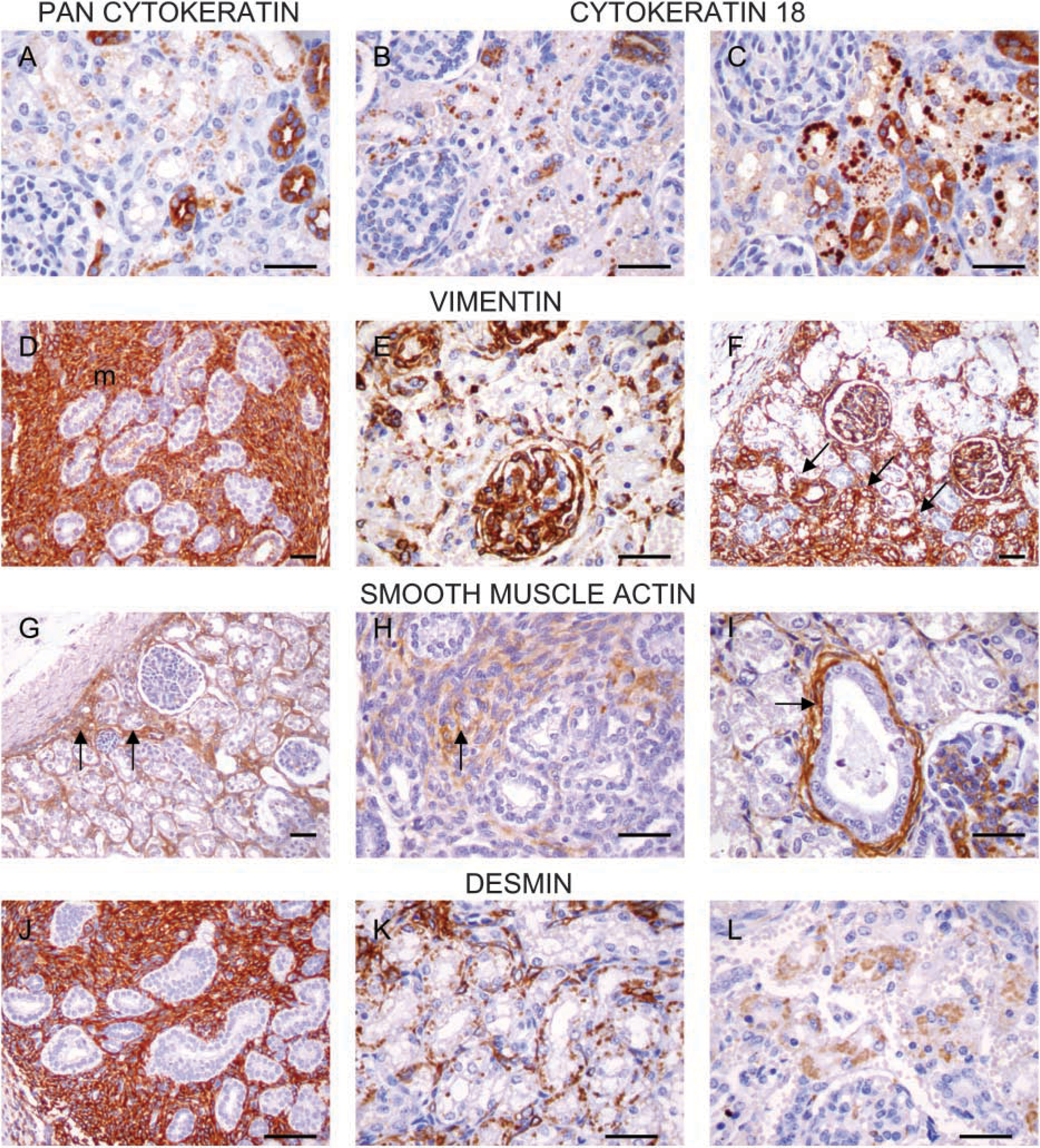
The results of the temporal study of intermediate filament expression in normal developing lamb kidneys are summarized in Table 1. Notable features are the consistent pattern of CK expression, with staining of cells of the developing glomeruli, distal convoluted tubules (DCTs) and collecting ducts (CDs) (Figures 2A and 2B), glomerular epithelium, loops of Henle, and transitional epithelium. The pan-CK reagent did not stain the cells of the proximal convoluted tubule (PCT). Therefore, a specific CK 18 antibody was used to stain these cells (Figure 2C).
Vimentin staining was evident in developing glomeruli (Figure 2D), mature glomeruli (Figure 2E), vessels, interstitial mesenchyme, and in some CDs and DCTs (Figure 2F).
Desmin expression was similarly observed in developing glomeruli (Figure 2G) and those in the outer cortex, but not in maturing glomeruli distant from the nephrogenic zone. Expression was also present in scattered epithelial cells at the corticomedullary junction up to day 84 (Figure 2H). In addition, staining of focal areas of interstitium was observed (Figure 2I).
Summary of marker expression on control kidneys throughout renal development a
aExpression detected by IHC in control fetuses. C, collecting ducts; D, distal convoluted tubules; G, glomeruli; L, loops of Henle; T, transitional epithelium; S, S-shaped bodies; V, vasculature; Ec, epithelial cells at corticomedullary junction; G1, immature outer cortical glomeruli; P, proximal convoluted tubules; Int, interstitial cells; MR, mesenchyme around medullary rays.
IHC on control renal tissue. Cytokeratin (CK) staining (
SMA expression was present in developing and mature glomeruli (Figures 2J and 2K), blood vessels (Figure 2K), and interstitial cells (Figure 2L).
In contrast, several striking alterations in intermediate filament expression were seen in the lesions from cloned lambs with nephropathy (summarized in Table 2). CK upregulation (identified using both pan-CK and CK 18) was evident in kidneys from lambs of 148-153 days (Figures 3A-3C). The pattern of this expression was notably punctate, in contrast to the usual uniform cytoplasmic staining observed in control sections (Figures 2A-2C).
Alterations in vimentin expression were also observed and were characterized by increased expression in the interstitium (Figure 3D, day 153) and also by reexpression on epithelial cells in four cases (Figures 3E and 3F; days 148 and 146, respectively).
SMA expression was increased in all cases with nephropathy. This was evident in association with prominent vasculature and in the interstitium (Figures 3G and 3H; days 118 and 153, respectively). In addition, SMA-positive muscular collars were present around some of the dilated tubules (Figure 3I; day 148).
Although these muscular collars were desmin-negative, desmin expression was both increased in the medullary interstitium (Figure 3J; day 153) and expressed on tubular epithelium in four cases (Figures 3K and 3L; day 148).
Discussion
Intermediate filament expression is tightly regulated in normal developing kidneys as the metanephric mesenchyme causes the ureteric bud to elongate and branch, while the tips of the branching ureteric bud induce the loose mesenchymal cells to form epithelial aggregates. There is therefore a tightly regulated developmental process involving reciprocal induction of epithelial and mesenchymal elements.
Cloned lambs frequently exhibit abnormal kidney development, which has features consistent with the presence of a degree of lower urinary tract obstruction. This results in architectural distortion of the developing kidney and interferes with the process of nephrogenesis described above. It is notable, however, that none of the kidneys contained evidence of any immature cartilage, as is often seen in human kidneys with cystic renal dysplasia.
This study was carried out to investigate both histological and IHC features of the entity in comparison with age-matched control tissues.
A panel of antibodies was used to investigate expression of cytokeratins, vimentin, desmin, and SMA. The pan-CK reagent was selected on the basis of presumed reactivity with all epithelial elements in the kidney. However, cells of the PCT did not stain with this antibody in control sections (Figure 2B). Therefore, a specific cytokeratin 18 antibody was used to demonstrate PCT epithelial cells (Figure 2C). CK 7 was used as a marker for transitional epithelium and collecting ducts.
The results from the study of control tissue show vimentin, cytokeratin, desmin, and SMA coexpression on the primitive glomerular structures. As the kidneys develop, the mutually exclusive nature of mesenchymal and epithelial staining patterns becomes more apparent. Notably, however, vimentin expression is evident in DCTs (Figure 2F) and in some collecting ducts but not in PCTs. In addition, desmin expression is evident in occasional tubules at the corticomedullary junction, although this feature is lost by day 98. An additional feature of desmin staining is the strong staining of cells in the interstitium that appears by day 112. It is interesting to speculate whether this population of cells represents some form of uncommitted precursor, especially given the prominent desmin expression in the increased mesenchymal tissue seen in CLN.
Histopathological and immunohistochemical features of CLN a
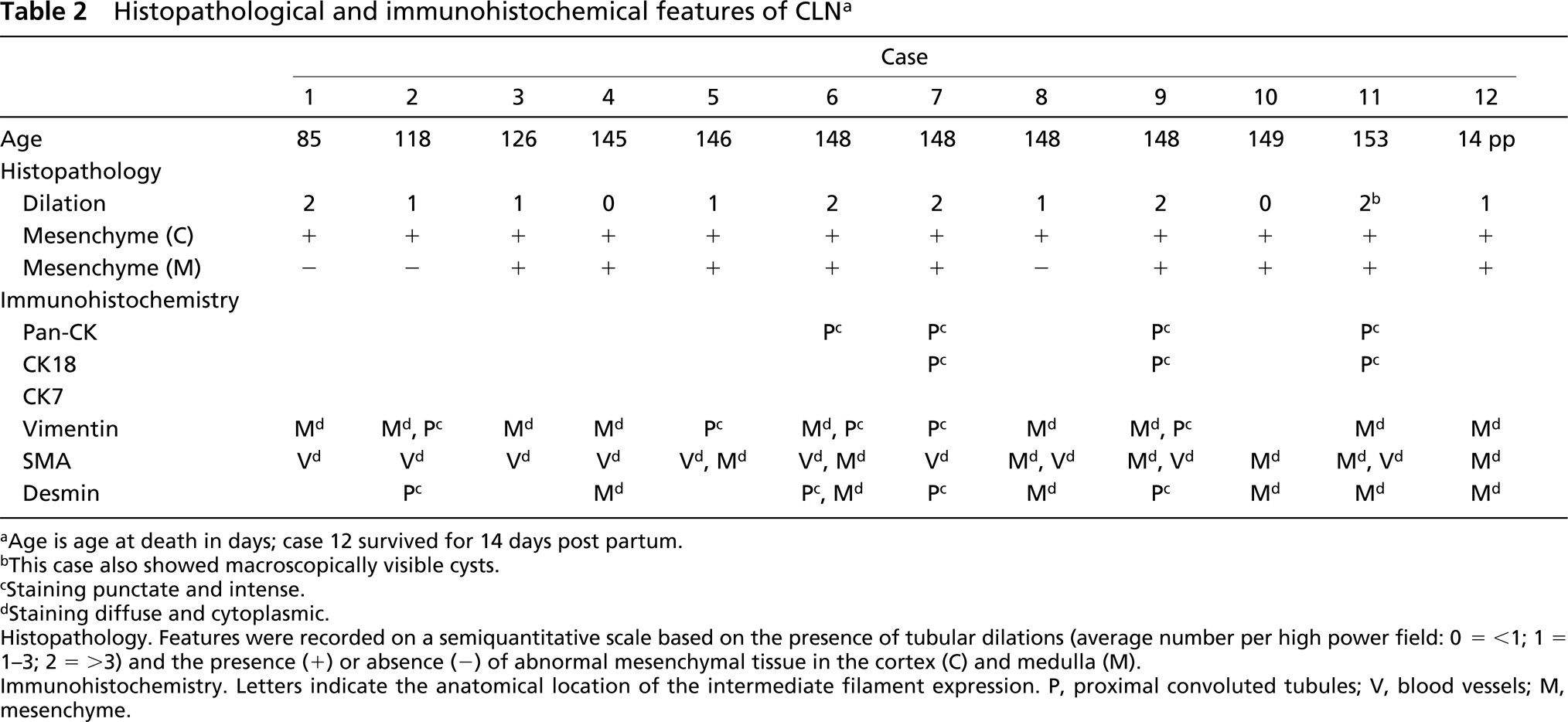
aAge is age at death in days; case 12 survived for 14 days post partum.
bThis case also showed macroscopically visible cysts.
cStaining punctate and intense.
dStaining diffuse and cytoplasmic.
Histopathology, Features were recorded on a semiquantitative scale based on the presence of tubular dilations (average number per high power field: 0 = <1; 1 = 1-3; 2 = >3) and the presence (+) or decrease (−) of abnomal mesenchymal tissue in the cortex (C) and medulla (M).
Immunohistochemistry. Letters indicate the anatomical location of the intermediate filament expression. P, proximal onvoluted tubules V, blood vessels; M, mesenchyme.
IHC features of CLN pan-CK and CK staining (
The major changes observed in the lambs with nephropathy were upregulation and reexpression of cytokeratins on proximal tubular epithelial cells in 4 of the 12 cases and reexpression of vimentin on the same cells in 4 of the 12 cases. In addition, four cases also revealed reexpression of desmin on proximal tubular epithelial cells. The second striking feature, in addition to the altered intermediate filament phenotype of the cells, was the presence of abundant and disorganized mesenchyme. This feature varied in extent from mild to severe.
That changes were seen in the youngest of the clones (day 85) is consistent with the nephropathy beginning at least before day 85. Investigation of the pathogenesis prior to this time point would require a prospective time-controlled study, because lambs dying prior to this stage are often not retrieved until a stage at which autolysis precludes meaningful histopathological analysis.
The presence of increased mesenchyme and the associated coexpression of vimentin and desmin (Figures 3D and 3J) is consistent with either a failure in the normal mesenchymal-to-epithelial transition or excessive proliferation of mesenchymal tissue. Furthermore, the reexpression of vimentin and desmin on proximal tubular epithelial cells suggests that alterations in the intermediate filament phenotype of the cells may be a response by these cells to stress. Similar changes have been reported in chronically damaged human kidneys, which have been shown to have increased or novel expression of certain cytokeratins, vimentin, and glial filament protein (Moll et al. 1991). It is additionally recognized that some renal cell carcinomas also express vimentin, and it has been considered that such vimentin expression represents a “reexpression” phenomenon that parallels a reduced degree of differentiation believed to result in greater mechanical stability of the cells (Moll et al. 1991).
In this study, the alterations in cytokeratin expression were not evident until day 148 and beyond, suggesting that this change either occurs as a consequence of more long-standing damage or is a response of more mature cells to injury. It is also notable that although there is a distinct pattern of histopathological and IHC features, the intermediate filament expression patterns are not identical among all clones and there appears to be a spectrum of alterations that appear to be linked, in most cases, to the severity of the underlying histopathological features. We hypothesize that this overall pattern reflects the severity of the presumptive functional urinary tract obstruction that underlies the genesis of these renal changes.
Previous studies in sheep investigating the changes associated with ureteral obstruction (by ligation of the ureter at day 70 of gestation) resulted in a more-severe phenotype with marked parenchymal and architectural disorganization (Matsell et al. 1996). This was characterized by an overly abundant mesenchyme and by the development of cystic structures thought to be derived from collecting ducts. Further typical features included primitive ductules surrounded by fibromuscular collars, primitive glomeruli, and abundant disorganized mesenchyme. These cortical features were present in association with an underdeveloped medulla. The study also revealed expression of SMA in the abnormally distributed smooth muscle and fibro-muscular collars of cysts and dilated collecting ducts, similar to that illustrated in Figure 3I. CK expression (using a pan-CK marker) was identified in the epithelia of tubular epithelial structures and most cysts.
In contrast to the study described above (by Matsell et al. 1996), the changes we report here associated with CLN are more subtle, presumably as a consequence of less severe urinary obstruction.
In conclusion, this study has defined the developmental patterns of intermediate filament expression in sheep kidneys. In comparing the kidneys of cloned lambs with nephropathy to these baseline data, it is clear that several striking alterations in expression patterns occur as a consequence of the nephropathy, with changes seen as early as day 85 of gestation.
Although the precise mechanisms involved in this presumptive obstructive nephropathy are currently unknown (because in the majority of cases, there is no gross evidence of obstruction in the lower urinary tract), the changes seen are consistent with some form of mechanical or functional obstruction occurring early in development, which has downstream consequences for renal development. In particular, there are alterations in the normal alignment of mesenchymal and epithelial elements. In addition, the insult causes alterations in the intermediate filament phenotype of individual cells, with reexpression and upregulation of cytokeratin, vimentin, and desmin on epithelial cells. Furthermore, in the more severely damaged kidneys there is the development of actin-positive smooth muscle collars around the dilated tubules.
In addition, this study has provided evidence for developmental plasticity in the kidney, with chronic damage resulting in alterations to the structural phenotype of individual cells.
Further characterization of the mechanisms involved in CLN will require tightly controlled prospective studies facilitating detailed analysis of the development of both the upper and the lower urinary tract.
Footnotes
Acknowledgements
Supported by funding from the Geron Corporation, California, the BBSRC, and the Department of Veterinary Pathology, R(D)SVS.