
Abstract
Aberrant mucin O-glycosylation is often observed in cancer and is characterized by the expression of immature simple mucin-type carbohydrate antigens. UDP-N-acetyl-D-galactosamine:polypeptide N-acetylgalactosaminyltransferase-6 (ppGalNAc-T6) is one of the enzymes responsible for the initial step in O-glycosylation. This study evaluated the expression of ppGalNAc-T6 in human gastric mucosa, intestinal metaplasia, and gastric carcinomas. Our results showed that ppGalNAc-T6 is expressed in normal gastric mucosa and in intestinal metaplasia. A heterogeneous expression and staining pattern for this enzyme was observed in gastric carcinomas. ppGalNAc-T6 was expressed in 79% of the cases, and its expression level was associated with the presence of venous invasion. Our results provide evidence that ppGalNAc-T6 is an IHC marker associated with venous invasion in gastric carcinoma and may contribute to the understanding of the molecular mechanisms that underlie aberrant glycosylation in gastric carcinogenesis and in gastric carcinoma.
M
In this study, we characterized the expression of ppGalNAc-T6 in normal gastric mucosa, intestinal metaplasia, and gastric carcinoma. We show that ppGalNAc-T6 is expressed in gastric mucosa and changes its expression during gastric carcinogenesis. Expression of ppGalNAc-T6 was found to be associated with a clinico-pathologic characteristic of gastric carcinoma.
Materials and Methods
Tissue Samples and Histological Classification
The study was performed using surgical specimens of gastric carcinomas and adjacent mucosa from patients operated at Hospital S. João, Porto, Portugal. The use of retrospective samples when informed consent cannot be obtained is authorized for research studies by Portuguese Law. Analysis of the expression of ppGalNAc-T6 (MAb T6.3) was performed in 76 tissue samples, fixed in 10% formalin, and embedded in paraffin. Serial sections were cut and used for conventional histo-pathological diagnosis. Carcinomas were classified according to Laurén (1965). The growth pattern was classified according to Ming (1977). Age, patient's survival, presence of lymphatic invasion, nodal metastasis and venous invasion, and tumor localization were also recorded in every case.
The pathological staging was achieved using the unified 1987 TNM system for gastric carcinoma (Pinto-De-Sousa et al. 2001). We evaluated MUC5AC, MUC6, and MUC2 mucin expression in 72, 44, and 55 cases, respectively. MUC5AC, MUC6, and MUC2 were detected using monoclonal antibodies CLH2 (Reis et al. 1997), CLH5 (Reis et al. 2000), and PMH1 (Reis et al. 1998b), respectively.
IHC
IHC evaluation of ppGalNAc-T6 was performed by the avidin-biotin-complex staining method (Hsu et al. 1981). All the formalin-fixed, paraffin-embedded samples were deparaffinated, rehydrated, and treated with 0.3% hydrogen peroxide in methanol for 30 min to block endogenous peroxidase. Sections were incubated with normal rabbit serum (DakoCytomation; Glostrup, Denmark) diluted 1:5 in PBS containing 10% of BSA for 20 min. After that, they were incubated overnight at 4C with the monoclonal antibody T6.3 for ppGalNAc-T6, diluted 1:400 in PBS containing 5% of BSA (Berois et al. 2006). The slides were washed in PBS and incubated for 30 min with biotinylated rabbit anti-mouse secondary antibody (DakoCytomation) diluted 1:200 in PBS containing 5% of BSA. Samples were washed with PBS and incubated with avidin-biotin peroxidase complex for 30 min (Vectastain Elite ABS kit; Burlingame, CA). Sections were stained with 3,3′-diaminobenzidine tetrahydrochloride (Sigma; St. Louis, MO) in a buffer containing 0.1% hydrogen peroxide, counterstained with Mayer's hematoxylin, dehydrated, and mounted. Negative controls were performed replacing primary antibody with PBS.
IHC for mucin expression and classification of intestinal metaplasia was performed as previously described (Reis et al. 2000).
Scoring of the Immunostaining and Statistical Analysis
A semiquantitative approach was used to score the immunostaining. Samples were scored as follows: low expression (<25% of positive staining cells) and high expression (>25% of positive staining cells) for IHC.
Statistical analysis was performed using the X 2 test with Yates correction using Statview 5.0 software. Fisher's test was applied whenever appropriate. Differences were considered statistically significant at p<0.05. Logistic regression was performed using Statview 5.0 software.
Results
Expression of ppGalNAc-T6 in Normal Gastric Mucosa
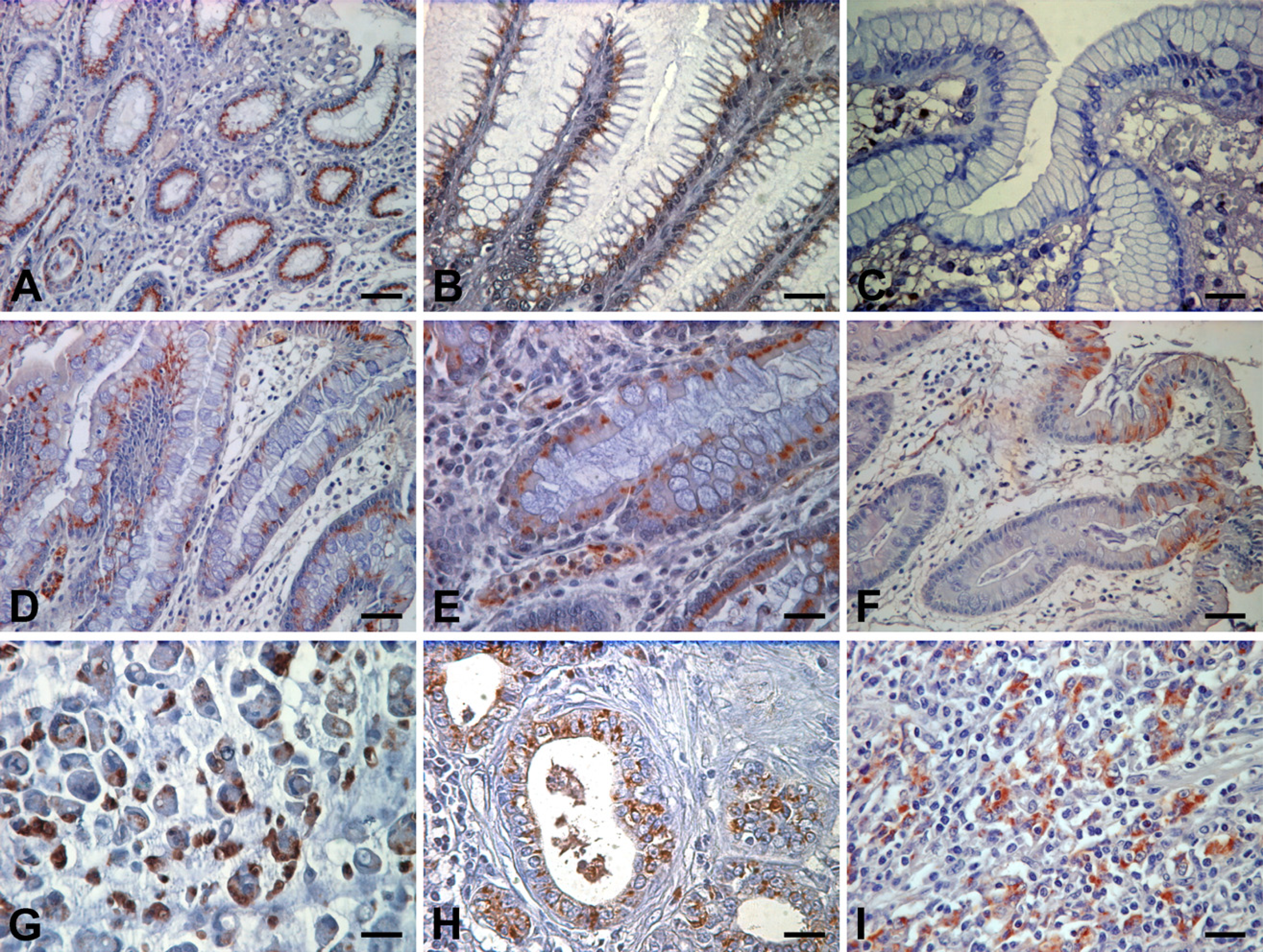
Normal gastric mucosa showed expression of ppGal-NAc-T6 in 25/36 (69%) cases. The expression was localized in the superficial foveolar epithelium and glandular cells of both antrum and body regions of the normal gastric mucosa (Figures 1A, 1B, and 2A). The staining pattern observed for ppGalNAc-T6 was always perinuclear and/or supranuclear characterizing a Golgi staining pattern.
Expression of ppGalNAc-T6 in Intestinal Metaplasia
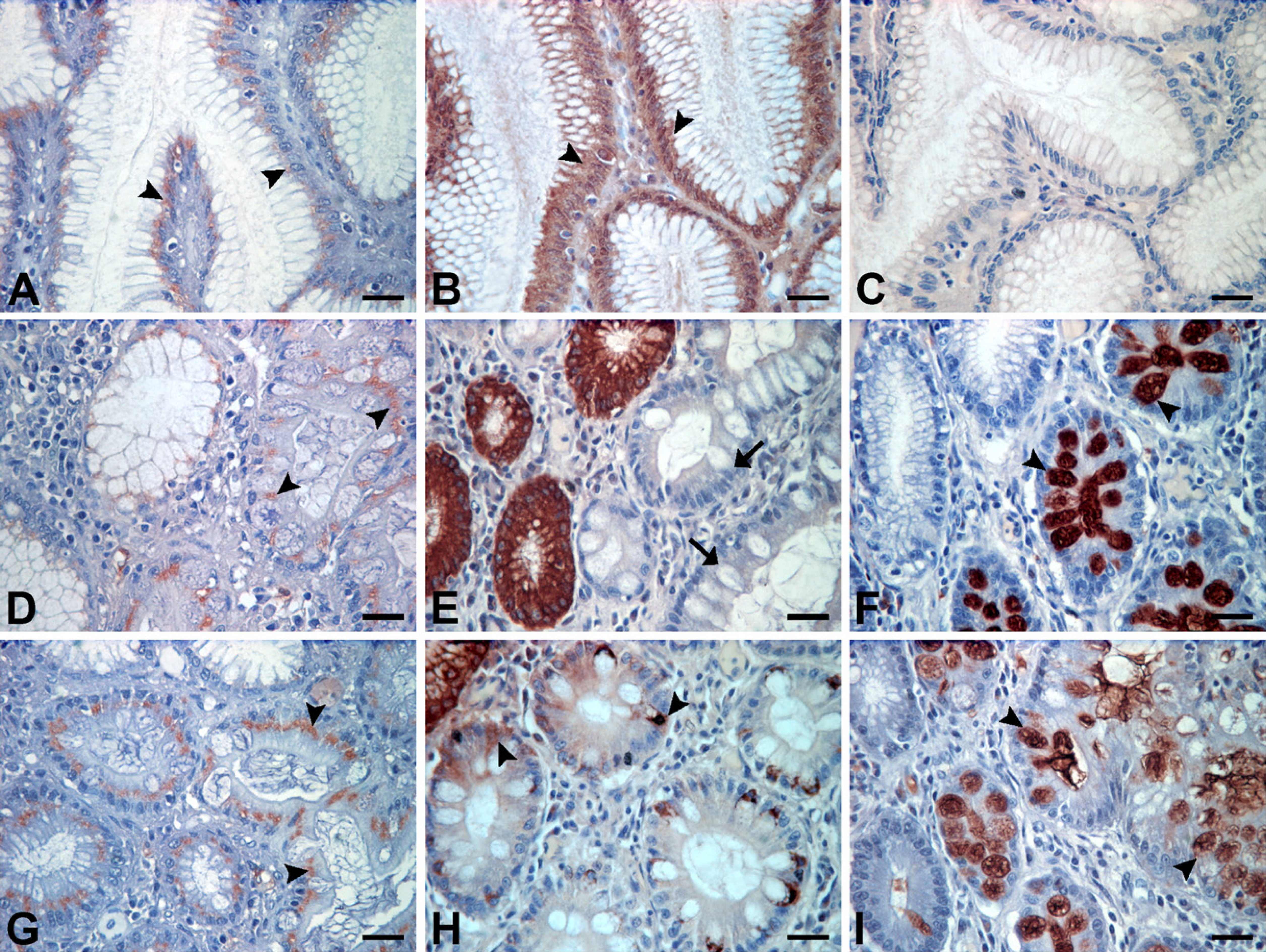
Intestinal metaplasia showed expression of ppGalNAc-T6 in 14/27 (52%) cases in both goblet and columnar cells of metaplastic glands. The staining pattern observed for ppGalNAc-T6 was always perinuclear and/or supranuclear characterizing a Golgi staining pattern (Figures 1D and 1E). Expression of ppGaNAc-T6 was observed in both complete and incomplete types of intestinal metaplasia (Figure 2).
Expression of ppGalNAc-T6 in Gastric Carcinomas
Evaluation of ppGalNAc-T6 using IHC in paraffin sections of a series of 76 gastric carcinomas showed expression of this enzyme in 60/76 (79%) of the cases. ppGalNAc-T6 expression was observed in 29/36 (81%) of intestinal type carcinomas, in 18/21 (86%) of diffuse type carcinomas, and in 13/19 (68%) of unclassified carcinomas, according to Laurén's classification (Table 1; Figures 1G and 1H). The immunostaining pattern of ppGalNAc-T6 was either perinuclear or diffuse cytoplasmic in gastric carcinoma cells.
A significant association was observed between the levels of expression of ppGalNAc-T6 and venous invasion (Table 1). Most cases with low levels of ppGal-NAc-T6 expression showed absence of venous invasion (p<0.03). In addition, analysis of contingency split by tumor localization showed that venous invasion was more associated with ppGalNAc-T6 in tumors localized in the body region of the stomach (Table 2). Further logistic regression showed no deterministic relation in which venous invasion was solely explained by ppGalNAc-T6.
IHC detection of UDP-N-acetyl-D-galactosamine:polypeptide N-acetylgalactosaminyltransferase-6 (ppGalNAc-T6) evaluated in normal gastric mucosa (
IHC of ppGalNAc-T6 and mucins MUC5AC and MUC2, in normal gastric mucosa (
The expression of ppGalNAc-T6 was not associated with other clinico-pathological characteristics of gastric carcinomas.
Coexpression of Mucins (MUC5AC, MUC6, and MUC2) and ppGalNAc-T6 in Gastric Carcinomas
Analysis of the coexpression of mucins (MUC5AC, MUC6, and MUC2) and ppGalNAc-T6 in gastric carcinoma is shown in Table 3. The expression of MUC5AC was inversely associated with the levels of expression of ppGalNAc-T6. Most cases positive for MUC5AC showed low expression of ppGalNAc-T6, whereas most cases negative for MUC5AC showed high expression of the enzyme (Table 3). No association was observed between the expression of ppGalNAc-T6 and the expression of mucins MUC6 and MUC2 (Table 3).
Discussion
Alterations in mucin-type O-glycans are associated with malignant transformation. This study evaluated the pattern of expression of ppGalNAc-T6, a key enzyme involved in O-glycan biosynthesis in normal, metaplastic, and neoplastic gastric tissues.
Our results showed that ppGalNAc-T6 was expressed both in the foveolar epithelium and glands of both antrum and body regions of the normal gastric mucosa.
Summary of data on the expression level of ppGalNAc-T6 and clinico-pathologic features in 76 gastric carcinoma cases
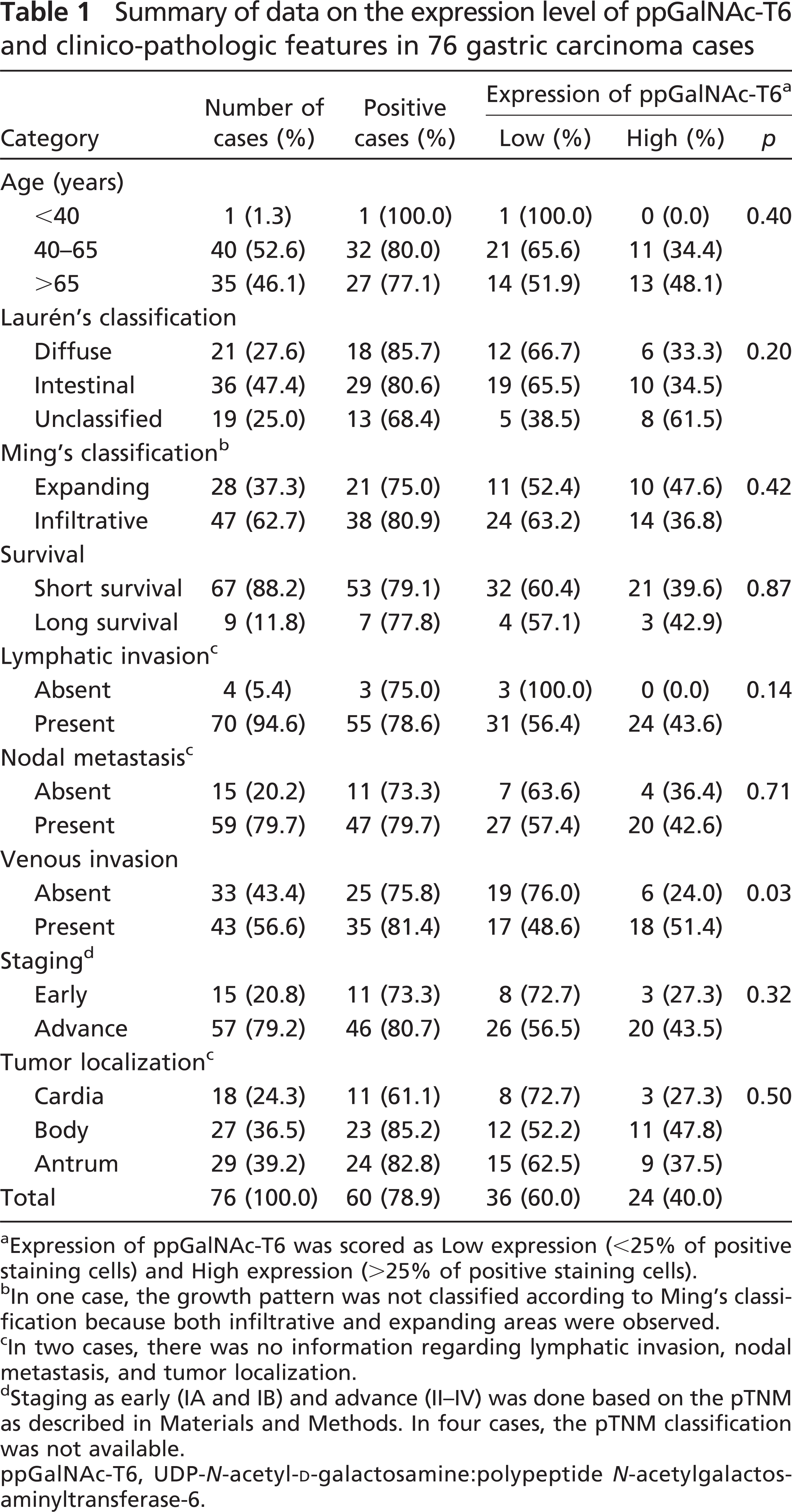
Expression of ppGalNAc-T6 was scored as Low expression (<25% of positive staining cells) and High expression (>25% of positive staining cells).
In one case, the growth pattern was not classified according to Ming's classification because both infiltrative and expanding areas were observed.
In two cases, there was no information regarding lymphatic invasion, nodal metastasis, and tumor localization.
Staging as early (IA and IB) and advance (II-IV) was done based on the pTNM as described in Materials and Methods. In four cases, the pTNM classification was not available.
ppGalNAc-T6, UDP-N-acetyl-D-galactosamine:polypeptide N-acetylgalactos-aminyltransferase-6.
In intestinal metaplasia, a precursor lesion of gastric carcinoma, we observed a slightly lower percentage of positive cases. Intestinal metaplasia shows a highly regulated pattern of expression of genes, reproducing the differentiation characteristics of intestinal cells. This is the case of mucins such as MUC2 (Reis et al. 1999) and a modified pattern of mucin-type O-glycans such as Sialyl-Tn. The percentage of cases expressing ppGalNAc-T6 observed in intestinal metaplasia may reflect the differentiation characteristics of these cells and may explain the modified pattern of mucin-type O-glycans observed in intestinal metaplasia (David et al. 1992; Ferreira et al. 2006; Mesquita et al. 2006).
Studies in vitro have shown that ppGalNAc-Ts display site specificity and different kinetic properties toward O-glycosylation sites in mucins, including the MUC1 tandem repeat (Wandall et al. 1997; Hassan et al. 2000b). These characteristics of ppGalNAc-Ts suggest that the members of this family of enzymes have different functions and that the repertoire of ppGalNAc-Ts expressed in a given cell may determine the pattern of O-glycan attachment, especially the density of glycosylation of the MUC1 tandem repeat (Bennett et al. 1998; Hassan et al. 2000b).
Expression level of ppGalNAc-T6 according to the presence of venous invasion and localization of the tumor in the stomach
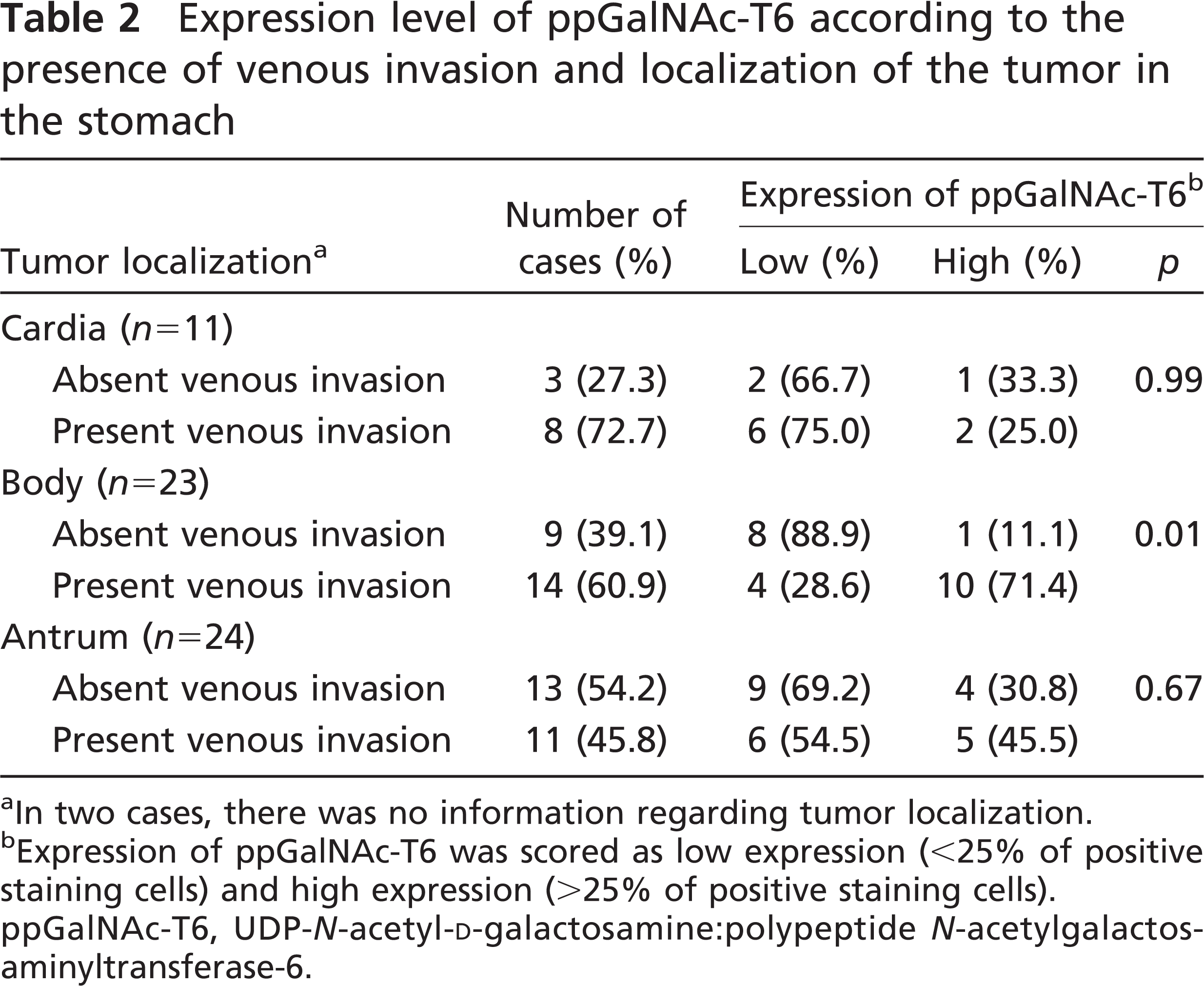
In two cases, there was no information regarding tumor localization.
Expression of ppGalNAc-T6 was scored as low expression (<25% of positive staining cells) and high expression (>25% of positive staining cells).
ppGalNAc-T6, UDP-N-acetyl-D-galactosamine:polypeptide N-acetylgalactos-aminyltransferase-6.
This study is, to the best of our knowledge, the first evaluation of the expression of ppGalNAc-T6 in gastric carcinomas. This evaluation of ppGalNAc-T6 of gastric carcinomas was possible because of the capability of MAb T6.3 to recognize formalin-fixed paraffin-embedded sections (Berois et al. 2006). The evaluation of ppGalNAc-T6 in 76 gastric carcinomas showed expression of this enzyme in 79% of the cases. Our results showed that the expression of ppGalNAc-T6 in gastric carcinomas was associated with the presence of venous invasion. In addition, our results also showed a particular association of venous invasion with ppGalNAc-T6 expression in tumors localized in the body region of the stomach.
Summary of data on the coexpression level of mucins (MUC5AC, MUC6, and MUC2) and ppGalNAc-T6 in gastric carcinoma cases
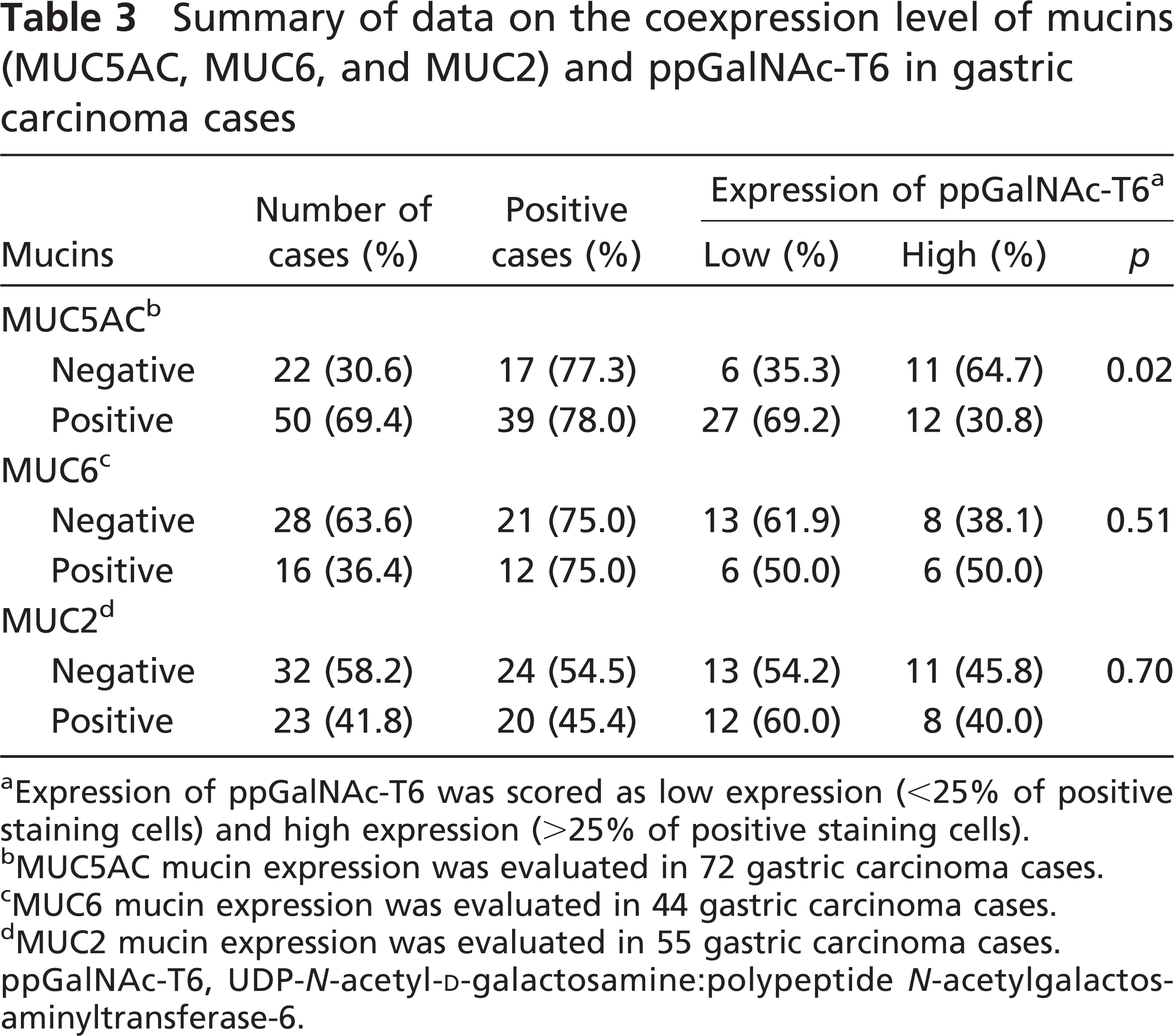
Expression of ppGalNAc-T6 was scored as low expression (<25% of positive staining cells) and high expression (>25% of positive staining cells).
MUC5AC mucin expression was evaluated in 72 gastric carcinoma cases.
MUC6 mucin expression was evaluated in 44 gastric carcinoma cases.
MUC2 mucin expression was evaluated in 55 gastric carcinoma cases.
ppGalNAc-T6, UDP-N-acetyl-D-galactosamine:polypeptide N-acetylgalactos-aminyltransferase-6.
Different levels of ppGalNAc-T3 have been detected in patients with various types of carcinomas (Shibao et al. 2002; Gu et al. 2004; Ishikawa et al. 2004; Miyahara et al. 2004; Yamamoto et al. 2004; Landers et al. 2005; Inoue et al. 2007), and this has been associated with a poor prognosis. ppGalNAc-T6 exhibits a high sequence homology to ppGalNAc-T3, and in vitro studies have shown that both enzymes display similar acceptor substrate specificities (Bennett et al. 1999b). Our results suggest that ppGalNac-T6 may also play a role in the biological characteristics of gastric carcinoma cells, most probably through the variation in mucin O-glycosylation. This is in line with previous studies showing that gastric carcinoma cell lines expressing different set of ppGalNAc-Ts are associated with variable patterns of mucin glycosylation (Marcos et al. 2003). The abnormal expression of ppGalNAc-T6 in gastric carcinomas, and the mucin glycoforms produced by this enzyme, could induce changes in cellular functions including adhesion and invasion. Although a preliminary evaluation of coexpression between ppGalNAc-T6 and simple mucin-type carbohydrate antigens (Tn and sialyl-Tn) in a subseries of gastric carcinomas did not show a direct association (data not shown), we cannot exclude that the expression of ppGalNAc-T6 contributes to a different level of O-glycan site occupancy in the mucin tandem repeats. Our results warrant further study on the relationship between ppGalNAc-T6 expression and alterations in the O-glycan density and expression of carbohydrate antigens in gastric carcinomas (Marcos et al. 2003). This evaluation should also include other glycosyltransferases that can contribute to the synthesis of these carbohydrate antigens. In addition, future functional in vitro studies will clarify the biological role of ppGalNAc-T6 overexpression in gastric carcinoma cells. Furthermore, the expression of the mucin protein core is tightly regulated in gastric mucosa. Our results showed that ppGalNAc-T6 is inversely associated with the expression of mucin MUC5AC. This result may stem from characteristics of differentiation of the cells expressing either protein. No association was observed with the other mucins evaluated. Finally, ppGalNAc-T6 expression was observed in both complete and incomplete intestinal metaplasia cases (Reis et al. 1999).
In summary, we showed that (a) ppGalNAc-T6 is expressed in normal gastric mucosa in both antrum and body regions; (b) in intestinal metaplasia, there is expression of ppGalNAc-T6 in 52% of the cases; (c) in gastric carcinomas, the expression of ppGalNAc-T6 is heterogeneous, with most cases (79%) expressing ppGalNAc-T6; and (d) ppGalNAc-T6 expression in gastric carcinomas is associated with venous invasion. Our results provide evidence that ppGalNAc-T6 is a novel IHC marker associated with venous invasion in gastric carcinomas and contributes to the understanding of the molecular mechanisms that underlie aberrant glycosylation during gastric carcinogenesis and gastric carcinoma.
Footnotes
Acknowledgements
This work was supported by Fundação para a Ciěncia e a Tecnologia (FCT) (PTDC/CTM/65330/2006 and PTDC/CVT/65537/2006); financiado no âmbito Programa Operacional Ciěncia e Inovação 2010 do Quadro Comunitário de Apoio III e comparticipado pelo FEDER; and Association for International Cancer Research (AICR Grant 05–088). J.G. (SFRH/BD/40563/2007), N.T.M. (SFRH/BD/11764/2003), and A.M. (SFRH/BD/36339/2007) acknowledge FCT for financial support.
We thank Leonor David and Ulla Mandel for suggestions, Mário Seixas for statistical analysis, and Nuno Mendes for technical assistance.