
Abstract
Severe destructive polyarthropathy with fibrillation and erosion of articular cartilage, deformation of articular surfaces, and proliferation of periarticular soft tissue was diagnosed in 5 bovine fetuses aborted in the last trimester. There was involvement of a single coxofemoral joint in 2 fetuses, both elbows, and a stifle in 1, both stifles and tarsal joints in another, and, in the most severely affected fetus, both hips, stifles, and shoulders as well as 1 carpus. In affected joints, the articular cartilage was irregularly reduced in thickness and contained fibrovascular tissue and, in some cases, inflammatory cells and fibrin. Four of 5 fetuses had microscopic placentitis; all had mild lymphoplasmacytic conjunctivitis. Ureaplasma spp. was identified in 4 cases by polymerase chain reaction or culture. Fetal arthropathy and Ureaplasma diversum-associated joint pathology have not been previously reported in the veterinary literature; however, other Ureaplasma spp. are known to cause reproductive disease and arthritis in humans.
Keywords
Members of the family Mycoplasmataceae, specifically Mycoplasma spp., are known to cause arthritis in a number of veterinary species, including cattle. 12 Although disease is more common in older animals, Mycoplasma spp.–associated arthritis has been observed in neonatal calves. 3 Joint swelling in a bovine fetus from a Mycoplasma spp.–associated abortion has also been reported. 3 Mycoplasmal arthritis often affects multiple joints and causes fibrinous exudation into the joint space with synovial inflammation and thickening. 12 Pannus and articular cartilage erosion have been described in chronic cases. 12 Despite the apparent tropism of Mycoplasma for joints, Ureaplasma, a closely related genus, has not been associated with articular disease in animals. Destructive polyarthropathy has not been described in fetuses of any species. This report describes destructive polyarthropathy in 5 bovine fetuses and its apparent association with Ureaplasma diversum.
Destructive arthropathy was first observed during routine examination of the coxofemoral joint in an aborted bovine fetus submitted to Prairie Diagnostic Services (PDS) in Saskatoon, Saskatchewan, in 2002. Since then, 4 additional cases have been identified, all of which showed evidence of U. diversum infection. To our knowledge, U. diversum has not been associated with articular disease in animals nor has destructive arthropathy been previously reported in fetuses of domestic species.
In all 5 fetuses, major appendicular joints (coxofemoral, stifle, shoulder, carpal, and tarsal) were evaluated. Tissues collected from all fetuses for histology included lung, heart, liver, spleen, diaphragm, adrenal gland, kidney, ileum, spiral colon, brain, jejunal lymph node, eyelid, thyroid, salivary gland, thymus, mediastinal lymph node, tongue, skeletal muscle, and placenta. Tissues from affected joints were collected in fetus Nos. 1 and 5, and periarticular soft tissue was collected from affected joints in fetus No. 2. Joints from fetus Nos. 3 and 4 were examined only macroscopically. Tissues were fixed in 10% neutral buffered formalin (with decalcification of bony tissue in 20% aqueous formic acid for 24 hours after initial fixation), processed routinely, and embedded in paraffin. Sections (4 µm) were stained with hematoxylin and eosin (HE).
Of the 5 fetuses (Table 1), the most severe articular lesions and the most complete information were from fetus No. 5. The source herd (which was also the source for fetus Nos. 2 and 3, submitted in 2005) had experienced 4 abortions over the course of 2 days, all in fully vaccinated heifers, in 2008. In fetus No. 5, there was marked intrauterine growth retardation (indicated by reduced weight and crown-to-rump length in relation to the stage of morphologic development, in terms of haircoat growth and tooth eruption). The shoulders, hips, stifles, and right carpus had irregularly distributed, marked fibrillation and erosion of articular cartilage, deformation of the articular surface, and thickening of periarticular soft tissue (Fig. 1). Both acetabulae were shallow and articulated loosely with the deformed femoral heads. Histologically, the articular cartilage in multiple joints was irregularly reduced in thickness. Fibrovascular tissue extended into the cartilage to form deep depressions and fingerlike projections that extended to the subarticular (epiphyseal) growth cartilage and subchondral bone in some areas (Fig. 2). The fibrovascular tissue contained neutrophils, lymphocytes, and plasma cells with fibrin deposits adjacent to the articular space (Fig. 2). At the periphery of the articular surfaces, the fibrovascular tissue was more organized and highly collagenous. There was loss of the synovial membrane with replacement by mature fibrous tissue, vessels, and myelinated nerves that extended into the joint space (Fig. 3). In the scapula, a 5-mm-diameter cystic structure within the subchondral bone was lined by fibrovascular tissue and contained neutrophils, fibrin, and synovial fluid. Although involved in the arthropathy, the articular-epiphyseal complex was not apparently targeted by the reaction. The physeal cartilage, as well as all other bone structures, was normal. There were no significant findings in any of the other tissues examined.
History, signalment, and lesions in bovine fetuses with Ureaplasma diversum-associated arthropathy.
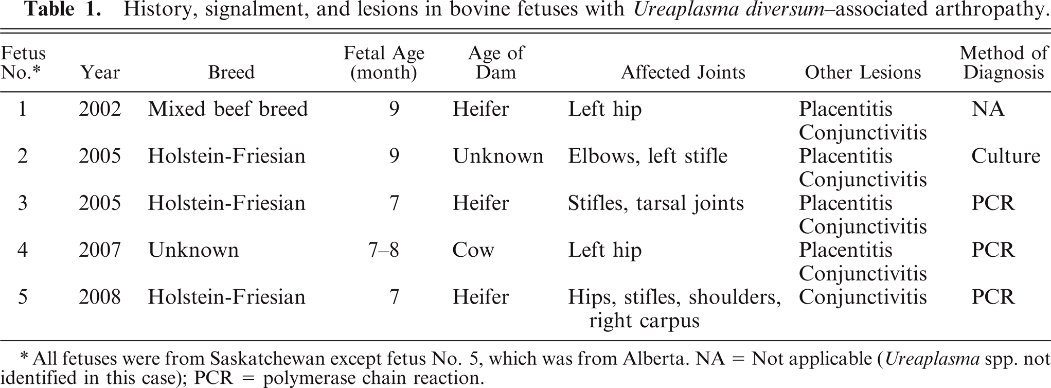
All fetuses were from Saskatchewan except fetus No. 5, which was from Alberta. NA = Not applicable (Ureaplasma spp. not identified in this case); PCR = polymerase chain reaction.
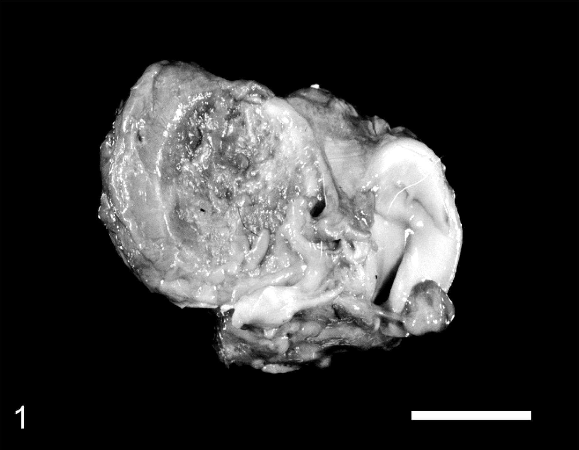
Tibial plateau; bovine fetus No. 5. Unilateral articular cartilage fibrillation and loss, with absence of one meniscus. Bar = 2 cm.
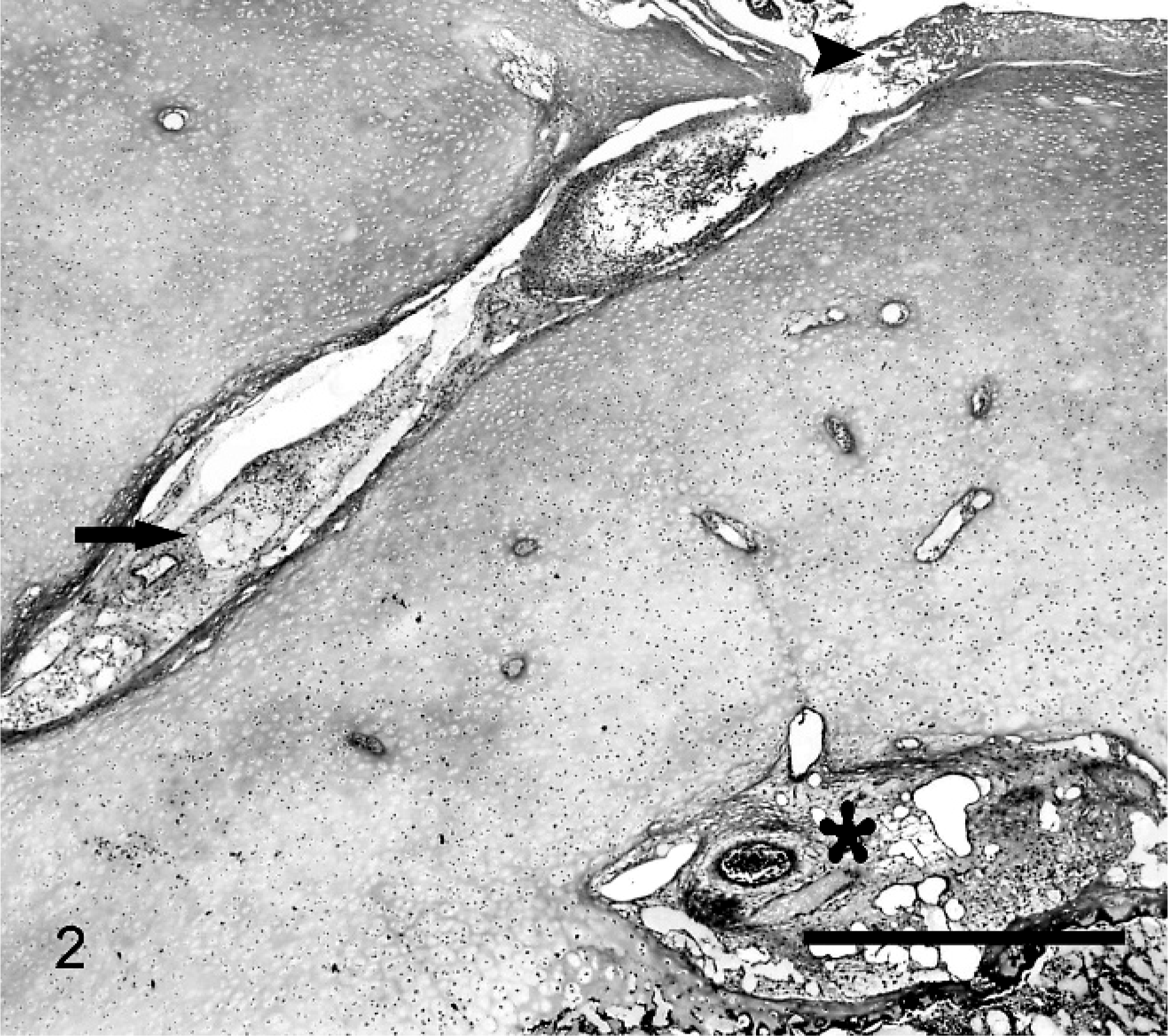
Articular surface of a femoral condyle; bovine fetus No. 5. The articular cartilage is irregularly thin, and there is fibrovascular tissue (arrow) extending to the epiphyseal cartilage and subchondral bone (asterisk). Deposits of fibrin are present adjacent to the articular space (arrowhead). HE. Bar = 1 mm.
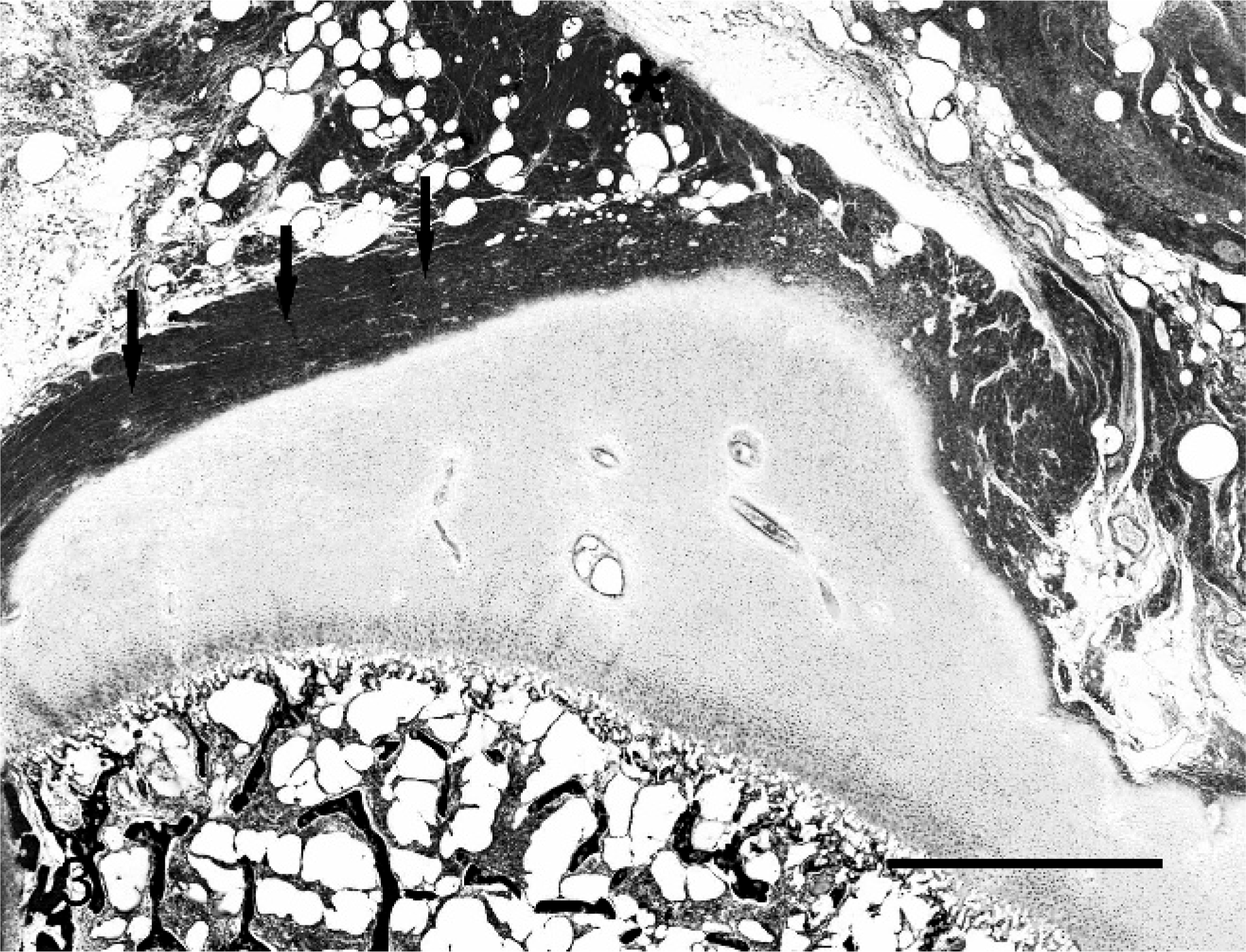
Femoral head; bovine fetus No. 5. Highly organized collagenous tissue covers the articular surface at the periphery of the joint (arrows). There is loss of the synovial membrane with replacement by mature fibrous and vascular tissue (asterisk). HE. Bar = 2 mm.
Gross changes in fetus No. 1, submitted in 2002, were limited to arthropathy and deformation of the left coxofemoral joint, loss of the synovial membrane, and proliferation of periarticular soft tissue (Fig. 4). Histologically, articular and epiphyseal cartilage in the acetabulum and femoral head were replaced by fibrous or fibrovascular tissue. Synovial membrane could not be identified.
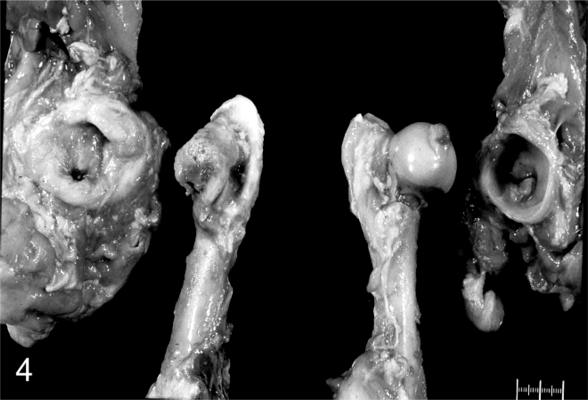
Coxofemoral joints; bovine fetus No. 1. Shallow acetabulum with periarticular soft tissue proliferation and deformed femoral head (left). The contralateral joint is normal.
Fetus Nos. 2 and 3, products of embryo transfer, originated from the same farm as fetus No. 5. Fetus No. 2 had arthropathy of both elbow joints and the left stifle joint. Fetus No. 3 had bilateral arthropathy of the stifle and tarsal joints. Both fetuses had mild thickening and opacity of the placenta. Articular cartilage was not examined microscopically in either fetus; however, there were irregularly distributed lymphocytes and plasma cells, with fewer neutrophils, within the fibrovascular periarticular tissue of the affected joints in fetus No. 2. Synovial membrane could not be identified.
Fetus No. 4 had severe destructive arthropathy of the left hip joint. It also had thickening and brown discoloration of the intercotyledonary placenta. The joint was not examined histologically.
All fetuses had mild lymphoplasmacytic conjunctivitis. Four had inflammation of the allantois with placental vasculitis in 3 cases. Pulmonary lesions were absent.
Microbiologic culture was performed on lung, abomasal fluid, and placenta from fetus Nos. 1, 2, and 4; lung, placenta and periarticular tissue from fetus No. 3; and abomasal fluid, placenta, and periarticular tissue from fetus No. 5. In all cases, except fetus No. 1, culture of Ureaplasma spp. was specifically attempted using Ureaplasma spp. media supplement (87% Fleichmann's yeast solution, 9% [v/v] 10% urea, 4% penicillin 200,000 IU/ml). Specific culture for Mycoplasma spp. was not performed. Ureaplasma and Salmonella spp. were isolated from the placenta of fetus No. 2. Organisms isolated from the other fetuses included Aeromonas spp., Escherichia coli, Proteus spp., alpha Streptococcus spp., Enterobacter spp., Citrobacter spp., Staphylococcus spp., and Bacillus spp., none of which were thought to have caused the abortion or arthropathy.
Genus-specific polymerase chain reaction (PCR) amplification of nucleotides 237–881 of the 16S rRNA gene of Ureaplasma spp. 6 was performed on DNA extracted from fresh synovial tissue from fetus Nos. 4 and 5, and on pooled placenta and synovium (homogenized and tested together) from fetus No. 3. All 3 cases were positive. To determine the species of Ureaplasma, the 645 base pair PCR product from fetus No. 5 was sequenced. High-quality sequence data for 625 base pairs had 99% homology with the published sequence from the type strain of U. diversum (strain A417) 2 and less than 97% homology with other Ureaplasma spp. The sequence amplified from fetus No. 5 has been deposited in Genbank, accession number EU714185. PCR, as described above, was also performed on pooled sections of formalin-fixed, paraffin-embedded placenta and articular cartilage from fetus No. 1 and placenta from fetus No. 2, both of which were negative.
U. diversum, a member of the family Mycoplasmataceae, 4, 10 colonizes cattle specifically, 9 with predilection for mucosal surfaces such as the nasal passages, vulva, vagina, and prepuce. 4, 10 It appears to be part of the normal flora of these surfaces 9, 10 and is most likely transmitted through coitus. 4, 10 U. diversum can also be found in bovine artificial insemination and embryo-transfer fluids, where it is able to survive freezing. 4, 10 U. diversum has been associated with several reproductive diseases of cattle, including granular vulvovaginitis and balanoposthitis, 4 abortion, and the birth of dead or weak calves at term. 4, 10 The conditions needed for the bacterium to produce disease, however, remain unclear. 4, 9 U. diversum–induced abortion occurs most commonly in the last trimester; fetal lesions include placental fibrosis, necrosis, vasculitis, nonsuppurative alveolitis and peribronchiolar lymphoplasmacytic cuffing, and lymphoplasmacytic conjunctivitis with goblet cell hyperplasia. 4, 10 All fetuses in this report had lymphoplasmacytic conjunctivitis; 4 had placentitis. Pulmonary lesions were not observed.
Although Ureaplasma spp., specifically U. urealyticum, are commensal organisms of the urogenital tract of sexually active women, 7 U. urealyticum is the most common microbe isolated from amniotic fluid of women at risk for preterm delivery 5 and has been associated with abortion, chorioamnionitis, low birth weight, pneumonia, and chronic lung disease in infants. 5, 7 As in cattle, the conditions needed for Ureaplasma spp. to produce reproductive disease in humans remain unclear. 5 Although U. urealyticum has not been associated with arthritis in human fetuses, it has been isolated from cases of arthritis and septic osteomyelitis in human neonates 7, 8, 11 and is a common cause of severe, chronic, erosive polyarthritis in humans with primary immune deficiencies. 1 This association with human arthritis suggests that Ureaplasma spp. has a tropism for joints and could cause articular lesions in other species.
This report describes the gross and histologic findings in 5 cases of bovine fetal polyarthropathy associated with U. diversum infection. Neither the fetal arthropathy nor its association with U. diversum has been previously reported. Bovine arthritis has, however, been associated with Mycoplasma spp. and may be erosive in its chronic form, secondary to pannus formation. 12 The fibrovascular tissue observed in the affected joints in these cases resembled pannus; however, the inflammatory response was minimal compared with the degree of cartilage loss. This may suggest a previous or underlying process such as vascular compromise and ischemia, with the inflammation being either a secondary or contributory effect.
Further study of additional cases is needed to elucidate the pathogenesis of this bovine fetal arthropathy and to determine whether it is caused by U. diversum. Because all joints are not consistently affected, thorough evaluation of multiple joints should be part of the routine necropsy examination of the bovine fetus to determine whether this condition is more widespread than previously recognized.