
Abstract
Following a routine necropsy of a bovine fetus aborted at 5 months of gestation, placenta, fetal tissue samples, and stomach contents were subjected to a number of laboratory tests. Staphylococcus warneri was isolated in pure culture from the lung, liver, and stomach contents, whereas the placenta yielded S. warneri and a number of contaminants. Gross evaluation of agar plates showed predominant colonies to be morphologically consistent with those of S. warneri and the identity of the agent was further confirmed on a Trek Diagnostic Systems Sensititre, gram-positive identification (GPID) plate. Microscopic evaluation of fetal tissue sections showed extensive necrotizing lesions of the tongue, lung, and placenta in which there were numerous coccoid shaped gram-positive bacteria with morphology consistent with Staphylococcus spp. These results provide strong diagnostic evidence of S. warneri as a possible cause of bovine abortion and suggest there should be further investigations into the abortivirulence of this agent.
Keywords
Determining the cause of abortion in livestock is often difficult as an etiologic agent is only identified in about 30–40% of livestock abortion cases submitted to diagnostic laboratories. 7 When a bacterial agent is isolated from fetal tissues, however, there often are no histologic lesions that prove the culpability of the isolated organisms. Staphylococcus warneri is a coagulase-negative member of Staphylococcus, a genus that consists of gram-positive coccoid bacteria. Although previously presumed to be innocuous, evidence now suggests a number of coagulase-negative staphylococci are important nosocomial pathogens in humans. 2,5,6,9,10 Among coagulase negative staphylococci, Staphylococcus epidermidis is the most commonly isolated species 5 with documented clinical significance.
The pathogenicity of S. warneri for humans and animals has been documented 8 ; recent literature has associated the agent with severe bacteremia and endocarditis in immuno-compromised patients. 6 Waites and coworkers 12 isolated S. warneri along with other organisms from the amniotic fluids of a 34-year-old pregnant woman who aborted at 16 weeks following conception with an intrauterine device in place. In domestic animals, S. warneri has been isolated from cerebrospinal fluids of a dog with a severe thrombotic meningoencephalitis. 4 In another study, S. warneri was isolated from bovine bulk tank milk in Norway; this isolate was resistant to quaternary ammonium compounds, which are used as disinfectants. 3 The present report describes evidence of S. warneri as a possible cause of bovine abortion.
A small piece of frozen placenta and a carcass of semifrozen male bovine fetus measuring about 60 cm (crown-rump length) were received for necropsy at the Veterinary Diagnostic Laboratory (VDL) of North Dakota State University (NDSU). The history indicated that the fetus had been aborted by a Holstein cow at approximately 5 months gestation. The pregnant cow belonged to a herd of 292 animals out of which 4 cows had spontaneously aborted. The herd was vaccinated for bovine virus diarrhea virus types I and II, infectious bovine rhinotracheitis virus, parainfluenza type 3 virus, bovine respiratory syncytial virus, and leptospirosis.

Placenta. Chorioallantoic membranes contain moderate to high numbers of degenerate neutrophils and many intralesional bacteria (white asterisks) are within the necrotic cellular debris. HE. Bar =100 μm.
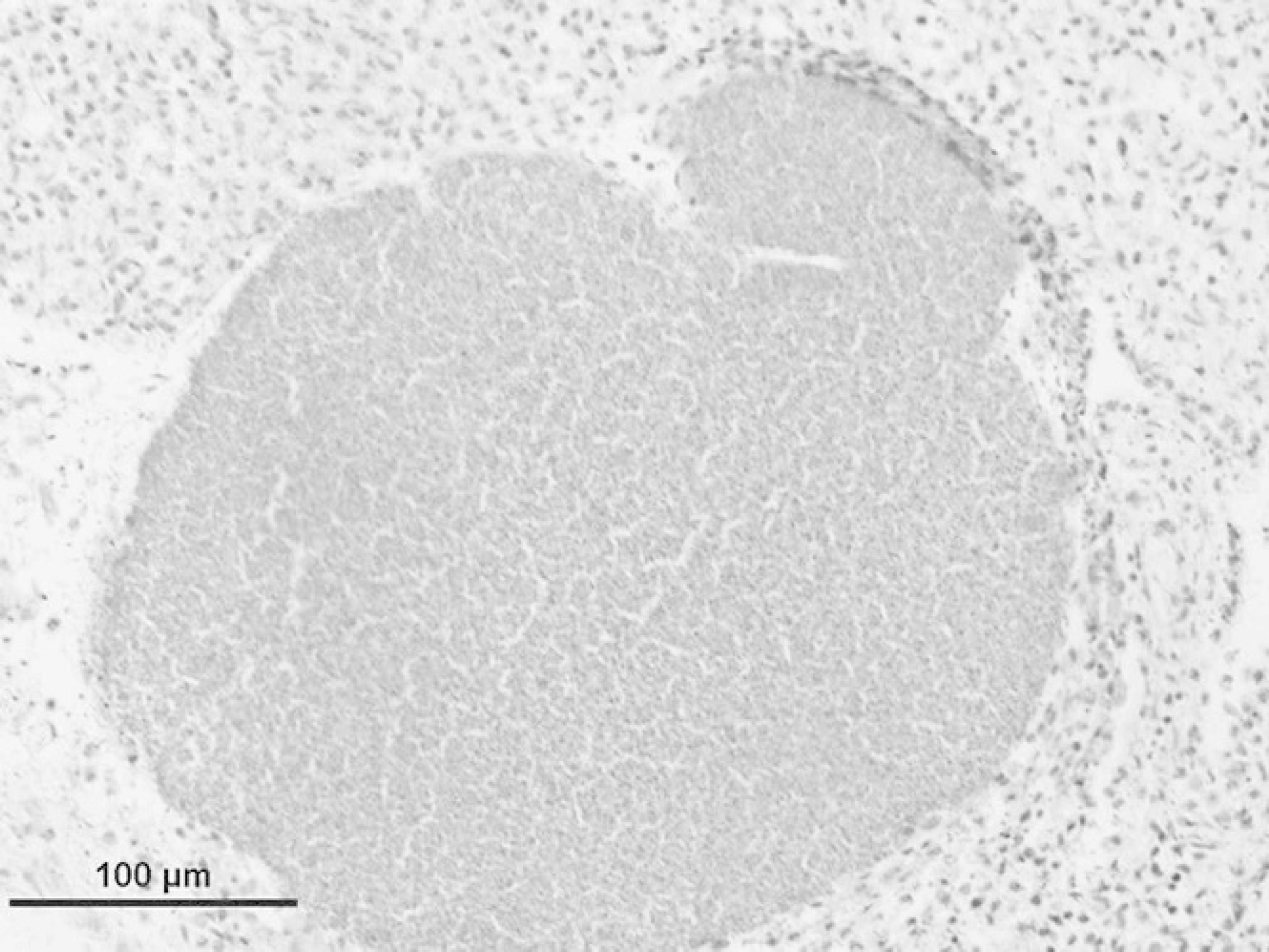
Lung. A massive colony of coccoid bacteria is present in the fetal lung. HE. Bar =100 μm.
A routine necropsy was conducted on the fetus with a detailed macroscopic evaluation of all organ systems. Fresh tissue samples, representative of all organs, were taken for bacteriology and virology. Two samples of stomach contents were taken; one for bacterial culture and the other for darkfield microscopy. Representative tissue samples from all organs plus skeletal muscle, tongue, and diaphragm were taken and fixed in 10% buffered formalin.
The formalin-fixed tissue samples were processed routinely, and paraffin embedded. Five-micron sections were deparaffinized and stained with hematoxylin and eosin (HE). a To further characterize the cytochemical characteristics of the intralesional bacteria, histological sections were stained with Brown and Brenn's modification of gram tissue stain a and then examined by light microscopy.
For bacteriologic culture, tissue samples and abomasal fluids were inoculated on a tryptose soy agar (TSA) II 5% sheep blood b and incubated in an aerobic incubator at 35°C for 12 hours. Samples were also inoculated on Skirrows media (Campylobacter) c and incubated in anaerobic conditions in a microaerophilic gas generating pack d at 37°C for 12 hours.
Tissue impression smears of fresh kidney tissue were tested by direct fluorescent antibody (FA) test for Leptospira antigens and a sample of stomach contents examined by darkfield microscopy for Leptospira-like structures. Frozen tissue sections were tested by direct FA test for bovine virus diarrhea virus antigens and bovine herpes virus type 1 (infectious bovine rhinotracheitis virus).
The only remarkable macroscopic findings were congested lungs and liver as well as large amounts of serosanguineous fluids in the thoracic and abdominal cavities. The small piece of fetal membrane showed abundant off-color necrotic debris on the cotyledons.
In the placenta, there were focally extensive areas of necrosis in which an extremely high number of coccoid shaped bacteria were present (Fig. 1). Many degenerate neutrophils admixed with necrotic tissue debris were seen in adjacent areas. Multiple sections of the lung contained massive colonies of cocci that were randomly distributed (Fig. 2). There were multifocal areas of necrosis in the tongue that predominantly involved the stratified squamous epithelium in which many coccoid bacteria were present. In sections stained with Brown and Brenn gram stain, the bacterial colonies were gram-positive and morphologically consistent with Staphylococcus spp. Apart from severe autolysis of the kidney, spleen, and liver, no remarkable changes were seen in sections of the skeletal muscle, diaphragm, and brain.
Staphylococcus warneri was recovered in pure culture from the lung, liver, and stomach contents. The placenta yielded a mixture of agents including S. warneri, α Streptococcus spp., Bacillus spp., Escherichia coli, Enterobacter spp., and a fungus. These organisms were identified on the Trek Diagnostic Systems Sensititre using gram-positive identification (GPID) or gram-negative identification (GNID) plates. e Morphologically, S. warneri colonies measured about 4–7 mm in diameter, were smooth and glossy, and were unpigmented to cream colored. Typical of this agent, the edges of individual colonies were rather flat but had dome-shaped centers.
Fresh kidney impression smears were negative for Leptospira antigens and no Leptospira-like structures were seen in a sample of stomach contents examined by darkfield microscopy. Bovine virus diarrhea virus and infectious bovine rhinotracheitis viruses were ruled out on the basis of negative FA test results.
This report suggests S. warneri as a potential cause of bovine abortion. A detailed diagnostic work-up on samples taken from a bovine fetus, aborted at 5 months of gestation, and placenta provided evidence that this agent was a probable cause of this abortion. It is important to note that various tests ruled out a number of abortifacient pathogens including bovine virus diarrhea virus, bovine rhinotracheitis virus, and Leptospira spp. An ascending infection of the genital tract cannot be ruled out in this case but the presence of microscopic placental lesions suggests that infection was most likely acquired from maternal blood and spread peripherally to involve various fetal tissues. Previous reports in humans indicate that most pathogenic coagulase-negative staphylococcal infections occur as bacteremias. 2,6,9 The microscopic lesions seen on the tongue mainly involved the superficial and middle layers of the stratified squamous epithelium and were characterized by multifocal areas of necrosis with massive intralesional colonies of gram-positive cocci. Staphylococcus spp. are known to have affinity for epithelial tissues 11 and the finding of lesions localized in the lingual epithelium is not surprising. Exfoliative toxins usually in the form of serine protease enzymes produced by Staphylococcus spp, have been shown to hydrolyze the desmosomal protein called desmoglein-1 1 found in epithelial tissues. The tongue lesions further suggest that the bacteria most likely entered the oral cavity through swallowing of amniotic fluids by the fetus. The human abortion case reported by Waites et al., 12 and the data in this report suggest that amniotic fluids may provide a favorable environment for amplification of a blood-borne S. warneri infection creating a springboard for a secondary reinvasion of the fetus via the oral cavity.
To the authors' knowledge, this is the first report in which S. warneri is linked to fetal loss in cattle. Plans are underway to characterize the S. warneri isolate to describe its antigenic profile and possible abortivirulence.
Footnotes
a.
VWR International, West Chester, PA.
b.
Becton Dickinson, Sparks, MD.
c.
Campylobacter Agar Skirrow, Vet Med Biological Media Services, University of California, Davis, CA.
d.
Mitsubishi Gas Chemical Co. Inc., New York, NY.
e.
Trek Diagnostic Systems, West Sussex, UK; distributed by Trek Diagnostic Systems, Cleveland OH.