
Abstract
An 11-year-old, Black and Tan Coonhound dog was presented with a history of lameness of the right hind leg for 2 months, osteolysis in the right distal femur, a pulmonary mass, and a presumptive diagnosis of osteosarcoma. By cytologic examination, neoplastic melanocytes were noted from fine needle aspirates of the femoral and pulmonary masses. Postmortem examination revealed a disseminated melanoma involving the right femoral bone marrow, lung, multiple lymph nodes, and adrenal gland, with diffuse infiltration of the leptomeninges of the brain and spinal cord. This case report describes a unique presentation of canine melanoma, which in some ways resembles leptomeningeal melanomatosis, a rare human melanoma variant.
Melanomas account for 7% of all malignant canine tumors. 15 Most canine melanomas involve the oral cavity, mucocutaneous junctions, nailbed, and eye. Metastasis is fairly common, especially to the lymph node and lung; however, it is not uncommon to find metastatic melanoma in other organs, such as the brain, heart, and spleen. 5 Leptomeningeal melanomatosis, a very rare neoplasm in humans, is characterized by diffuse infiltration of melanocytic tumor cells in the leptomeninges of the brain and spinal cord. 4, 7– 10, 12, 17 Only a few cases have been reported in humans since it was first recognized by Virchow in 1859. Metastases of melanomas to the bone marrow are rare, because melanomas often spread via lymphatics. 6 To our knowledge, no case of melanoma diffusely involving the leptomeninges or bone marrow has been reported in the veterinary literature. Here, we report a disseminated melanoma involving the leptomeninges of the brain and entire spinal cord, bone marrow of the right femur, lung, multiple lymph nodes, and adrenal gland.
An 11-year-old, Black and Tan Coonhound dog was referred to the Veterinary Medical Teaching Hospital (VMTH), University of Missouri, with a 2-month history of lameness of the right hind leg and a large lipoma on the chest. Radiographic examination by the referring veterinarian had revealed a moth-eaten pattern of osteolysis in the distal aspect of the right femur. A presumptive diagnosis of osteosarcoma was made at that time. At the VMTH, additional imaging by thoracic radiographs and computed tomography (CT) revealed a mass in the right middle lung lobe as well. Fine needle aspirates were taken from the lytic femoral lesion and the pulmonary mass for cytologic examination.
Cytologically, Wright-Giemsa-stained aspirates of the femoral lesion were moderately cellular with a population of pleomorphic cells arranged individually and in small clusters. These cells ranged in shape from polygonal to spindloid. The nuclei were round to ovoid, varied from approximately 10 to 18 μm in diameter, had coarsely stippled chromatin, and often had one or more variably sized and shaped nucleoli. Cytoplasm was ample to abundant, lightly basophilic, and contained markedly variable amounts of fine dark green pigment consistent with melanin (Fig. 1). Binucleation, moderate anisocytosis, and moderate anisokaryosis were evident in some cells. The CT-guided aspirates from the pulmonary mass contained a similar population of cells as seen in the femoral aspirates. Based on the cytologic findings, melanoma was diagnosed. Due to the poor prognosis, the owner elected euthanasia.
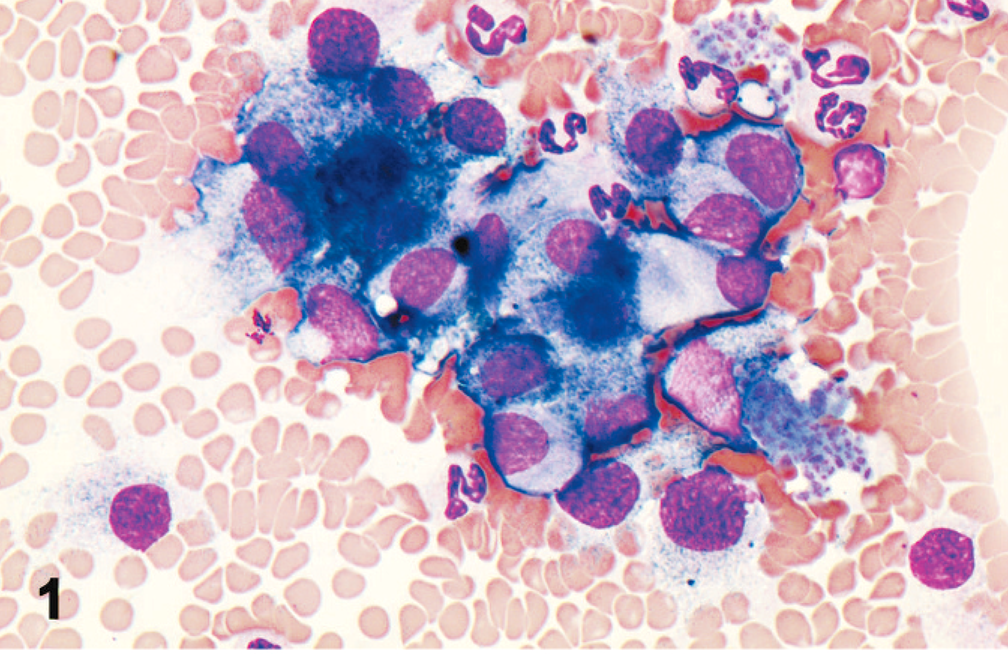
Bone marrow aspirate; dog. Cohesive neoplastic melanocytes with moderate anisocytosis and anisokaryosis and fine green cytoplasmic granules. Wright-Giemsa.
At necropsy, a 4-cm-diameter brown-black mass involved the right distal epiphysis and metaphysis (Fig. 2). The right middle lung lobe contained a single, 5-cm-diameter, black, friable, nodular mass. Multiple slightly raised black plaques in the parietal pleura extended into the intercostal muscles. Several mediastinal lymph nodes, the right popliteal lymph node, and multiple mesenteric lymph nodes were enlarged (2–4 times expected diameter) and diffusely black when incised. Diffuse black discoloration was in the leptomeninges covering the brain (especially at the base of the brain), around cranial nerves, and circumferentially around the entire surface of the spinal cord (Fig. 3). In addition, a 15-cm-diameter, well-defined lipoma was in the subcutis of the right ventrolateral thoracic wall.
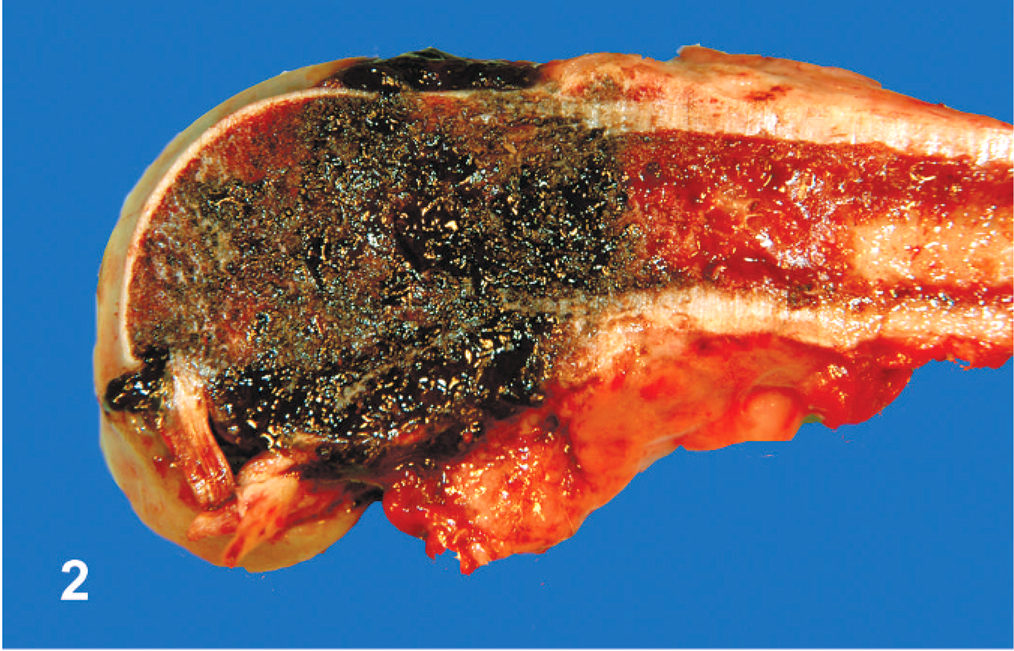
Femur; dog. A pigmented melanoma infiltrates medulla and cortex in the distal epiphysis and metaphysis.
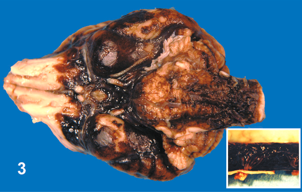
Brain and spinal cord (inset); dog. Disseminated brown-black discoloration and thickening of the leptomeninges.
Microscopically, all of the neoplastic masses were similar in character. Invasive tumors in the lung, lymph nodes, intercostal muscle, and adrenal gland consisted of heavily pigmented, polyhedral epithelioid cells or spindloid cells, which were arranged in solid nests or interwoven fascicles. Mitoses were common (up to 4 mitotic figures per 400× field). There were multiple foci of necrosis within the tumor. The bone marrow of the right distal femur was replaced by similar cells (Fig. 4). Pigmented cells also separated the leptomeninges throughout the cerebrum, cerebellum, brainstem, medulla oblongata, and spinal cord. Despite the extent of dissemination, the tumor was limited to the leptomeninges and did not extend into the neuropil or form a discrete mass (Fig. 5). Aside from the main pulmonary mass, multiple patches of melanoma cells were embedded in the visceral pleura.
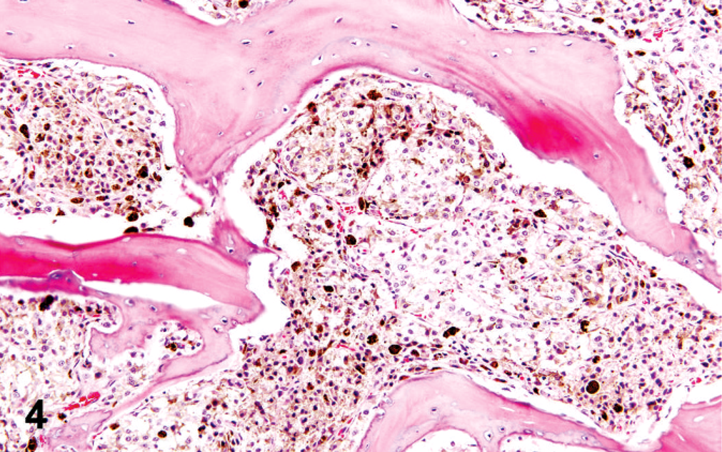
Femur; dog. Pigmented neoplastic melanocytes infiltrate the bone marrow. HE.
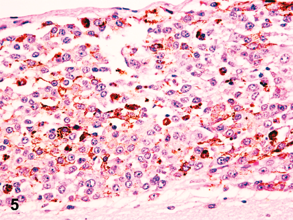
Cerebrum, leptomeninges; dog. Diffuse infiltration of leptomeninges by neoplastic melanocytes without involvement of the cerebral cortex. HE.
Melanomas are the third most common metastatic central nervous system (CNS) tumor in humans. Approximately 10–40% of human melanomas metastasize to the CNS. 1, 11, 18 According to a recent survey, only 3% (6 of 177) of canine metastatic brain tumors were melanomas. 16 Primary CNS melanomas are rare in humans 3 and, to our knowledge, have not been reported in animals. Melanocytes are normally found throughout the meninges, especially ventrolateral to the medulla oblongata. 12 There are two different types of meningeal melanomas. One invades the leptomeninges diffusely (leptomeningeal melanomatosis), similar to the present case, and the other forms nodular lesions. 7, 8, 12 Leptomeningeal melanomatosis occurs mainly in adults, but several childhood cases have been reported, often as part of a rare congenital syndrome neurocutaneous melanosis (NCM), which is characterized by proliferation of melanocytes in the skin (one large or many smaller nevi) and leptomeninges. 10 Approximately 40–60% of children with NCM develop leptomeningeal melanomatosis, which warrants a poor prognosis. 4, 9, 17
Distinguishing primary from metastatic melanoma in the CNS can be difficult and depends largely on negative findings in expected primary sites. Even so, uncertainty remains, because regression of primary melanoma after metastasis has been documented in humans. 2 The distinction has prognostic implications, however, because appropriate treatment results in long-term survival for patients with primary CNS melanoma, whereas most patients with metastatic melanoma of the CNS survive less than 1 year after diagnosis. 3
In the present dog, the entire body (especially the oral cavity, skin, eyes, and footpads) was thoroughly examined at necropsy and no mass was found aside from the aforementioned lesions. In addition, the history was negative with respect to prior tumor removal. In light of the lesion distribution, primary leptomeningeal melanomatosis with multiorgan metastasis was considered likely in this case. Although a few cases of primary CNS melanomas with distant metastases to the lung, 9 liver, 13 and vertebra 14 have been reported in humans, we failed to find any reports of disseminated metastatic melanoma of CNS origin in domestic animals.
Despite disseminated involvement of the leptomeninges, this patient had no neurologic deficits or appreciable spinal discomfort. Instead, the chief complaint on presentation was lameness, which was attributed to metastatic melanoma in the right femur. Bone marrow is an uncommon site of melanoma metastasis in humans, reported in only 5–7% of metastatic cases. 6 No report of melanoma in the bone marrow of animals was found. Thus, this report describes a unique presentation of disseminated canine melanoma with disseminated involvement of the leptomeninges and focal bone marrow metastasis.