
Abstract
A total of 90 tumors from 85 domestic hamsters (70 Djungarian hamsters and 15 Syrian hamsters) were examined by histology. In the Djungarian hamsters, 64 neoplastic and 11 non-neoplastic lesions were identified, whereas 14 Syrian hamsters showed neoplastic disease, and one showed non-neoplastic disease. The Djungarian hamsters showed a high prevalence of neoplastic disease, similarly to laboratory Djungarian hamsters. In the Djungarian hamsters, almost all tumors were integumental, whereas hematopoietic tumors were the most common type in the Syrian hamsters. The most common integumental tumors in the Djungarian hamsters were mammary tumors, atypical fibromas and papillomas, and a spectrum of integumental tumors that have not been reported in laboratory Djungarian hamsters were identified. Most mammary tumors occurred in the females, whereas all atypical fibromas were observed in the males. In the Syrian hamsters, plasmacytomas and lymphomas were the most common tumors. The small number of Syrian hamsters in this study may reflect the low prevalence of spontaneous tumors seen in laboratory Syrian hamsters. The mean age of the affected hamsters was 19.8 months, which is relatively advanced. To our knowledge, this is the first comprehensive study of tumors in domestic hamsters.
Hamsters are popular as pets, with Syrian (Golden) hamsters (Mesocricetus auratus), Djungarian (Siberian) hamsters (Phodopus sungorus), Roborovski hamsters (Phodopus roborovskii), Campbell hamsters (Phodopus campbelli), and Chinese hamsters (Cricetulus griseus) being the one most commonly kept; their chromosome numbers are 44, 28, 28, 28, and 22, respectively. However, the majority of reports and studies on neoplastic diseases in these species were related to spontaneous and induced tumors in laboratory hamsters.
Laboratory Syrian hamsters have been used in various medical research fields, particularly in carcinogenesis studies. 9 Thus, there are numerous reports on tumors induced by carcinogens and oncogenic viruses in this species. According to Pogosianz, 15 the spectrum of spontaneous tumors in Djungarian hamsters differs from that in Syrian hamsters. However, reports on spontaneous tumors in domestic hamsters are scarce, and most are individual case reports. We thus performed a comprehensive study on spontaneous tumors that occur in hamsters, particularly Syrian and Djungarian hamsters.
Materials and Methods
A total of 90 biopsy specimens from 85 domestic hamsters submitted to the Laboratory of Veterinary Pathology, Nihon University, between 1994 and 2007 were studied. All the hamsters were kept as pets in the metropolis of Tokyo, Saitama Prefecture, and Kanagawa Prefecture. In some cases, internal examination and surgical removal were performed when the practitioner identified the presence of internal masses by palpation and/or radiographic imaging. All specimens were fixed in 10% neutral buffered formalin, embedded in paraffin, sectioned at 5 µm, and stained with hematoxylin and eosin (HE) according to standard histopathologic methods. Additional sections were prepared in some cases, and these were stained with Masson's trichrome, periodic acid–Schiff reaction, Watanabe's silver impregnation method, and Grimelius's method.
Diagnoses were made by using HE-stained sections and supplemental sections with special stains. The tumor type was classified by organ. Mammary glands were included in the integumental system, and integumental tumors other than mammary tumors were subdivided into epithelial and mesenchymal tumors based on the World Health Organization classification. 6, 8 Data on species, age, and sex of affected hamsters and localization of integumental tumors were also obtained. Tumors derived from ganglion cell–like (GL) cells that characteristically occur in Djungarian hamsters were classified as atypical fibroma according to criteria previously described by Baba et al. 1
For atypical fibromas, immunohistochemistry was performed by using the EnVision System (horseradish peroxidase enzyme-labeled polymer method; Dako, Tokyo, Japan). The primary antibodies used were mouse monoclonal antibody anti-human cytokeratin (diluted 1 : 100; Dako, Tokyo, Japan), mouse monoclonal antibody anti-pig vimentin (diluted 1 : 100; Dako, Tokyo, Japan), rabbit polyclonal antibody anti-human androgen receptor (AR) (diluted 1 : 50; Ylem S.R.L, Roma, Italy), rabbit polyclonal antibody anti-bovine S-100 protein (diluted 1 : 1000; Dako, Tokyo, Japan), mouse monoclonal antibody anti-human neurofilament (diluted 1 : 50; Dako, Tokyo, Japan), mouse monoclonal antibody anti-bovine synaptophysin (diluted 1 : 10; Dako, Tokyo, Japan). High-temperature antigen retrieval was performed for all primary antibodies by using commercially available retrieval solution (Target Retrieval Solution, Dako, Tokyo, Japan).
Results
A total of 75 tumors, including neoplastic and non-neoplastic lesions, were seen in the 70 Djungarian hamsters, and 15 tumors were observed in the 15 Syrian hamsters. Five Djungarian hamsters had 2 types of tumor (atypical fibroma/trichoepithelioma; atypical fibroma/papilloma; papilloma/epidermal cyst; trichoblastoma/epidermal cyst; atypical fibrosarcoma/pheochromocytoma). The diagnosis, number, organ, age, and sex distribution of the 75 tumors, including non-neoplastic lesions, in the 70 Djungarian hamsters are summarized in Table 1, and the same information in the Syrian hamsters is given in Table 2. Of the 75 tumors from the Djungarian hamsters, 64 were neoplasms, and 11 were non-neoplastic lesions. In the Syrian hamsters, 14 neoplasms and 1 non-neoplastic lesion were observed. The mean age of the affected hamsters was 19.8 months (range, 5–36 months), although age was unknown in 27 hamsters. Of 85 hamsters, 43 were male and 28 were female, whereas sex was not recorded for 14.
Organ, diagnosis, number, sex, and age distribution of 75 tumors, including non-neoplastic lesions from 70 Djungarian hamsters.∗
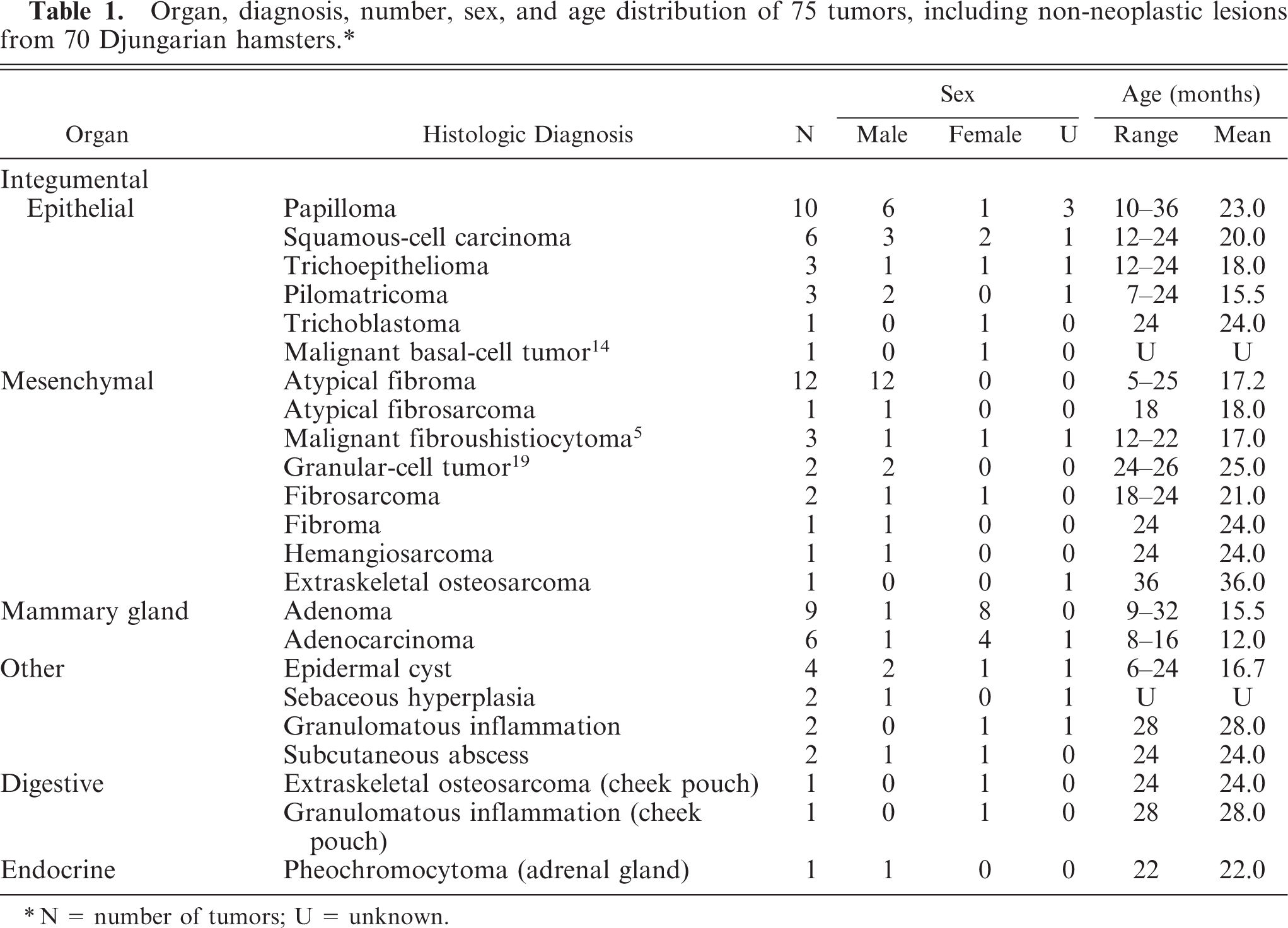
N = number of tumors; U = unknown.
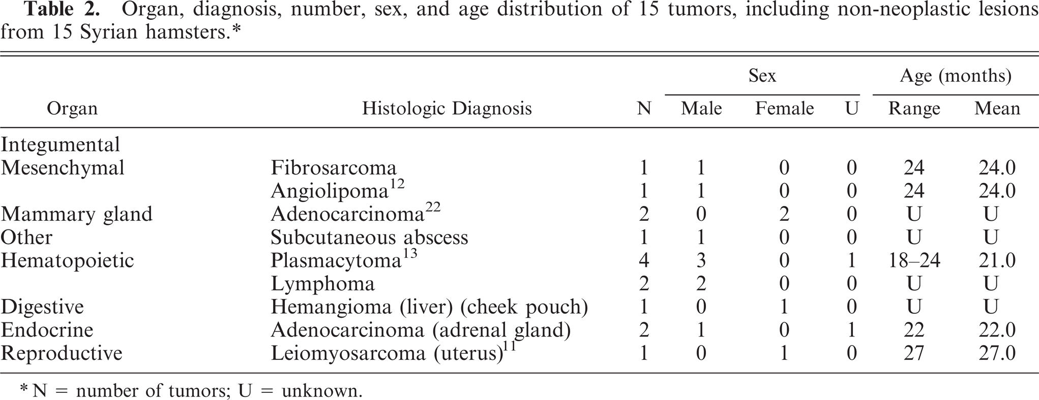
Organ, diagnosis, number, sex, and age distribution of 15 tumors, including non-neoplastic lesions from 15 Syrian hamsters.∗
N = number of tumors; U = unknown.
Djungarian hamsters
Integumental neoplasms were the most frequent, and various types were present. The most common integumental neoplasms were mammary tumors (n = 15; including benign and malignant) (Fig. 1), atypical fibroma (13, including atypical fibrosarcoma), papilloma (10) (Fig. 2), and squamous-cell carcinoma (6) (Fig. 3). Anatomical locations of integumental neoplasms are given in Table 3. Atypical fibromas were composed of proliferation of neoplastic GL cells, with various amounts of collagen fibers (Fig. 4). Cellularity of neoplastic GL cells varied by case. Local invasion, high cellularity, and prominent pleomorphism of the nucleus and the cytoplasm of neoplastic GL cells were observed in one case. On immunohistochemical examination, most neoplastic GL cells had intense cytoplasmic staining for vimentin, and the nuclei were positive for AR. Neoplastic GL cells were negative for cytokeratin, S-100 protein, neurofilament, and synaptophysin. Non-neoplastic lesions of the integumental system included epidermal cysts (4), sebaceous hyperplasia (2), subcutaneous abscess (2), and granulomatous inflammation (2). Neoplasms that occurred in internal organs were rare, and one osteosarcoma of the cheek pouch and one pheochromocytoma were observed.
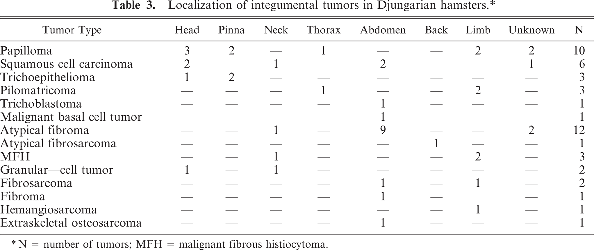
Localization of integumental tumors in Djungarian hamsters.∗
N = number of tumors; MFH = malignant fibrous histiocytoma.
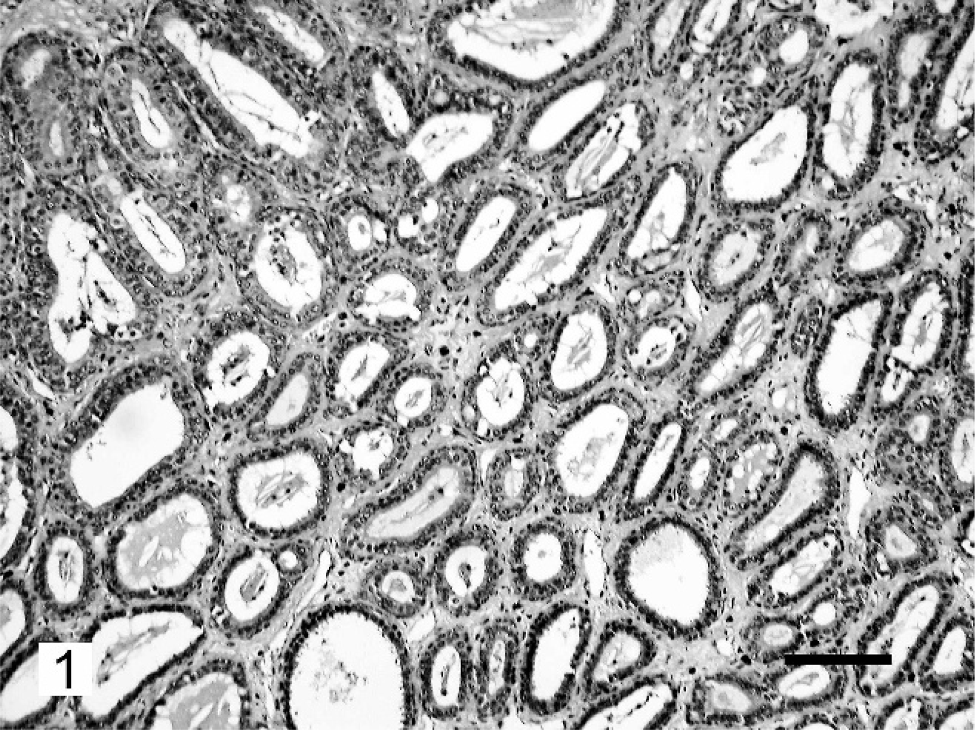
Mammary adenoma; Djungarian hamster. Proliferation of well-differentiated luminal epithelial cells. HE. Bar = 100 µm.
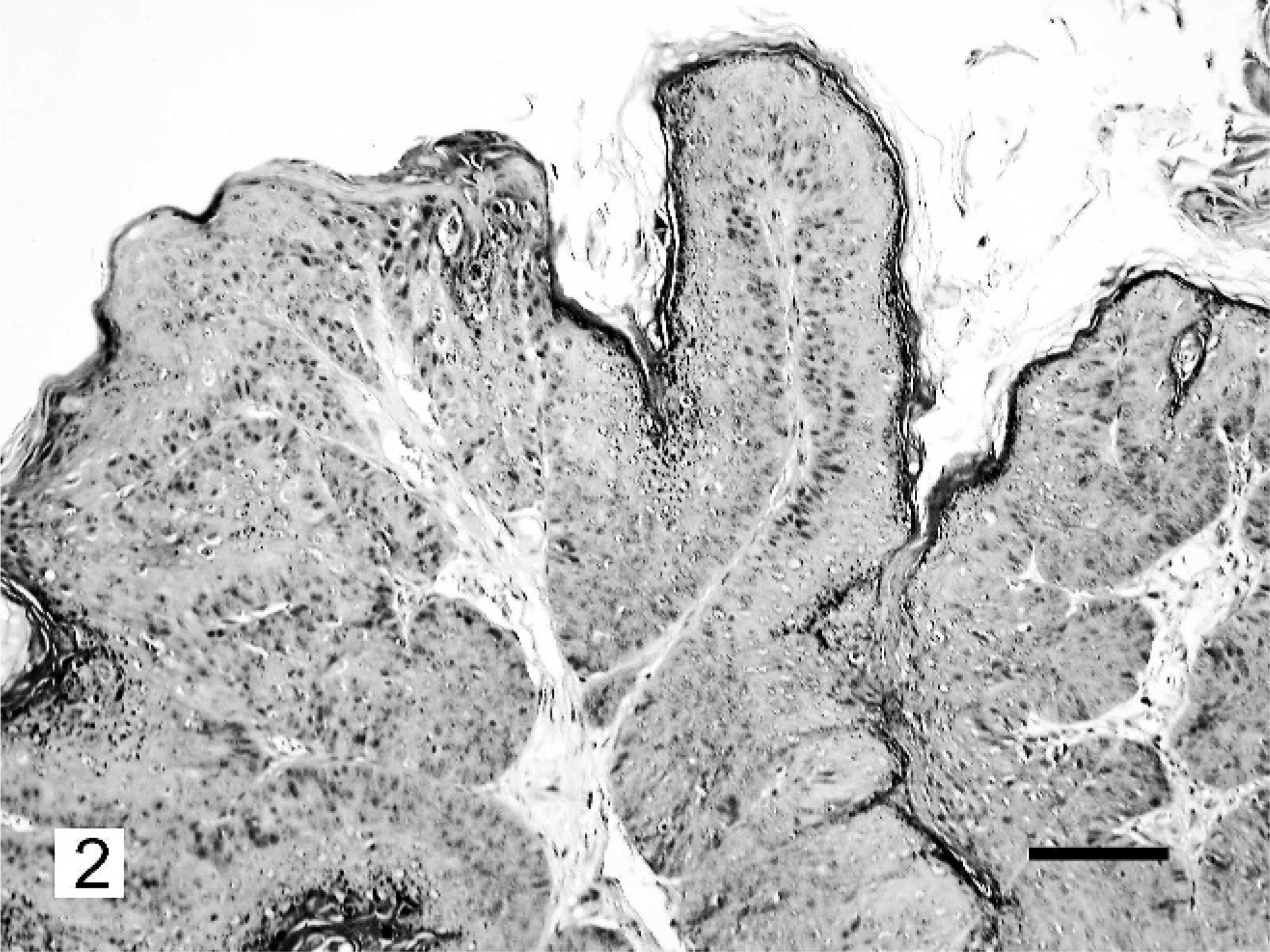
Papilloma; Djungarian hamster. Exophytic neoplastic proliferation of the squamous epithelium. HE. Bar = 100 µm.
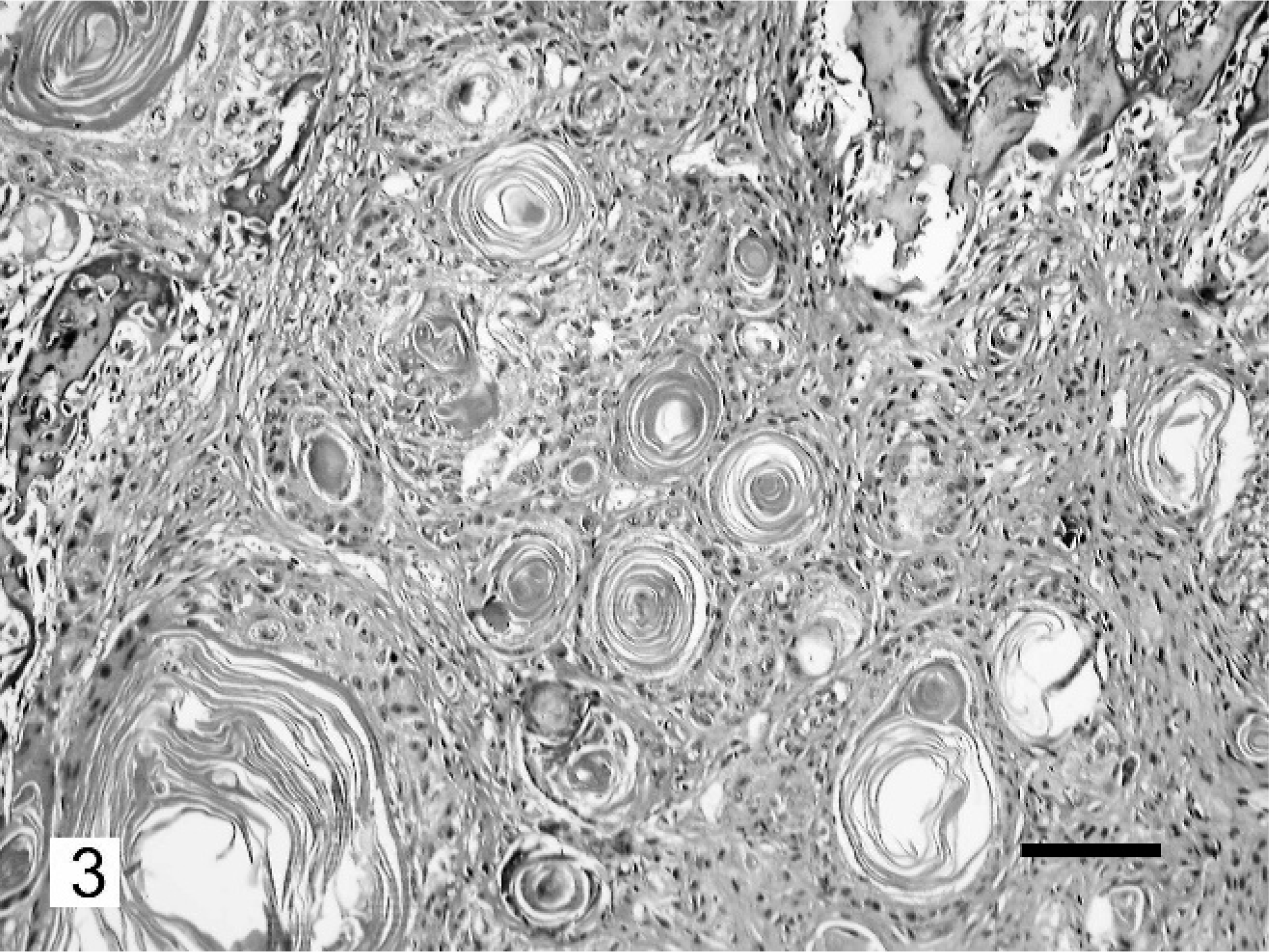
Squamous-cell carcinoma; Djungarian hamster. Trabeculae of neoplastic epithelial cells with formation of keratin pearls. HE. Bar = 100 µm.
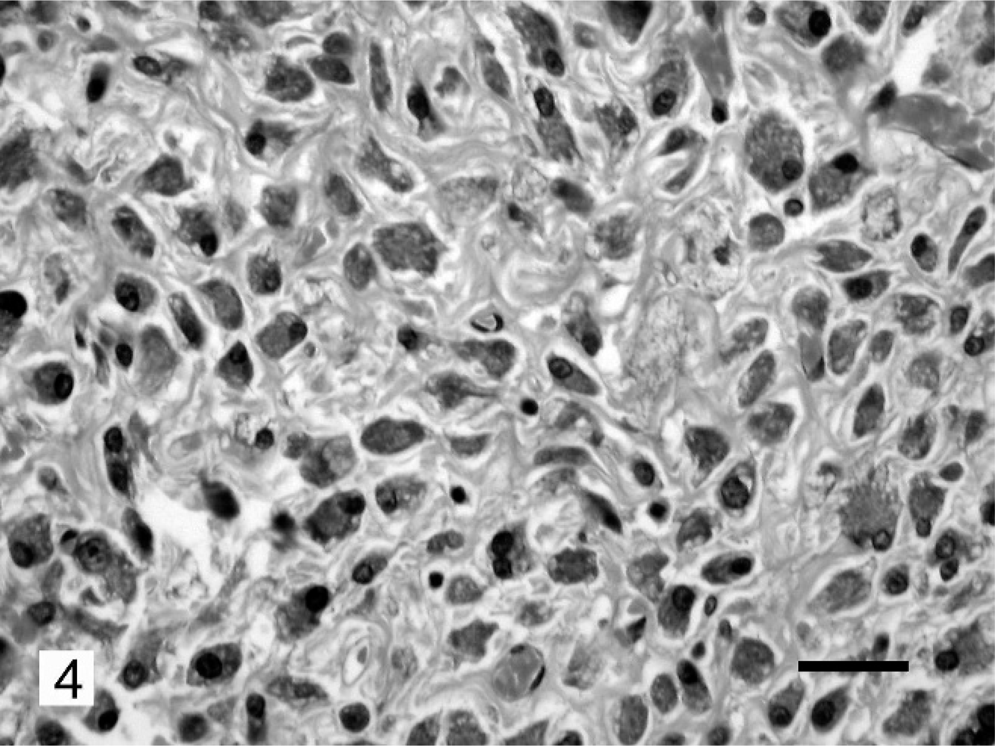
Atypical fibroma; Djungarian hamster. Proliferation of neoplastic GL cells with various amounts of collagen fiber. HE. Bar = 25 µm.
Syrian hamsters
The prevalence of integumental tumors in the Syrian hamsters was low in comparison with that in the Djungarian hamsters. Hematopoietic tumors, including plasmacytomas (4) (Fig. 5) and lymphomas (2) (Fig. 6), were the most common. All hematopoietic tumors in Syrian hamsters were found as cutaneous masses and were located on the neck. Neoplasms that occurred in internal organs included hemangioma of the liver (1), adrenocortical adenocarcinomas (2), and uterine leiomyosarcoma (1). Only one non-neoplastic tumor was found, a subcutaneous abscess.
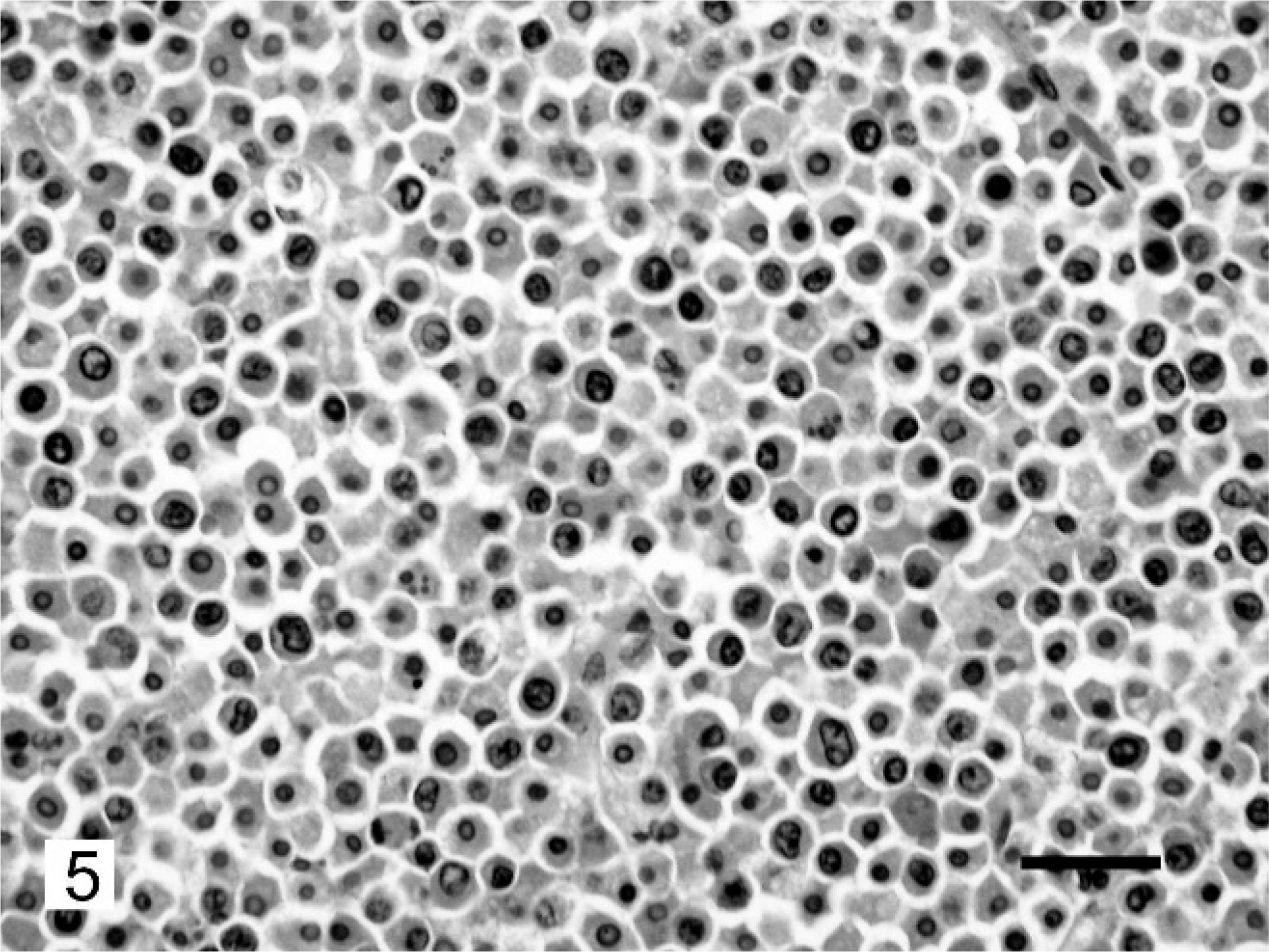
Plasmacytoma; Syrian hamster. Diffuse proliferation of relatively uniform plasmacytoid cells with eccentrically placed nuclei. HE. Bar = 25 µm.
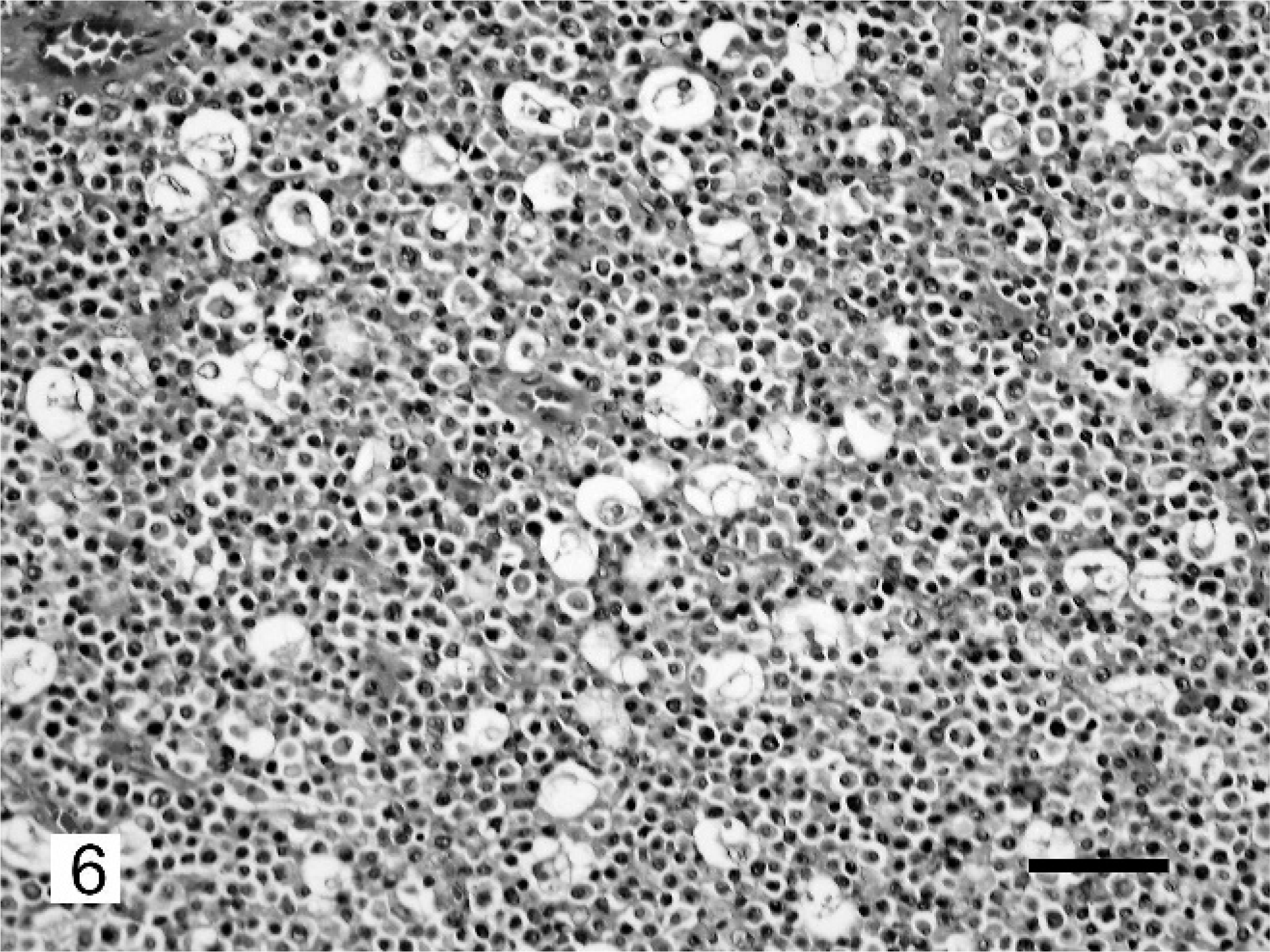
Lymphoma; Syrian hamster. Solid sheets of neoplastic lymphocytes and starry-sky appearance. HE. Bar = 50 µm.
Discussion
In the present study, the total number of spontaneous tumors in Djungarian hamsters was markedly greater than in Syrian hamsters. According to Kamino et al., 9 laboratory Syrian hamsters have a low prevalence of spontaneous neoplastic lesions, whereas Cooper et al. 4 reported that laboratory Djungarian hamsters show a high prevalence of neoplastic disease. The results of our study support the previously reported prevalence of spontaneous tumors in laboratory Djungarian and Syrian hamsters. These species have different numbers of chromosomes, thus resulting in differences in the prevalence and frequency of spontaneous tumors.
According to Pogosianz, 15 the spectrum of spontaneous tumors in laboratory Djungarian hamsters differs from that in Syrian hamsters. The spectrum of affected sites and tumor types also differed markedly between the Djungarian and Syrian hamsters in the present study on domestic hamsters. The majority of tumors in the Djungarian hamsters were integumental, and various types of tumor were identified. In the Syrian hamsters, hematopoietic tumors, which were not seen in the Dungarian hamsters, were the most common. The age of hamsters with neoplasms varied widely. The normal life span of hamsters is 18 to 24 months, and older individuals are common. 7 In our study, the mean age of hamsters with neoplasms was considered to be relatively old compared with the life span.
In laboratory Djungarian hamsters, tumors of the mammary glands, skin, and lungs were predominant. 15, 16 Although no tumors of the respiratory system were observed in domestic hamsters, various neoplasms were seen in other body systems, which is in agreement with reports in laboratory Djungarian hamsters. Integumental neoplasms, including mammary tumors, were the most common in our study. Sex predilection was prominent in the occurrence of mammary tumors and atypical fibromas.
Most mammary tumors were seen in the female Djungarian hamsters; however, 2 cases were in the male animals. Although there are no reports on mammary tumors in male domestic and laboratory Djungarian hamsters, the prevalence of mammary tumors may not be rare. Interestingly, the mean age of Djungarian hamsters with mammary tumors was lower than for all other tumors.
Atypical fibromas are characteristically composed of proliferating GL cells of the skin. Atypical fibromas are reported predominantly in male Djungarian hamsters and are seldom seen in females, because they are androgen-dependent tumors. 1 Kashida et al. 10 suggested that GL cells in Djungarian hamsters are derived from intrinsic undifferentiated mesenchymal cells in the dermis or subcutaneous adipose tissue. Cooper et al. 4 reported fibromas and fibrosarcomas in laboratory Djungarian hamsters, but whether they were atypical fibromas is unknown. There are no reports about the occurrence of atypical fibromas in domestic or laboratory Syrian hamsters. In our study, all atypical fibromas occurred in males, which confirmed the reported sex predilection. Neoplasms composed of GL cells with malignant findings were provisionally diagnosed as atypical fibrosarcomas. According to Baba et al., 1 predilection sites of atypical fibromas are the thoracoabdominal area. In our study, anatomical localization of most atypical fibromas and immunohistochemical characteristics were consistent with previous reports.
Papillomas have been reported in laboratory Djungarian hamsters. 15, 16 There are no previous reports of papillomas in domestic Djungarian hamsters. We believe that the prevalence of papillomas is high, because 10 cases were observed in our study. The head area, including the pinna, is an apparent predilection site for papillomas.
In laboratory Syrian hamsters, hematopoietic tumors, particularly lymphomas, are the most common type of spontaneously occurring tumor, and plasmacytomas are relatively rare. 2 Lymphoma that resembles mycosis fungoides was also reported in domestic Syrian hamsters. 20 Plasmacytomas were more frequent than lymphomas in our study, and our results were different from those in laboratory Syrian hamsters. Tumors of the adrenal cortex are one of the most common types of spontaneous neoplasm in laboratory Syrian hamsters and have also been reported in domestic Syrian hamsters. 3, 21 We diagnosed 2 adrenocortical adenocarcinomas. Although the population of domestic Syrian hamsters is unknown, the small number of Syrian hamsters in our study may reflect the low prevalence of spontaneous tumors in this species. However, further studies and a large numbers of cases are necessary to identify predilection site, sex, and age.
The prevalence, spectrum, and site of spontaneous tumors varied among different hamster strains. 9, 17, 18 Although it is difficult to clarify the origins and total populations of domestic hamsters, regional differences in tumor incidence and spectrum may be present. Epidemiologic studies on tumor prevalence in domestic Syrian and Djungarian hamsters in different regions are thus necessary. To our knowledge, this is the first comprehensive study of tumors in domestic hamsters.
We examined biopsy specimens from 2 species of domestic hamster, and the spectrum of tumors was identified. In particular, domestic hamsters primarily developed integumental tumors rather than the multiorgan tumors reported in laboratory hamsters. However, if complete necropsy examinations were performed, various other tumors may have been found.
Footnotes
Acknowledgements
This study was supported by a Grant-in-Aid-for JSPS Fellows (195323) from the Japan Society for the Promotion of Science.