
Abstract
We induced hypothyroidism in rats by conducting a thyroidectomy (TD) and investigated subsequent changes in the morphology of the skin, especially that of the epidermis and hair follicles. The 6 rats in the TD group seemed less active than the 3 rats in the control group and had cold, dry paws. All of the rats in the TD group exhibited retarded hair growth 12 weeks after surgery. Histologically, all of the rats in the TD group exhibited epidermal thinning from 12 weeks after surgery. Many hair follicles were in the telogen phase: the bulbs and papillae were involuted and had migrated towards the epidermis. Hair follicle atrophy involving thinning of the outer root sheath and the inner root sheath was often observed. The immunoreactivities of antithyroid hormone receptors α and β in the outer root sheaths of 5 of the TD rats were weaker than those of control rats. Cell proliferation in hair follicles of TD rats was weaker than in follicles of control rats 4 weeks after surgery. It is suggested that decreased expression of TRs and decreased cell proliferation activity in the hair follicles of rats is associated with a lack of thyroid hormone and results in retardation of hair growth.
Hypothyroidism is a common cause of alopecia in humans and animals, especially dogs. 8, 12, 13 Thyroid hormone affects organs throughout the body because it regulates the activity of enzymes and proteins that participate in metabolism at the cellular level. In the human hair follicle, the thyroid hormone receptor (TR) participates in hair development. 1, 2 However, little research has been conducted on TRs and hair in animals or on the pathologic mechanism responsible for alopecia caused by hypothyroidism.
Thyroidectomized animals are widely used to study hypothyroidism and thyroid hormone function. Although there are many reports on the effects of thyroidectomy (TD) on organs such as the anterior pituitary, liver, and kidneys, 7, 14 little research has been conducted on the effects of TD on the skin. 3
In this study, we induced hypothyroidism in rats by removing both lobes of the thyroid and investigated the subsequent morphologic changes in the skin, especially those in the epidermis and hair follicles.
Materials and Methods
Nine female Wistar rats (7 weeks old, 170–200 g) were kept under normal housing conditions (21°C ± 1°C, 50–60% humidity, light from 6:00 a.m. to 6:00 p.m.) with free access to food and water. Six rats were thyroidectomized (TD group) under diethyl ether as previously described, 20 and 3 rats underwent a sham operation (control group). Thyroidectomized rats were given drinking water containing 1% CaCl2 to maintain calcium balance because the parathyroid gland was removed together with the thyroid gland. At 4, 8, 12, 16, and 32 weeks after surgery, skin samples were obtained under ether anesthesia from the dorsal trunk of the animals. The skin was repeatedly sampled from the same animal. Dorsal trunk skin was classified into five places (the most rostral part for 4 weeks, the most caudal part for 32 weeks), and the skin was removed every 4 weeks after shaving an approximately 2-by-2-cm area. The skin was routinely sutured after sampling of the skin. Skin samples were fixed in 10% formalin and used for histologic and immunohistochemical analyses.
Blood samples for assay of thyroid hormonal levels were collected under anesthesia from the heart at 32 weeks after surgery, after which the animals were killed by severing the abdominal aorta. Blood samples were centrifuged at 3,000 × g for 15 minutes, and serum total thyroxine (T4) and free thyroxine (FT4) concentrations were determined using an electrochemiluminescence immunoassay and a radioimmunoassay, respectively (SRL Co. Ltd., Tokyo, Japan). Data were expressed as means plus or minus standard deviations (SDs). The significance of differences between means was determined by using Student's t-test. A null hypothesis probability of less than 0.05 was considered statistically significant.
Tissue samples that had been fixed in 10% formalin were dehydrated and embedded in paraffin. Tissue sections were cut to a thickness of 4 µm and stained with HE. For morphometric study of epidermis thickness (from the bottom of the corneal layer to the basal membrane) using HE staining specimen, 25 epidermis thickness of each groups were measured by using Motic images plus 2.0S (Shimazu Co., Kyoto, Japan) and the means plus or minus SDs was calculated. The significance of differences between means was determined by using Student's t-test. A null hypothesis probability of less than 0.05 was considered statistically significant. For the immunohistochemical study, deparaffinized sections were rinsed with phosphate buffered saline and endogenous peroxidase activity was blocked by using 3% hydrogen peroxide diluted in methanol. The sections were incubated for 24 hours at 4°C with a primary antibody: antithyroid hormone receptor α1/2 (TRα) rabbit serum (Abcam Ltd., Cambridge, UK), antithyroid hormone receptor β1 (TRβ) rabbit serum (Santa Cruz Biotechnology, Inc., Santa Cruz, CA, USA), or antiproliferating cell nuclear antigen (PCNA) mouse serum (Dako Cytomation, Inc., CA, USA). The sections were then incubated for 1 hour at room temperature with the secondary antibody, peroxidase-conjugated anti-rabbit or anti-mouse IgG (Nichirei Biosciences Inc., Tokyo, Japan). The reactions were visualized by using 3, 3′-diaminobenzidine tetrahydrochloride (DAB) (Nichirei Biosciences Inc.). The percentage of PCNA immunoreactive nuclei in 3,000 hair follicle cells (outer root sheath, inner root sheath, connective root sheath, and hair matrix) per skin section was calculated and used for analysis of PCNA-positive cells in hair follicles. The significance of differences between means was determined by using Student's t-test. A null hypothesis probability of less than 0.05 was considered statistically significant.
The experimental procedures and care of animals were in accordance with the guidelines of the Animal Care and Use Committee of Kitasato University.
Results
Gross inspection
On gross inspection, the TD rats seemed less active than the control rats and had cold, dry paws. In the control rats, the parts that had been shaved and sampled at 4 and 8 weeks after surgery were once again covered with hair at 12 weeks after surgery (Fig. 1). In contrast, hair growth was not observed in the TD group and shaved part in 8 weeks had been bald (Fig. 2). Thirty-two weeks after surgery, the control rats had normal skin and hair, but in the TD rats, hair growth was not observed in the sections that had been sampled at 16 weeks.
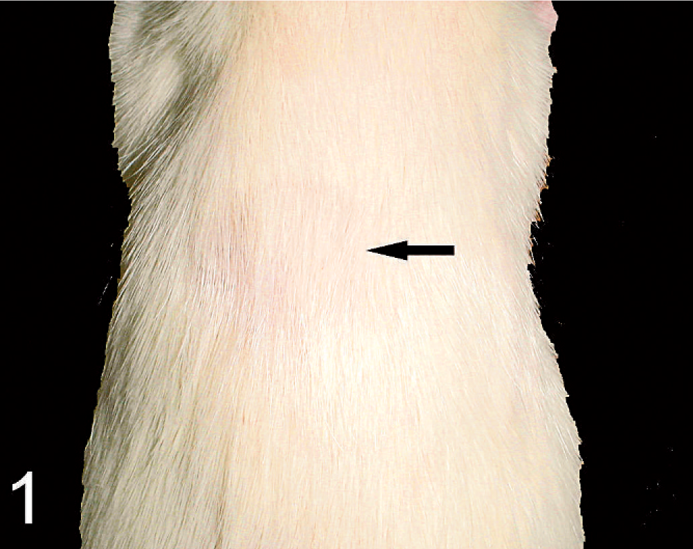
Control rat. Twelve weeks after a sham operation. The shaved areas at 4 and 8 weeks (arrow) are again covered with hair.
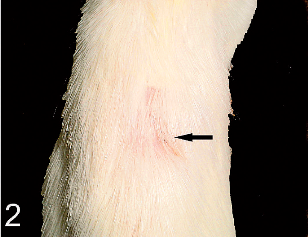
TD rat. Twelve weeks after TD. Hair regrowth is not complete 8 weeks after shaving (arrow).
Serum analysis
T4 and FT4 levels at 32 weeks after surgery were significantly lower in the TD rats than in the control rats (P < .01) (Table 1).
Result of serum analysis in TD rats and control group rats.
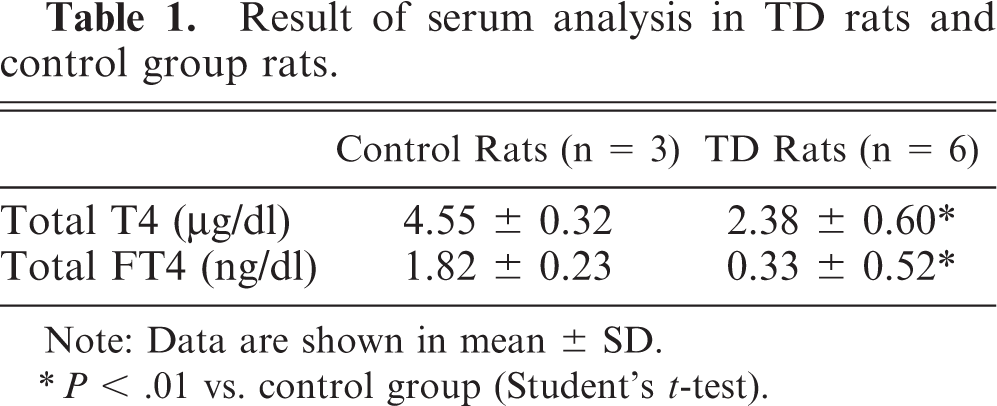
Note: Data are shown in mean ± SD.
P < .01 vs. control group (Student's t-test).
Light microscopic analysis
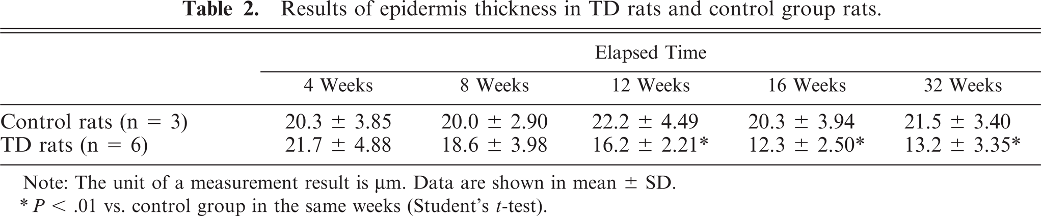
At all sampling time points, the epidermis of the control rats was composed of a basal layer, a spinous layer, a granular layer, and a corneal layer. The cells of the granular layer, containing many keratohyalin granules, were observed clearly (Fig. 3). In contrast, in the TD rats the epidermal layer was composed of only the basal layer and the corneal layer. The basal cells were flattened. The epidermis thinning of the TD rats, as compared with that of the control rats, was observed at 12, 16, and 32 weeks after surgery (Table 2, Fig. 4).
Results of epidermis thickness in TD rats and control group rats.
Note: The unit of a measurement result is μm. Data are shown in mean ± SD.
P < .01 vs. control group in the same weeks (Student's t-test).
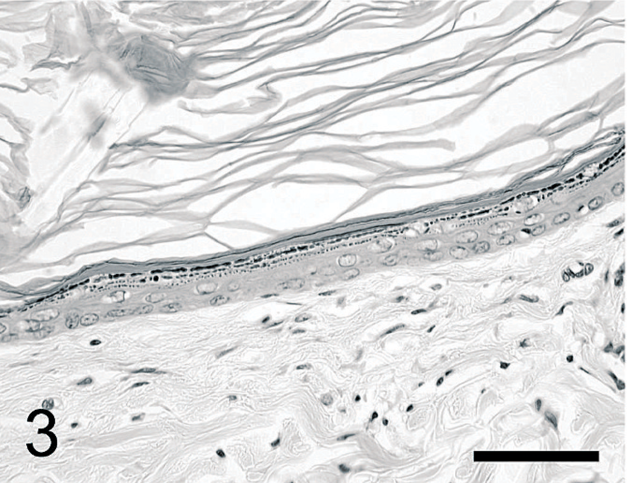
Epidermis; control rat. Thirty-two weeks after a sham operation. The epidermis consists of a basal layer, a spinous layer, a granular layer, and a corneal layer. HE. Bar = 50 µm.
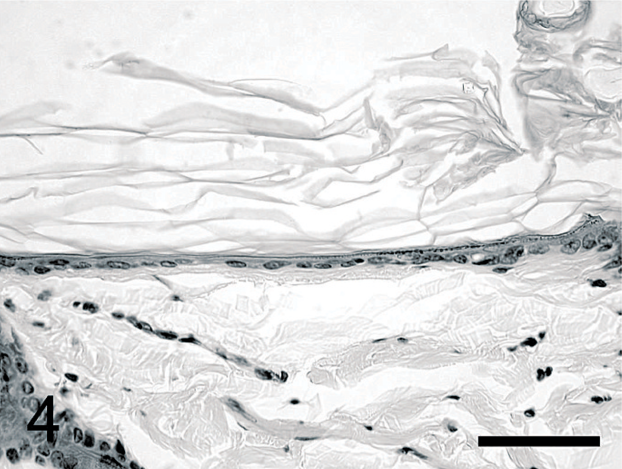
Epidermis; TD rat. Thirty-two weeks after TD. The epidermis consists only of the basal and corneal layers. The nuclei of the basal cell layer are dark. HE. Bar = 50 µm.
At 4 weeks, the hair follicles of the control rats were deeply invaginated into the skin and subcutaneous hair bulbs in the anagen phase were often observed (Fig. 5). These hairs had well-developed outer root sheaths, inner root sheaths, hair papillae, and hair matrixes (Fig. 6). At 12 weeks, many follicles were in the telogen phase: the hair bulbs and hair papillae were involuted and had migrated towards the epidermis. At 16 weeks, anagen-phase hair follicles were observed again. Thus, the hair cycle of control, anagen, and telogen phases was repeated in about 8 weeks. However, the hair follicles of the TD rats showed the telogen phase in all stages (Fig. 7), and hair follicle atrophy involving reduction of the thickness of the outer root sheath and the inner root sheath was often observed (Fig. 8).
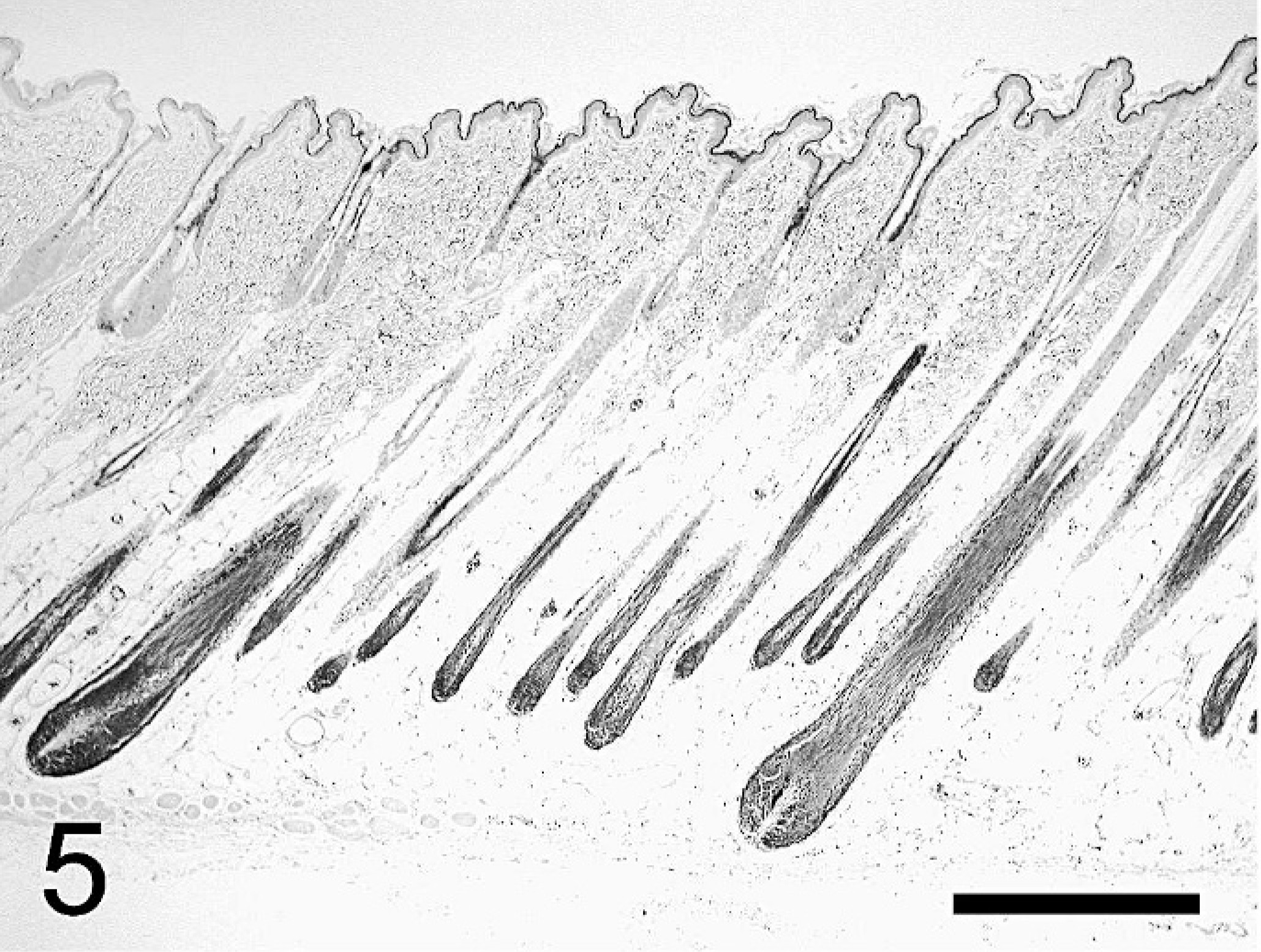
Hair follicle; control rat. Thirty-two weeks after a sham operation. Many hair follicles are in the anagen phase. HE. Bar = 500 µm.
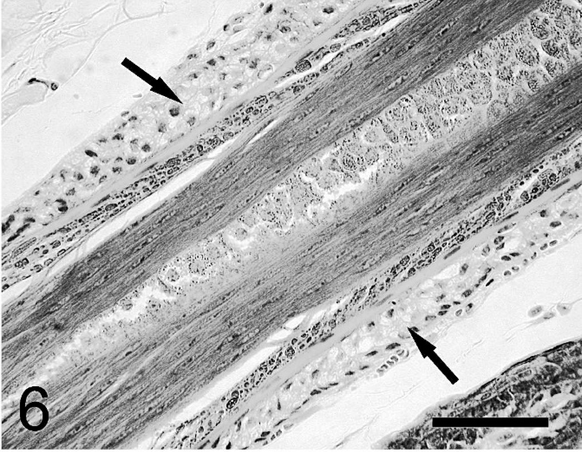
Hair follicle; control rat. Thirty-two weeks after a sham operation. The outer root sheath (arrows) is well developed. HE. Bar = 50 µm.
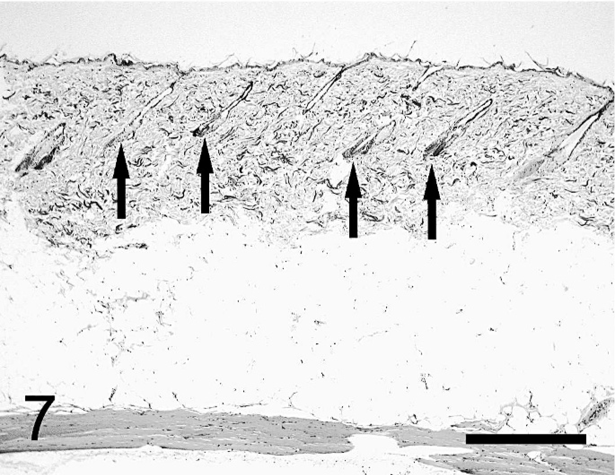
Hair follicle; TD rat. Thirty-two weeks after TD. TD rats exhibit many follicles in the telogen phase (arrows). HE. Bar = 500 µm.
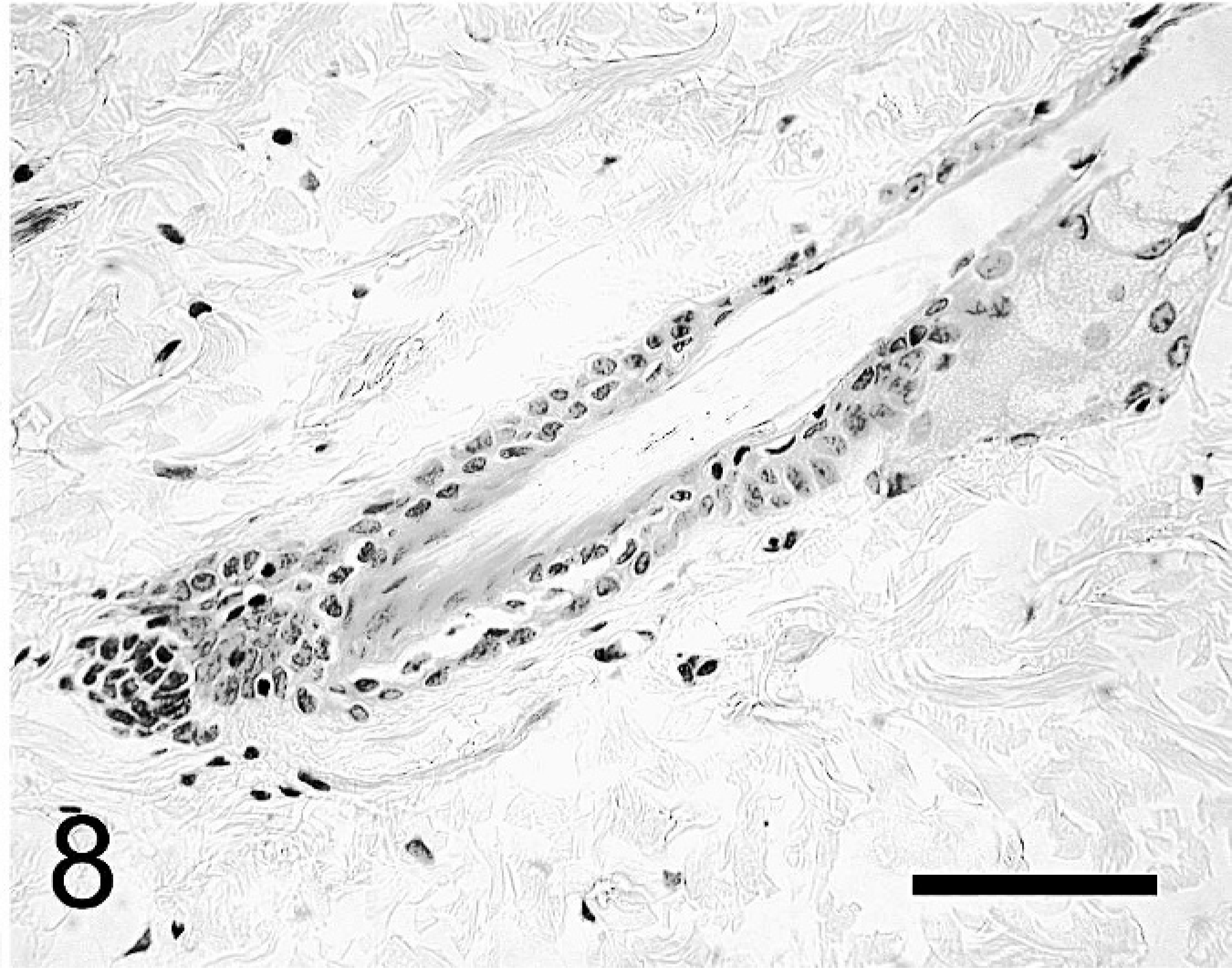
Hair follicle; TD rat. Thirty-two weeks after TD. Few outer root sheaths and inner root sheaths of follicles are evident. HE. Bar = 50 µm.
Immunohistochemistry
TRα-, TRβ-, and PCNA-positive reactions were observed in the cell nuclei of both groups.
TRα-positive reactions. In the control group, anti-TRα was expressed in all epidermal layers except the corneal layer, parts of the dermis containing fibroblasts, and subcutaneous tissue. Hair matrices and outer root sheaths stained strongly for anti-TRα, but the inner root sheath and the connective tissue sheath did not stain positively (Fig. 9). Positive reactions for anti-TRα were observed in sebaceous glands and arrector pili muscles. In skin tissue from TD rats, regions that stained positive for anti-TRα were similar to those in control rats. However, in 5 of the 6 TD rats, the intensity of staining weakened with time, becoming barely visible in the outer root sheath at 32 weeks after surgery (Fig. 10).
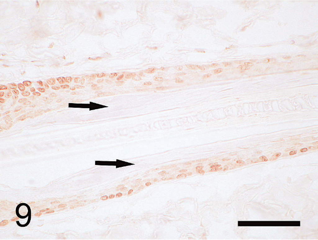
Hair follicle; control rat. Thirty-two weeks after a sham operation. The hair matrices and outer root sheaths were strongly stained, but the inner root sheaths (arrows) and connective tissue sheaths did not stain positively. Immunohistochemistry for TR α, DAB staining. Bar = 50 µm.
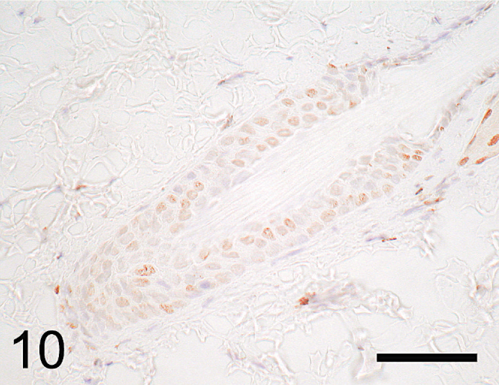
Hair follicle; TD rat. Thirty-two weeks after TD. Stained regions are similar to those of the control rats, but the intensity of staining weakened. Immunohistochemistry for TR α, DAB staining. Mayer hematoxylin counterstain. Bar = 50 µm.
TRβ-positive reactions. Immunoreactivity to anti-TRβ was observed in subcutaneous adipose cells, cutaneous muscles, vascular endothelial cells, and all epidermal layers except the corneal layer. The outer root sheath reacted strongly, and the inner root sheath, connective tissue sheath, and hair matrix were stained (Fig. 11). Positive reactions to anti-TRβ as well as anti-TRα were observed in sebaceous glands and arrector pili muscles. In the TD rats, skin regions that stained positive for anti-TRβ were similar to those in the control rats, but the intensity of staining weakened with time in a manner similar to that observed for anti-TRα (Fig. 12).
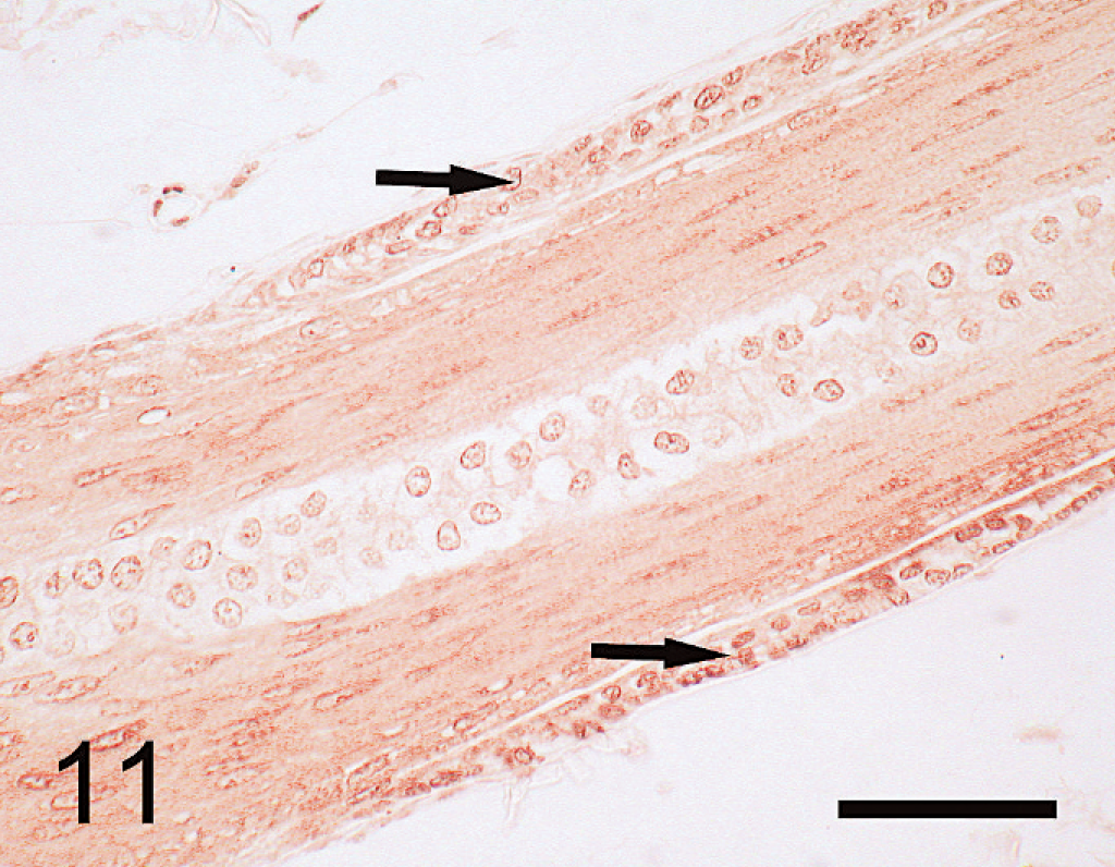
Hair follicle; control rat. Thirty-two weeks after a sham operation. The outer root sheath stained strongly (arrows), and inner root sheath, connective tissue sheath, and hair matrix stained moderately. Immunohistochemistry for TR β, DAB staining. Bar = 50 µm.
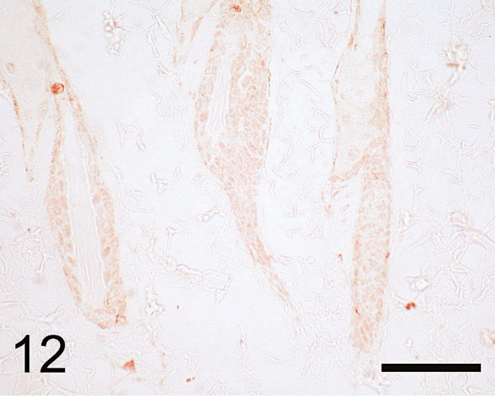
Hair follicle; TD rat. Thirty-two weeks after TD. Stained regions are similar to those of the control rats, but the intensity of staining is weaker. Immunohistochemistry for TR β, DAB staining. Bar = 50 µm.
Statistical analysis of PCNA. In the control rats, many PCNA-positive cells were observed in the outer root sheaths and hair matrix cells in anagen-phase hair follicles. In telogen-phase hair follicles of the TD rats, fewer cells were positive for PCNA than in those of the control rats. The percentage of PCNA-immunoreactive nuclei in hair follicle cells of rats in the TD group was significantly less than that of the control group at 4 weeks after surgery (P < .01) (Fig. 13).
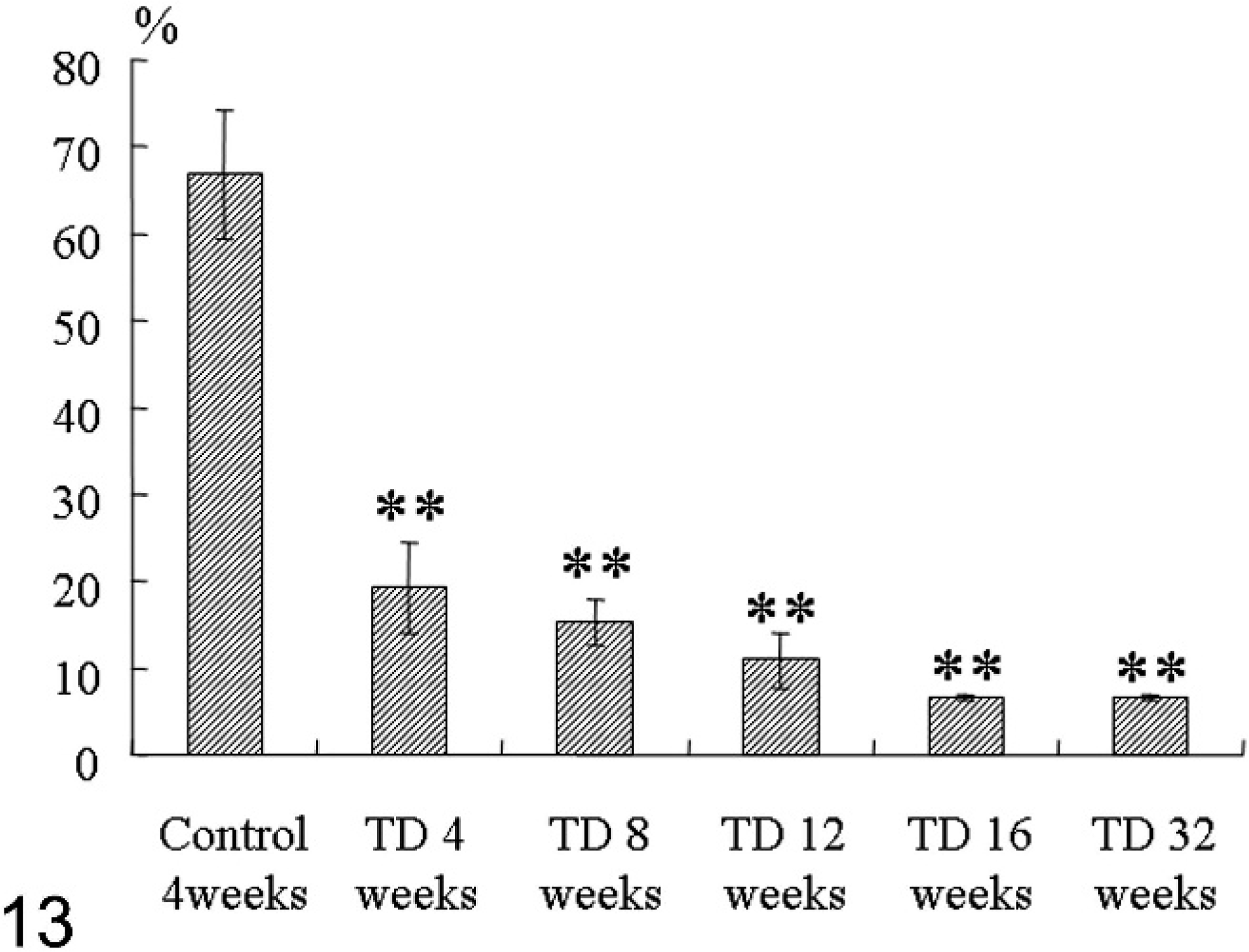
Results of statistical analysis of the percentage of PCNA-positive cells in hair follicles of rats subjected to TD or a sham operation (control group). The significance of differences between the means for the control and TD groups was determined using Student's t-test. ∗∗P < .01 was considered statistically significant at 4 weeks.
Discussion
The clinical signs observed in the TD rats are similar to those of primary hypothyroidism in dogs and humans. We thought that epidermal thinning was a characteristic finding of the TD rat. The epidermal thinning is described by human hypothyroidism. 10, 11 Also, it is reported that changes in the thickness of the epidermis are variable in dogs with hypothyroidism. 12 Hypothyroidism is a common cause of alopecia in humans and animals, especially dogs. 8, 12, 13 However, in the TD rats, alopecia was not observed, but hair growth retardation was observed. It is reported that although the epidermis of rats is thinner than in humans, there is a greater density of hair follicles, overwhelmingly higher than in humans and marmosets. 18 Such a density of hair follicles may be related to the differences in morphology of the skin of the TD rats and the skin in spontaneous cases of hypothyroidism in other species. Additionally, the removal of the parathyroid gland together with the thyroid gland may have influenced the outcome.
The growth retardation and morphologic changes that occurred in the hair of the TD rats indicate that thyroid hormone is important for development of hair follicles and a normal hair cycle. 4, 5, 16, 17 Thyroid hormone affects all organs of the body. In hair follicles, thyroid hormone may initiate the anagen phase and promote hair growth. Thus, a thyroid hormone deficiency may delay the anagen phase and promote the telogen phase. Most cases of hypothyroidism in dogs are caused by thyroid disease, and skin abnormalities are observed in 80% of cases. 12, 13 These skin abnormalities are characterized by delayed hair growth and an increased numbers of follicles in the telogen phase. 12 In humans, a similar abnormality of the hair cycle is associated with decreased thyroid hormone levels. 8 It is thus reasonable to assume that the observed morphologic changes in the hair follicles of the TD group were caused by the absence of thyroid hormone consequent to TD.
The results of immunohistochemical analyses indicated that TRα and TRβ are widely distributed throughout the skin of the rat. In the hair follicle, the outer root sheaths and hair matrix cell nuclei were positive for both anti-TRα and anti-TRβ; in contrast, the inner root sheath and dermal papilla cells were positive for anti-TRβ only. In humans, most cells of the hair follicle express TRβ and few express TRα. 1, 2 Several isoforms of the TR have been identified (α1, α2, β1, and β2), and it is known that these are organ specific. 9, 15, 19 Although TRα knockout mice differ little from wild-type mice, TRβ knockout mice exhibit thyroid hormone resistance syndrome. 6, 21 It is suggested that the roles of TR isoforms may differ, respectively. 6, 21 Consequently, the effect of thyroid hormone on hair growth and the hair follicle in rats may differ from that in humans.
The decrease in immunoreactivity to anti-TRα and anti-TRβ over time in 5 of the 6 TD rats indicates that down-regulation of TRs by decreased thyroid hormone. In addition, the paucity of PCNA-positive cells in the hair follicles of the TD rats appears to be associated with the expression of TR and decreased cell proliferation. Thyroid hormone exerts its effects by binding to TRs in the cell nucleus. The level of expression of TR mRNA in the heart, liver, and other organs depends on the level of thyroid hormone in serum. 15 Safer et al. 17 reported that topical triiodothyronine stimulates epidermal proliferation, dermal thickening, and hair growth in mice and rats, and suggested that thyroid hormone promotes proliferation of the epidermis and hair directly. Therefore, the decrease in cell proliferation activity in the TD rats appears to have been caused by a lack of thyroid hormone, as was the case with the elevated numbers of follicles that had atrophied or were in the telogen phase in these rats.
In conclusion, it is suggested that decreased expression of TRs and decreased cell proliferation activity in the hair follicles of TD rats is associated with a lack of thyroid hormone and results in retardation of hair growth.