
Abstract
A progressive wart-like syndrome in both captive and wild populations of the Western barred bandicoot (WBB) is hindering conservation efforts to prevent the extinction of this endangered marsupial. In this study, 42 WBBs exhibiting the papillomatosis and carcinomatosis syndrome were examined. The disease was characterized by multicentric proliferative lesions involving cutaneous and mucosal surfaces, which were seen clinically to increase in size with time. Grossly and histologically the smaller skin lesions resembled papillomas, whereas the larger lesions were most commonly observed to be squamous cell carcinomas. Large amphophilic intranuclear inclusion bodies were observed in hyperplastic conjunctival lesions of 8 WBBs under light microscopy. Conjunctival lesions from 2 WBBs examined using transmission electron microscopy contained a crystalline array of spherical electrondense particles of 45-nm diameter, within the nucleus of conjunctival epithelial cells, consistent with a papillomavirus or polyomavirus. Conjunctival samples from 3 bandicoots that contained intranuclear inclusion bodies also demonstrated a positive immunohistochemical reaction after indirect immunohis-tochemistry for papillomavirus structural antigens. Ultrastructural and/or immunohistochemical evidence of an etiologic agent was not identified in the nonconjunctival lesions examined. Here we describe the gross, histopathologic, ultrastructural, and immunohistochemical findings of a papillomatosis and carcinomatosis syndrome recently identified in the WBB.
Infectious diseases have the potential to drive population declines and contribute to species extinction. 14,15 The Western barred bandicoot (WBB), Perameles bougainville, is an endangered Australian marsupial that was once widespread across western and southern Australia. Now extinct on the mainland, wild populations are known only to exist on Bernier and Dorre Islands, in the World Heritage Area of Shark Bay Western Australia. 37,41,42,49 Conservation efforts to prevent the extinction of this species are currently hampered by a progressively debilitating cutaneous and muco-cutaneous papillomatosis and carcinomatosis syndrome observed in both captive and wild WBBs. Affected animals develop multicentric papillomatous lesions, which appear clinically to progress to larger sizes. These lesions are extremely debilitating. Involvement of the feet, eyes, and mouth leads to problems with ambulation, vision, and eating. Affected animals often die due to secondary complications or are euthanatized on welfare grounds. Awareness of the papillomatosis syndrome first arose in 1999, when lesions were noticed developing on a captive adult female housed at Kanyana Wildlife Rehabilitation Center, Perth. 48 Within a year, additional WBBs housed at the facility were noticed to have developed lesions. Similar cutaneous lesions were also noticed on captive WBBs at another breeding colony in Shark Bay and identified on wild WBBs on Bernier Island by the Western Australian Department of Environment and Conservation (DEC) in 2001. A historic trace back performed by one of the authors (SH) on 93 museum specimens from 5 museums in Australia found similar lesions to be present on Western Australian Museum WBB specimens dating back to 1982. 22 The purpose of this report is to describe the gross, histologic, ultrastructural, and immunohistochemical features of a multicentric papillomatosis and carcinomatosis syndrome identified in 42 WBBs.
Materials and Methods
Study population
Between 2000 and 2006, submissions from 42 WBBs displaying focal or multifocal cutaneous and/or muco-cutaneous thickenings or masses were received by the pathology section at the Murdoch University School of Veterinary and Biomedical Sciences. Essential criteria for inclusion of WBBs in this study were epithelial lesions with gross and histologic characteristics consistent with either papilloma formation and/or squamous cell carcinomas, as classified by Goldschmidt et al. 1998. 18 All animals in the study were sourced from captive populations of WBBs held at either Kanyana Wildlife Rehabilitation Center, Gooseberry Hill, Western Australia; Dryandra Field Breeding Center, near Narrogin, Western Australia; the Peron Captive Breeding Center, Shark Bay, Western Australia; or the Adelaide and Monarto Zoological Gardens, South Australia.
Submissions
Submissions consisted of either surgical biopsies collected into 10% neutral buffered formalin or entire deceased WBBs for postmortem examination. Entire animals submitted for postmortem were subjected to a complete postmortem examination; tissue samples were collected from skin lesions and all the major organ systems into 10% neutral buffered formalin. Samples from skin lesions were also trimmed to <3 mm in thickness and immersed in 5% glutaraldehyde for the purpose of transmission electron microscopy. Surgical biopsies and postmortem samples were formalin-fixed for at least 24 hours before processing for routine light microscopy.
Clinical records
Clinical records relating to skin disease onset and progression in cohort individuals were sourced from the aforementioned captive facilities as well as VetPath Diagnostic Laboratory Western Australia, the Western Australian Department of Agriculture and Food, and the Western Australian Department of Environment and Conservation.
Sample processing and evaluation
Tissues fixed in 10% neutral buffered formalin, collected as described above, were routinely processed for examination by light microscopy. 4 After fixation for a minimum of 24 hours, tissues were trimmed to 5-mm-thick sections and placed into labeled cassettes. Sections were dehydrated through graded ethanol concentrations, cleared with xylene, and embedded with paraffin wax in a Leica EG 1150C automated processor (Leica Microsystems, Nussloch, Germany). Tissue blocks were sectioned at 5 μm using a Leica 2135 microtome (Leica Microsystems). Slides were stained with Harris's hematoxylin and 1% eosin (HE), dehydrated, cleared, and mounted with a cover-slip using dibutyl phthalate pix xylene (DPX).
For ultrastructural examination using transmission electron microscopy, tissue sections were immersed in glutaraldehyde solution (5% vol/vol) for at least 24 hours prior to processing. For tissues fixed in 10% neutral buffered formalin, the formalin was removed and the tissue resuspended in Sorenson's buffer for a maximum of 30 minutes, following which the buffer was removed and discarded. This process was repeated 3 to 4 times. Tissues were then placed in glutaraldehyde solution (5% vol/vol). Tissues were postfixed in Dalton's chrome osmic acid (aqueous osmium tetroxide, potassium dichromate, aqueous sodium chloride), dehydrated in sequential ethanol concentrations, followed by treatment with propylene oxide and embedded in TAAB 812 epoxy resin. Sections were cut to 1 μm in thickness using a Reichart OM U3 ultramicrotome (Reichart, Vienna, Austria) and stained with 1% toluidine blue for examination by light microscopy to select appropriate resin-embedded tissue blocks for transmission electron microscopy. Ultrathin (90 nm) sections from selected blocks were cut onto grids, stained with lead citrate (0.1–0.4% wt/vol) and uranyl acetate (2% saturated aqueous wt/vol, pH 4.5), and examined using a transmission electron microscope (Philips CM100 BioTwin transmission electron microscope, Eindhoven, The Netherlands). Tissue sections were then photographed (Kodak electron microscope film 4489, 6.5 × 9 cm, New Haven, CT).
Indirect immunohistochemistry using a polyclonal antibody recognizing conserved papillomavirus capsid antigens (rabbit anti-bovine papillomavirus [BPV-1], B0580, Dako Corporation, Carpinteria, CA) 29 was performed on 5-μm paraffin sections according to a modified manufacturer's protocol. Sections from a known papillomavirus-infected canine oral papilloma were used as a positive control. In brief, 5-μm paraffin sections were deparaffinized in xylene. Sections were then subjected to antigen retrieval. The sections were placed in a pH 9 Tris-EDTA-sucrose (TES) buffer solution and microwaved twice on the reheat setting and twice on the low setting: each cycle was for 4 minutes. The sections were then cooled by a gradual addition of de-ionized water. Following cooling, the sections were treated with 3% hydrogen peroxide for 5 minutes then washed with distilled water followed by pH 7.8 Tris/HCl-buffered washing solution. Sections were ringed with a wax pencil and treated with serum-free protein block (Dako Corp., X0909) for 10 minutes. Protein block was poured from the test slides without washing, and the primary antibody diluted 1 : 600 with Dako or Chemicon (Temecula, CA) antibody diluent was applied for 10 minutes. The negative control slides remained in the protein block for this 10-minute period. The slides were then washed in pH 7.8 Tris/HCl-buffered washing solution, and then treated with anti-rabbit antibody (ENVISION+, K4003, Dako Corp.) for 30 minutes. Following this step, they were treated with DAB+ substrate-chromogen system (K3468, Dako Corp.) solution for 3.5 minutes, changing the solution twice. Slides were counterstained with Harris's hematoxylin, dehydrated, cleared, and mounted with a cover-slip using DPX.
Results
Disease onset, duration, and age affected
The study cohort comprised 15 affected males and 25 affected females; the sex of 2 individuals was not recorded. The average age of onset of lesions was 3 years and 2 months of age (median, 3 years; range, 1 year to 5 years and 9 months). The time elapsed between the onset of skin lesions and euthanasia or death due to other causes averaged 1 year and 4.5 months (median, 1 year; range, 2 weeks to 4 years and 6 months). The average age at death of WBBs in the cohort was 4 years and 6 months (median, 4 years and 9 months; range, 2 years to 6 years and 6 months). The maximum life expectancy of WBB in captivity is estimated at 6 years of age, whereas in wild populations it is estimated at only 4 years of age (J. Butcher and N. Thomas, personal communication.) The age of 2 individuals was unknown as these animals were captured in the wild as adults.
Gross morphologic appearance and lesion distribution
The cutaneous and/or muco-cutaneous lesions in affected animals were variable in size and either solitary or multicentric. They ranged from regions of alopecia and small (1–2 mm in diameter and height) nodular papillomatous thickenings to large exophytic masses (>5 × 5 × 5 mm) often with an exudative crust or ulceration. Sites at which lesions were observed included skin of the digits (64% of study cohort); haired skin of the neck, thorax, and abdomen (55%); the periaural region (52%); and muco-cutaneous junctions of the lips (64%), conjunctiva (50%), and pouch/inguinal region (26%) (Figs. 1, 2). Within the cohort, 7 (16.7%) animals displayed a lesion at a single anatomic location only. The remaining 35 (83.3%) affected WBBs displayed lesions with a multicentric distribution.
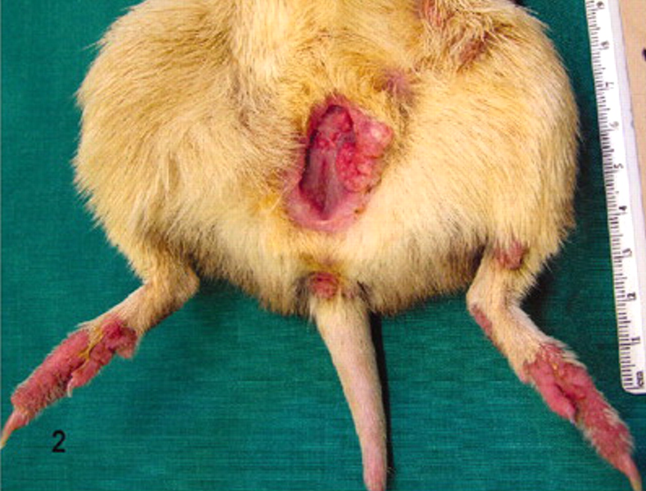
Ventral abdomen; Western barred bandicoot. Irregular papillomatous thickening of the dorsal skin of the hind digits and muco-cutaneous junctions of the pouch and cloacal opening. A large exophytic papillomatous mass is seen arising from the muco-cutaneous junction of the pouch.
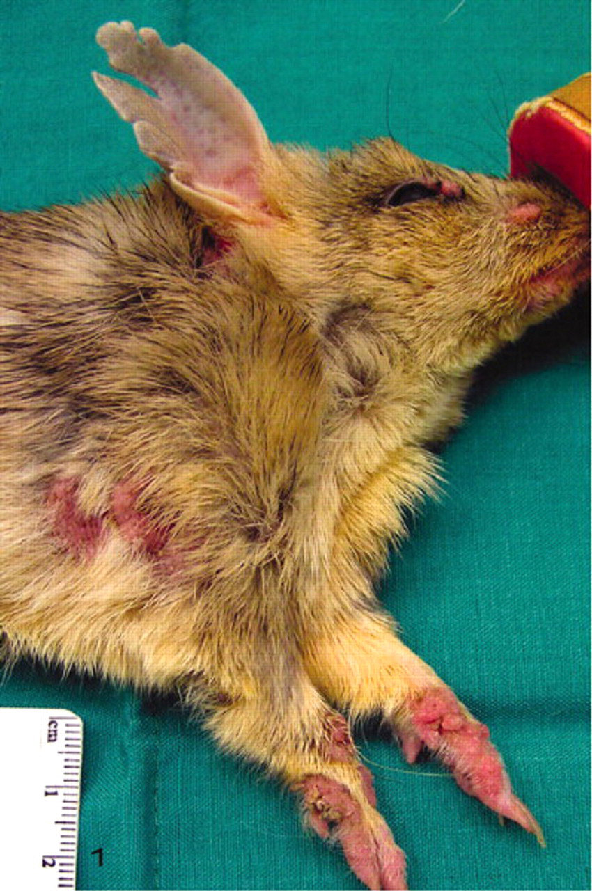
Skin of the eyelids, lips, forelimbs, and digits; Western barred bandicoot. Demonstrates nodular and irregular papillomatous thickening.
Histopathology of skin lesions
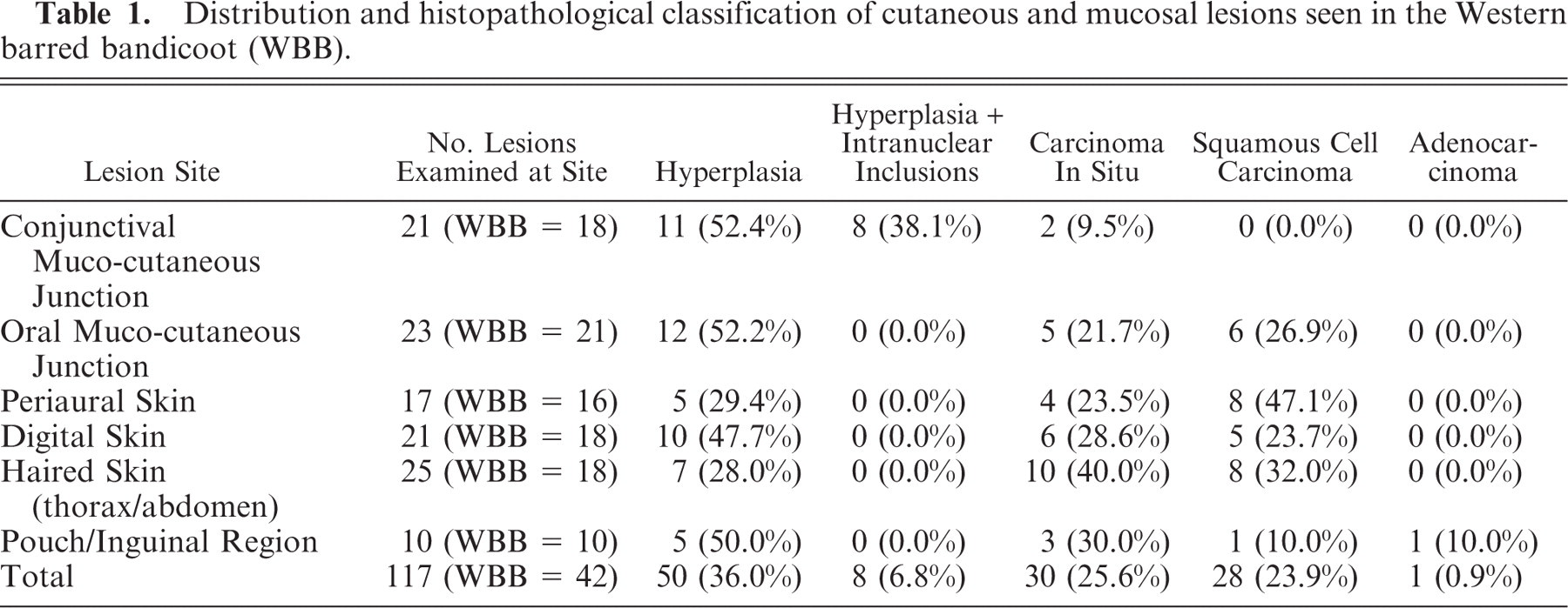
One hundred and seventeen skin and/or muco-cutaneous lesions were examined by light microscopy. Of these, 58 were classified as epithelial hyperplasia (49.6%), 30 as carcinoma in situ (25.6%), and 28 as squamous cell carcinoma (SCC) (23.9%). Adenocarcinoma of the pouch was recorded in one WBB.
There were 35 WBBs in the study cohort with multicentric lesions. Purely multicentric hyperplastic epithelial lesions were seen in 11 WBBs (31.4%), entirely malignant lesions were seen in 9 WBBs (25.7%), and both malignant and benign lesions were seen concurrently in 15 WBBs (42.8%). Solitary lesions were seen in 7 animals and consisted of either epithelial hyperplasia (4/7) or SCC (3/7); they were seen on the oral mucosa (3/7), the digital skin (3/7), or the haired skin of the body (1/7).
Thirty WBBs displayed 1 or more lesions classified as epithelial hyperplasia (n = 58). These lesions were characterized by mild, moderate, or marked irregular hyperplasia of the stratifying epithelium. The hyperplasia was attributable to an increase in the number of suprabasilar epithelial cells at mucosal sites and an increase in the number of keratinocytes within both the stratum spinosum and stratum granulosum of the epidermis. Hyperplasia of the outer root sheath of hair follicles, and the glandular elements of sebaceous and sweat glands, was also seen. Koilocytes and epithelial cells with marginated nuclear chromatin and large amphophilic inclusions (Fig. 3A) were evident in 8 cases with hyperplastic conjunctival lesions (Table 1).
Distribution and histopathological classification of cutaneous and mucosal lesions seen in the Western barred bandicoot(WBB).
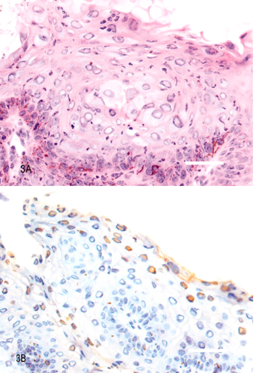
Conjunctival epithelium; Western barred bandicoot.
Lesions categorized as carcinoma in situ (n = 30) were seen in 17 individuals. These lesions showed an expansion of the stratum spinosum and stratum granulosum and mild to moderate dysplastic changes in keratinocyte morphology and epidermal architecture, such as disturbed polarity of keratinocyte differentiation and suprabasilar mitoses. Individual cellular characteristics included hyperchromasia of nuclei, prominent nucleoli, clear or vacuolated cytoplasm, and mild anisocytosis and anisokaryosis. In 1 conjunctival lesion classified as carcinoma in situ, the nuclei of some keratinocytes in the stratum granulosum displayed margination of nuclear chromatin and large amphophilic inclusions.
Lesions categorized as SCC (n = 28) were located at cutaneous and muco-cutaneous sites in 20 individuals (Fig. 4). Histologically, the carcinomas consisted of invasive cords, islands, and trabeculae of neoplastic epithelial cells showing varying degrees of differentiation. In well-differentiated tumors, there was formation of keratin “pearls” circumscribed by neoplastic keratinocytes. The neoplastic epithelium extended into the dermis, with or without association with the overlying epidermis. The neoplastic cells themselves showed abundant eosinophilic cytoplasm with distinct cell borders, intercellular desmosomes, and varying degrees of anisocytosis and anisokaryosis. Nuclei were often large, ovoid, and vesicular with prominent nucleoli. The squamous cell carcinomas were locally aggressive, invading into the surrounding dermis, subcutis, and even bone (n = 1). Inflammatory infiltrates were frequently observed within SCCs or in the surrounding subcutis, comprising neutrophilic, histiocytic, lymphoplasmacytic, or mixed cellular populations. In 3 animals in this study with cutaneous and muco-cutaneous SCC, metastases were observed in a lymph node (n = 1) and the lungs (n = 2).
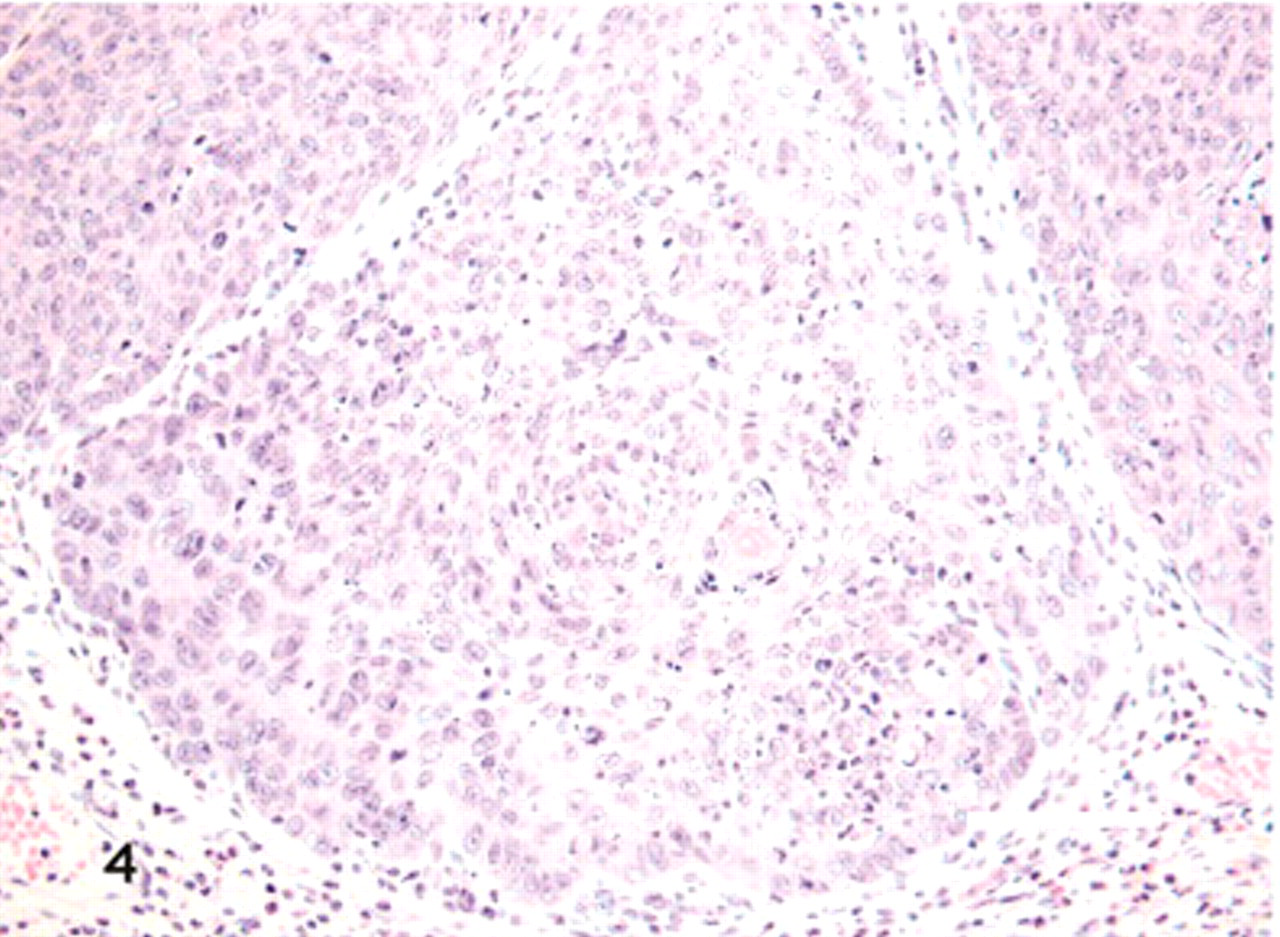
Cutaneous squamous cell carcinoma; Western barred bandicoot. Islands of neoplastic epithelial cells containing central cores of keratin invade the dermis. Cells are closely apposed with intercellular desmosomes visible and have abundant eosinophilic cytoplasm and basophilic staining nuclei with prominent and often multiple nucleoli. There is moderate to marked anisokaryosis and anisocytosis and mitotic figures are observed frequently. A mild neutrophilic infiltrate is seen in the subcutis and neoplastic epithelium. HE. Bar = 200 μm.
The distribution and histologic classification of lesions according to their anatomic site is detailed in Table 1. SCCs were seen most commonly over the haired skin of the thorax and abdomen and in the periaural region. Adenocarcinoma of the pouch mammary glands was observed in 1 female affected by papillomatous and carcinomatous lesions at other anatomic sites. Conjunctival lesions were least frequently observed to undergo malignant transformation, with only 2/21 lesions designated carcinoma in situ and no cases of SCC observed at this site. However, these lesions exclusively displayed hyperplastic epithelium (n = 8) or carcinoma in situ (n = 1) in which keratinocytes demonstrated nuclear chromatin margination and amphophilic intranuclear inclusions.
Ultrastructural examination of skin lesions
Ultrathin sections from a range of hyperplastic and malignant lesions from 11 WBBs were examined by transmission electron microscopy. Two hyperplastic conjunctival lesions from 2 individuals that displayed keratinocyte intranuclear inclusions visible on the toluidine-blue-stained slides displayed a crystalline array of spherical electron dense particles, approximately 45 nm in diameter (Fig. 5), within keratinocyte nuclei. The morphology and size of these particles were consistent with that of a papillomavirus or a polyomavirus.
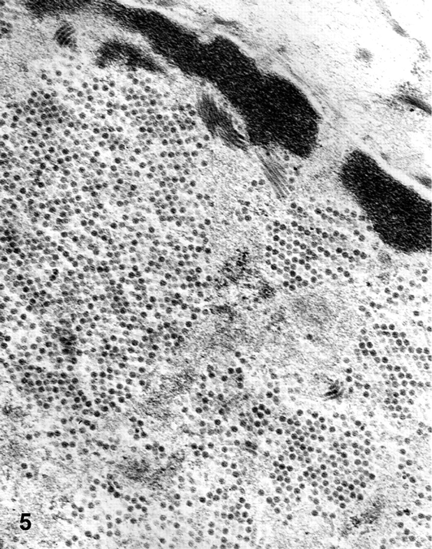
Conjunctival keratinocyte nuclei; Western barred bandicoot. Crystalline array of spherical electron dense virus-like particles ∼45 nm in diameter seen within hyperplastic keratinocyte nuclei. Bar = 500 nm.
Immunohistochemistry
Indirect immunohistochemistry using a polyclonal antibody against BPV-1 (Dako Corporation) was performed on skin lesions from 14 individuals. Positive staining was observed in conjunctival epithelial lesions from 3 WBBs in which keratinocytes displayed margination of nuclear chromatin and large glassy amphophilic inclusions (Fig. 3B).
Discussion
This report describes the pathologic features of a recently recognized papillomatosis and carcinomatosis syndrome in 42 WBBs. The spectrum of cutaneous and muco-cutaneous lesions recorded resembles the stepwise progression of epithelia from premalignant stages into invasive squamous cell carcinomas. A single case of adenocarcinoma of the pouch mucosa was observed; however, the significance of this tumor in relation to the otherwise exclusively squamous carcinomatosis syndrome is still unclear. This disease occurs in adult individuals, lesions increase in size with age, and malignant transformation occurs in those lesions >5 × 5 × 5 mm. The discernable stages of lesion development and the progression in lesion size and malignancy seen with increasing age is consistent with a multistage model of carcinogenesis, reflecting the acquisition of genetic alterations by tumor cells driving progressive transformation into highly malignant derivatives. 21,53
The development of malignancy involves complex interactions between several factors, both exogenous (environmental) and endogenous (genetic, hormonal, immunologic). 53 Certain viruses are known to play key roles in carcinogenesis in humans and various animal species. 23 Gross and histopathologic features of the skin and mucosal lesions combined with the ultrastructural and immunohistochemical features of conjunctival lesions in affected WBBs in this study are supportive of the involvement of either a papillomavirus, or less probably a polyomavirus, in the pathogenesis of this disease. In addition, the observation of skin lesion development in seemingly healthy, wild WBBs introduced into captive colonies of WBBs affected with the skin lesions provides further support that the syndrome may be caused by an infectious agent. Fight wounds and incidental trauma including loss of tails, ears, and eyes have been observed by the authors in both captive and wild WBBs. These incidents may facilitate the entry of an epitheliotropic agent such as a papillomavirus or polyomavirus, and thus it must be considered that a multicentric lesion distribution may be related to sites of epithelial trauma. Alternatively, the multicentric distribution of lesions may have arisen secondary to autoinoculation or perhaps viremia as has been alluded to in other species infected with papilloma- or polyomaviruses. 2,3,6,17,40
In general, papillomaviruses are species specific and induce excessive irregular proliferation of cutaneous and mucosal epithelia in both humans and a range of animal species. 46,54 Over 100 papillomavirus types have been completely characterized in humans alone. 16 Cutaneous papillomatosis is not always associated with papillomavirus infection, however, with a range of infectious as well as noninfectious agents also recognized to induce proliferative and papillomatous lesions in animal species. 46
Despite often appearing early in the course of disease (author observation, L. Woolford) and an increase in lesion size seen clinically, conjunctival lesions were least frequently observed to undergo malignant transformation. In addition, all lesions that demonstrated ultrastructural (n = 2) or immunohistochemical evidence (n = 3) of papillomavirus virions were derived from the conjunctiva only. Nonconjunctival lesions examined in this study may have lacked ultrastructural and immunohistochemical evidence for papillomavirus infection due to being intermittently infected, a condition described in papillomavirus-induced lesions in other species. 44 Alternatively, nonconjunctival lesions may have lacked evidence of virions if sampled early in the course of development. Experimental infections in other species have shown that lesions do not express the capsid antigen (L1) until 7 weeks or more after infection. Consequently, early proliferative lesions often prove negative by immunohistochemistry for L1 capsid antigens and do not contain viral particles on ultrastructural examination. 32 Furthermore, epithelia undergoing malignant transformation in association with papillomavirus infection may be negative for structural antigens by immunohistochemistry and be unremarkable on ultrastructural examination if viral DNA has become integrated into the host genome, leading to a nonproductive infection. Integration of papillomavirus DNA into host genomic DNA has been well documented in malignant lesions associated with high risk human papillomavirus infections. 12,24,26,27,36
The development of malignancy involves a complex interaction between both exogenous and endogenous factors. Indeed the outcome of a papillomavirus infection and the role of the virus in carcinogenesis may be influenced by factors such as the virulence of the particular papillomavirus type, 8,43,45,54 compromise of the host immune system, 7,33,34 host genetics, 1,9,20,30,51 and the presence of various chemical cofactors. 10,25,38 Immunodeficiency and/or immunocompromise in the WBB are considered as potential risk factors for development of this disease. Assessment of immunocompetence in the WBB and the factors by which it may be influenced are being investigated concurrently by the authors. An increased incidence of papillomavirus-related disease is being reported in other endangered animal populations 6,28 with decreases in genetic diversity a proposed cause of increased susceptibility to disease agents in some endangered species. 47 In attempts to elucidate the pathogenesis of this disease in the WBB, it must be considered that a complex interaction between viral and other factors may be driving the development of malignant epithelial tumors.
Immunohistochemical staining of WBB conjunctival lesions in this study led us to consider the involvement of a papillomavirus in the pathogenesis of this disease. However, we did not feel we could rule out the involvement of a polyomavirus on the basis of immunohistochemical results alone. Significant antigen retrieval techniques were required to demonstrate papillomavirus structural antigens, which in the authors' experience have not been required for papillomavirus-associated epithelial lesions from other species using an otherwise identical technique. In addition, gross, histologic, and ultrastructural features of lesions did not discount a polyomavirus as an alternative etiologic agent. The mammalian polyomaviruses display a narrow host range and do not productively infect other species, typically causing apathogenic subclinical natural infections in immunocompetent hosts. These viruses may cause severe disease in the immunocompromised host or can cause tumor formation when they are introduced into an unnatural host. 11,31 The hyperplastic changes seen affecting the root sheath epithelium in WBB lesions from areas of haired skin was reminiscent of the appearance of polyomavirus-associated trichoepitheliomas seen in hamsters. 19 These tumors of hamsters occur primarily on the skin of the head, chin, neck, back, and frequently around the eyes and external ears; histologically they demonstrate proliferation of the hair root epithelium, forming cyst-like masses filled with cornified material. 39 Definitive evidence to support or refute the presence of a specific etiologic agent in this syndrome is also lacking at this stage of investigations as nonconjunctival epithelial lesions did not demonstrate intranuclear inclusion bodies nor ultrastructural or immunohistochemical evidence of viral infection.
The papillomatosis and carcinomatosis syndrome observed in the Western barred bandicoot is a novel and emerging disease in this species and, to the authors' knowledge, is unlike skin diseases of Australian marsupials previously documented. No specific skin diseases of Australian bandicoot species have been documented to date; however, a conjunctivitis syndrome has been seen in captive and wild WBBs associated with novel Chlamydiales sp. isolated from the conjunctiva. 5,52 Papillomatous lesions have been described in a common brushtail possum (Trichosurus vulpeca) in association with a papillomavirus infection, this being the only papillomavirus-associated lesions documented in an Australian marsupial to the authors' knowledge. 35 Papillomatous lesions have also been recorded in a North American opossum (Didelphis virginiana) associated with a papillomavirus. 46
Captive breeding and reintroduction programs form the basis of many animal species conservation efforts, and vigilance must be maintained for the emergence of new diseases: these form a major threat to biodiversity and have been shown to undermine the success of breeding and reintroduction programs. 13–15,50 The papillomatosis and carcinomatosis syndrome in captive and wild WBBs is a pertinent example of disease threatening the survival of a species. Future investigations into the papillomatosis and carcinomatosis syndrome of the Western barred bandicoot will involve pursuing detection of an etiologic or contributing viral agent using molecular techniques, studying the transmissibility of the disease, and examining the patterns of disease in wild and captive populations to enable further elucidation of the pathogenesis of this novel and debilitating disease.
Footnotes
Acknowledgements
We thank Gerald Spoelstra of the Murdoch University histopathology laboratory for his skillful production of histology slides, and James Poynton for his technical assistance in the Murdoch University necropsy suite. We are grateful to June Butcher and the volunteers at Kanyana Wildlife Rehabilitation Center for their care and monitoring of the Western barred bandicoots housed there, and the Denham and Narrogin Department of Environment and Conservation Western Australia (DEC) officers for the care and monitoring of Western barred bandicoots in the Peron and Dryandra captive breeding facilities, respectively. This project is funded by the Australian Research Council in partnership with Murdoch University and the Western Australian Department of Environment and Conservation (DEC) under Linkage Project LP0455050. Lucy Woolford is funded by the Lorna Edith Murdoch Veterinary Trust Scholarship.