
Abstract
Necropsy of a 17-month-old male common marmoset (Callithrix jacchus) with a history of increased abdominal girth resulted in the finding of a unilateral polycystic renal neoplasm. Detailed histopathologic and immunohistochemical investigations revealed different tissue types within the tumor including stromal connective tissue and fusiform mesenchymal cell formations surrounding blastemal cells as well as different developmental stages of organ-specific epithelial cells accompanied by extensive cyst formation. Metastases were not observed. In consideration of the macroscopic, histologic, and immunohistochemical findings, the tumor was classified as a nephroblastoma closely resembling the so-called Wilms' tumor, a malignant embryonic renal tumor frequently observed in humans, especially in young children. In contrast, this tumor entity has rarely been observed in nonhuman primates. This report represents the first documented case of a cystic variant of nephroblastoma in a nonhuman primate.
Malignant nephroblastoma, also referred to as adenomyosarcoma or Wilms' tumor, is a renal neoplasia primarily occurring in children under 10 years of age and represents the second-most common visceral neoplasm in this age group with a frequency of 20% to 30% of all malignant tumors. In adults, the prevalence is still approximately 5% to 10% of all malignant tumors. 9
Nephroblastomas are embryonic neoplasms composed of several cell types within the kidney arising from metanephric blastema, and thus occur in young individuals. 11 They often become clinically evident by abnormal intra-abdominal masses and anemia. The affected tissues usually express several lines of differentiation, including blastemal, epithelial, and stromal cells and, therefore, replicate the histology of various stages in normal or abnormal nephrogenesis. However, a great structural and histologic diversity may exist in these tumors. 9
Blastemal parts consist of round, densely packed cells with a serpentine, diffuse, nodular, or basaloid growth pattern. Epithelial parts may be arranged in glomerular, tubular, or papillary formations. A variety of stromal patterns may occur, predominantly spindle and myxoid cells but also smooth and skeletal muscle cells. Fibroblasts as well as cartilage, adipose, bone, and neuroglial tissue may be observed. In most cases, nephroblastomas are unilateral and rarely appear in association with other neoplasms, whereas pulmonary metastasis is a common event in children. 9
The etiology and pathogenesis of nephroblastoma has not been fully clarified so far. In children, the neoplasm is frequently associated with congenital abnormalities or documented syndromes, such as cryptorchism, hemihypertrophy, hypospadias, and sporadic aniridia. Two loci on chromosome 11, locus 11p13 (WT1 gene) and locus 11p15 (WT2 gene), have been implicated in the genesis of Wilms' tumors in children with developmental disorders. An abnormal WT1 gene is present in patients with WAGR syndrome (Wilms' tumor, aniridia, genitourinary abnormalities, mental retardation) or Denys-Drash syndrome (Wilms' tumor, progressive glomerulonephritis, male pseudohermaphroditism). A mutated WT2 gene can be observed in patients with Beckwith-Wiedemann syndrome or hemihypertrophy. However, the genetics of Wilms' tumor appear to be multifactorial and probably include further chromosomal abnormalities. Familial Wilms' tumor is very rare, occurring in about 1% of cases and is not associated with mutations in chromosome 11. 3,7,9
While nephroblastoma is a relatively common neoplasm in swine and chickens, it has rarely been reported in dogs, sheep, and cattle. 11 Concerning laboratory animals, nephroblastoma represents a common renal tumor of rats, which can be experimentally induced by prenatal exposure to the carcinogen N-ethylnitrosouria (ENU). Monophasic spindle-shaped patterns of nephroblastoma in rats have to be carefully differentiated from mesenchymal tumors of the kidneys, such as mesenchymal renal tumor or polyoma virus sarcoma. Renal mesenchymal tumor, a malignant heterogeneous mesenchymal neoplasm of the young rat, histologically displays a wide spectrum of cell and tissue types. It is often misdiagnosed for nephroblastoma as it may contain remnants of epithelial profiles from the preexisting parenchyma, which can be mistaken for the expression of bipotential differentiation into neoplastic epithelium. In addition, condensations of anaplastic mesenchymal cells in fibrosarcomatous sheets bear resemblance to blastemal clusters of nephroblastoma. In contrast, nephroblastoma in rats has a rather uniform histologic pattern, in which neoplastic blastema is the hallmark, along with the presence of tubular and primitive glomerular structures. Further diagnostic criteria of nephroblastoma include the expansive growth and the outer cortex as the site of origin. Polyoma virus sarcoma is a malignant mesenchymal neoplasm induced by the polyoma virus, which is usually bilateral, appears to arise in the outer medulla, and is characterized by the absence of neoplastic tubules or glomeruli. 2,5,8
In domestic animals, an extrarenal manifestation of nephroblastoma has been observed in dogs, which develops hind limb paresis due to spinal nephroblastoma arising from ectopic embryonal remnants of renal tissue. 11
In nonhuman primates, primary neoplasms of the kidney have been reported infrequently not only as spontaneous cases but also in association with exposure to radiation, chemical carcinogens, and parasites. Most of the renal tumors were incidental findings at necropsy after the animals had died spontaneously or had been euthanatized because of an unspecific debilitated or moribund condition. In only a few cases, the tumor could be directly related to the death. Far more frequently, the neoplasms were considered to be contributory factors in combination with other neoplasms or nonneoplastic diseases. 6
The most commonly described renal neoplasms in nonhuman primates include carcinomas and adenomas. 6 Only a few reports exist about nephroblastomas in Old World and New World monkeys; among them 1 case in a cynomolgus macaque (Macaca fascicularis), 1 2 recent cases in baboons (Papio sp.), 4 and 1 case in a cotton-top tamarin (Saguinus oedipus). 6 To the knowledge of the authors, this report documents the first description of a cystic variant of malignant nephroblastoma in a common marmoset (Callithrix jacchus).
A facility-born, 17-month-old male common marmoset (Callithrix jacchus) became clinically suspicious by abdominal distension and hematuria. An ultrasonic investigation revealed remarkable hypodense spaces expanding parts of the left kidney (Fig. 1). Euthanasia was elected due to a poor prognosis. A complete necropsy was performed and representative tissue samples of various organs, including the kidneys, were fixed in 10% and 4% phosphate-buffered formaldehyde for histologic and immunohistochemical investigations. Following fixation in 10% phosphate-buffered formaldehyde for at least 24 hours, tissue samples were automatically paraffin-embedded, sectioned at 3 μm, and stained with hematoxylin and eosin (HE) and periodic acid Schiff (PAS) for light microscopy. Immunohistochemical investigations (IHC) were carried out on paraffin-embedded tissue sections of the kidney using the streptavidin-biotin-complex (SABC) method with the chromogen DAB (diaminobenzidine tetrahydrochloride, iView DAB detection kit, Ventana, Illkirch, France). The primary antibodies used for immunohistochemistry included anti-vimentin antibody (monoclonal mouse-anti-human-vimentin, clone V9, Dakocytomation, Hamburg, Germany) at a dilution of 1 : 100, anti-desmin antibody (monoclonal mouse-anti-human-desmin, clone D33, Dakocytomation) at a dilution of 1 : 100, anti-S-100 antibody (polyclonal rabbit-anti-S-100, clone Dakocytomation) at a dilution of 1 : 1,000, anti-smooth muscle actin (SMA) antibody (monoclonal mouse-anti-human-smooth muscle actin, clone 1A4, Dakocytomation) at a dilution of 1 : 200 and anti-neuron-specific enolase (NSE) antibody (monoclonal mouse anti-human-neuron-specific enolase, clone BBS/NC/VI-H14, Dakocytomation) at a dilution of 1 : 200. Choice of the secondary antibody depended on the comments of the manufacturer (Ventana enhanced biotin-conjugated secondary antibody, affinity purified goat-anti-mouse and goat-anti-rabbit immunoglobulin G, Ventana iView DAB detection kit, Ventana). In addition, bacterial culture of the left kidney was performed on a blood agar plate incubated at 37°C for 24 hours (Columbia-Agar Basis, Merck, Darmstadt, Germany).
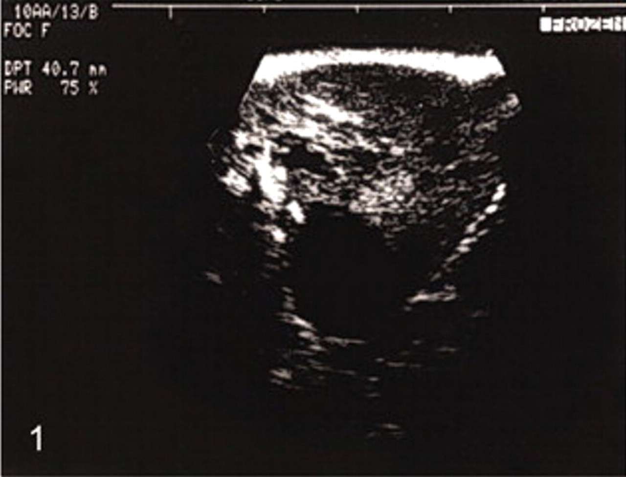
Kidney; Callithrix jacchus. Ultrasonographic examination of the left kidney shows multiple hypoechogen structures indicating cyst formation within the organ.
At necropsy, the left kidney was markedly enlarged and edematous and showed a multicystical transformation (Figs. 2, 3). The capsule was detached from the organ surface and contained 45 ml of a brownish fluid.
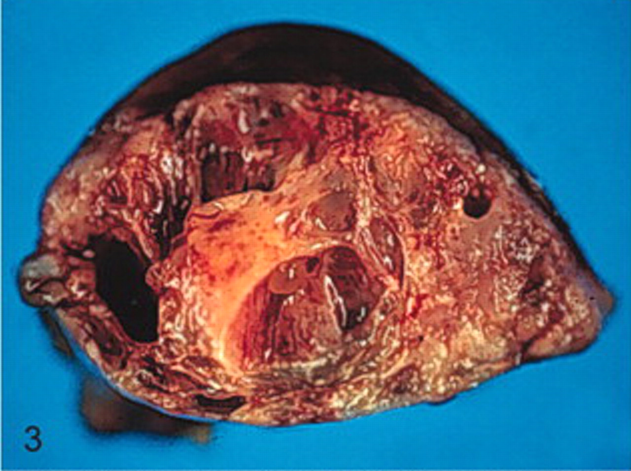
Kidney; Callithrix jacchus. Cross-section of the left kidney reveals multiple cystic spaces surrounded by an edematous parenchyma.
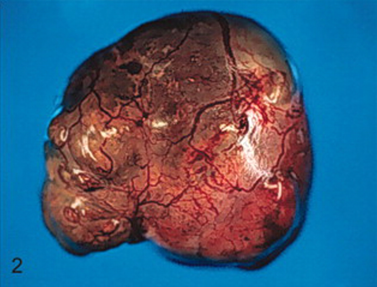
Kidney; Callithrix jacchus. Macroscopic image of the altered organ in toto: the transformed shape indicates the presence of prominent cysts.
The right kidney was unremarkable except for a circumscribed subcapsular hemorrhage. Extensive hemorrhages were also found in the right ureter. The urinary bladder contained moderate amounts of a bloody fluid.
Histopathology of the left kidney revealed an unencapsulated neoplasm completely replacing the preexistent renal parenchyma. Parts of the renal mass were characterized by the development of numerous cysts mainly filled with a protein-rich homogeneous eosinophilic fluid, cellular debris, as well as calcareous deposits and lined with a flattened epithelium (Fig. 4). The solid parts of the neoplasm were composed of different tissue types. Stromal collagenous connective tissue formed the matrix for densely packed, small, round blastemal cells with round-to-oval hyperchromatic nuclei. These blastemal components were arranged in clusters mainly showing serpentine and nodular growth patterns (Fig. 5). In the periphery of the blastemal nests, the cells showed a palisade-like arrangement, whereas there was multifocal necrosis with randomly distributed dystrophic calcification within the centers of the lobulated blastema (Fig. 6). Immature epithelial structures resembling early stages of glomerulogenesis (Fig. 7) and abortive tubule formation were randomly distributed throughout the tumor, often surrounded by spindle-shaped cells with moderate anisocytosis and anisokaryosis, which were partly arranged in interwoven fascicles and exhibited a moderate mitotic rate. Inflammatory reactions were absent.
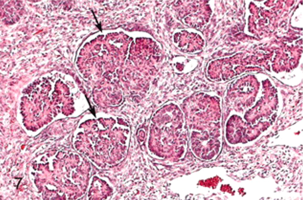
Kidney; Callithrix jacchus. Various stages of glomerular differentiation with poorly formed Bowman spaces (arrows). HE. Bar = 100 μm.
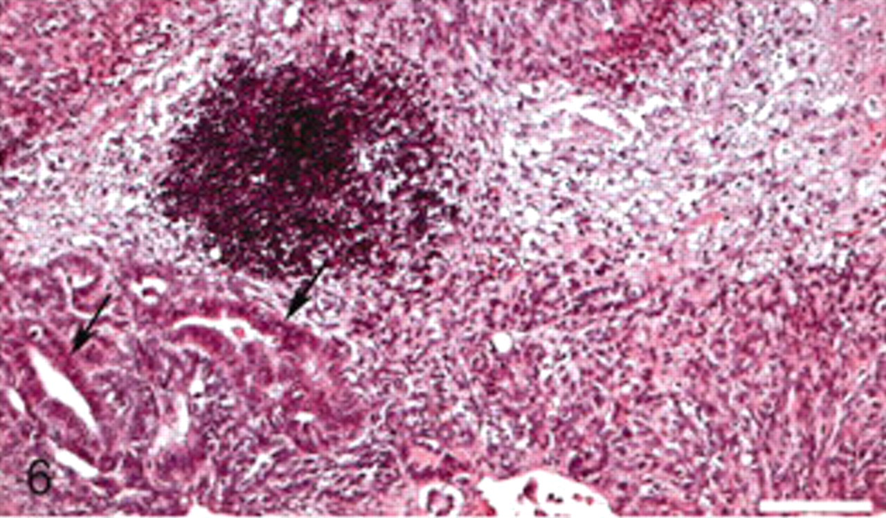
Kidney; Callithrix jacchus. Dystrophic calcification of a blastemal tumor cell nest. Tubular differentiation is present in the periphery of the blastema (arrows). HE. Bar = 200 μm.
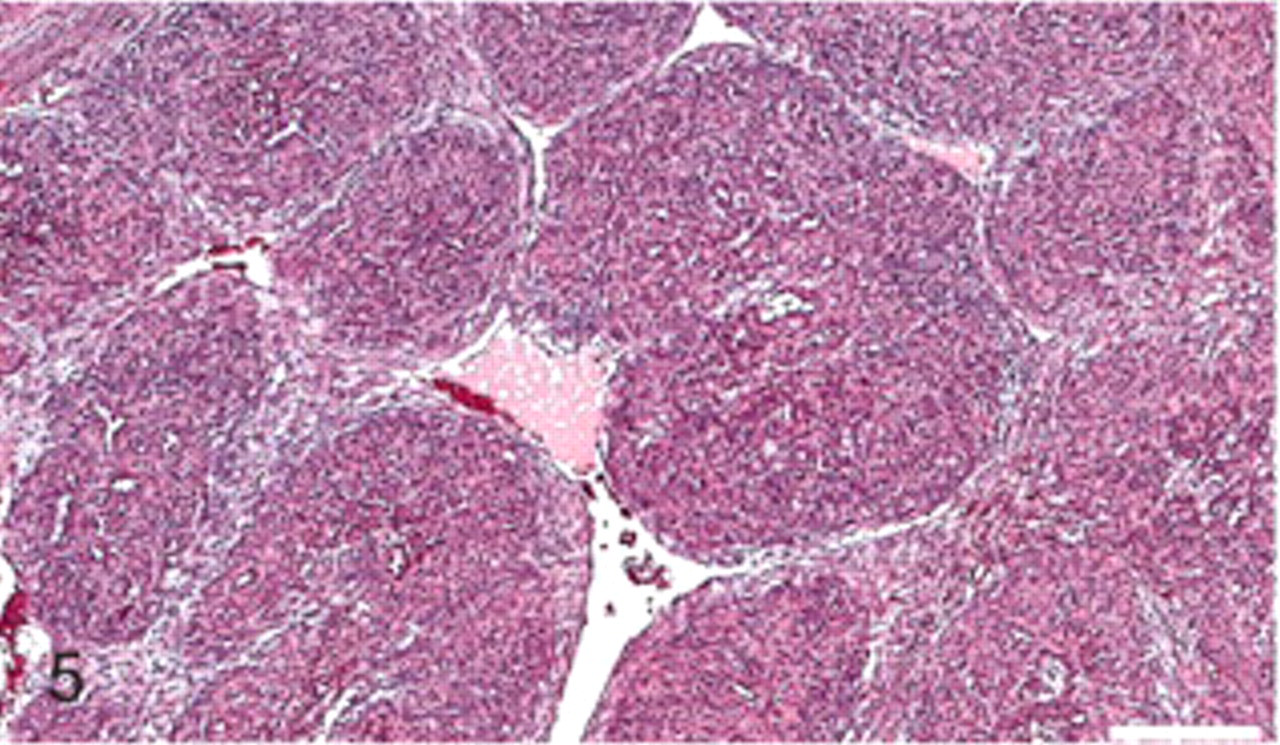
Kidney; Callithrix jacchus. Serpentine blastemal cell clusters surrounded by a sparse fibromyxomatous stroma. HE. Bar = 200 μm.

Kidney; Callithrix jacchus. Cysts lined by a flattened epithelium contain protein-rich fluid. Focal mineralization (arrows) is evident within the separating stroma. HE. Bar = 200 μm.
As macroscopic and light microscopic examinations did not reveal evidences of neoplastic growths in other organs, metastasis from the nephroblastoma could be excluded. Immunohistochemical examinations revealed a variable Ki-67 expression throughout the tumor. The percentage of Ki-67 positive cells was highest in blastemal parts, but considerable proliferative activity could also be observed in the stromal and epithelial tumor parts (Fig. 8). Immunohistochemical labeling with antidesmin and anti—smooth muscle actin antibodies revealed the presence of smooth muscle cell differentiation within one focus in the periphery of the tumor. Vimentin expression was correlated with localizations of interwoven fascicles of spindle-shaped cells confirming the mesenchymal origin of these cells. These spindle-shaped mesenchymal cells also showed moderate diffuse smooth muscle actin immunoreactivity. In contrast, the glomerular and tubular formations as well as the blastemal cells were vimentin negative. Staining for NSE and S100 revealed the presence of multifocal neural structures, probably representing remaining elements of the renal innervation system.
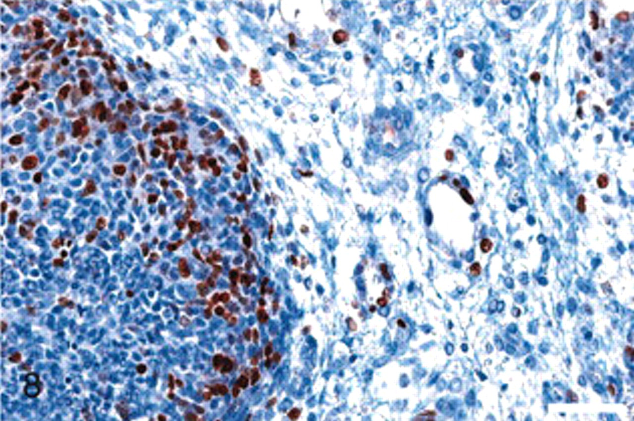
Kidney; Callithrix jacchus. Blastemal and myxomatous stromal cells show variable mitotic activity with accentuation on the blastemal cells. IHC—SABC, Ki-67. Bar = 50 μm.
Microbiologic examinations of the left kidney were unsuggestive of bacterial pathogens.
The solitary renal tumor identified in this Callithrix jacchus was diagnosed as a malignant nephroblastoma on the basis of the clinical history and the macroscopic and microscopic pathology, including immunohistochemical examinations. Previous cases of nephroblastomas in nonhuman primates have been described for both New World and Old World monkeys, among them a cynomolgus macaque, 2 baboons and 1 cotton-top tamarin. 1,4,6 In the 3 cases from Old World monkey species, the tumors exclusively affected juvenile animals, whereas the description for Saguinus oedipus referred to an adult cotton-top tamarin. The common marmoset, which also belongs to the New World monkeys, was 17 months old when the nephroblastoma was detected and was therefore close to sexual maturity. However, the comparatively late clinical onset of the disease may be attributed to the high compensatory capacity of the contralateral kidney. Therefore, it can be assumed that the nephroblastoma had developed in early childhood and persisted for several months without remarkable clinical signs. Concerning the macroscopic picture of nephroblastomas in nonhuman primates, solid or lobulated grey-yellow encapsulated renal masses with hemorrhagic areas and central necrosis represented the major gross findings in the previously reported cases. In contrast, the macroscopic appearance of nephroblastoma in the common marmoset was dominated by cyst formations, as it is occasionally seen in human nephroblastomas. It is postulated that the cysts originate from primary epithelial parts of the kidney and represent dilated tubules or collecting ducts. In human medicine, the presence of multiple cysts besides solitary portions in a nephroblastoma is referred to as cystic Wilms' tumor, which has to be distinguished from cystic nephroma, cystic partially differentiated nephroblastoma (CPDN), the cystic variant of mesoblastic nephroma, and cystic renal cell carcinoma. 10 In domestic animals, cystic nephroblastoma has been reported once in an aged dog. 12
Histologically, the nephroblastoma of the common marmoset was characterized by a triphasic growth pattern including mesenchymal, epithelial, and blastemal components. The triphasic pattern is the most frequently observed histologic manifestation of nephroblastoma in humans and animals, but biphasic and monophasic lesions also occur in variable compositions. 9,11 Concerning the biologic behavior of nephroblastomas, the classic triphasic tumor is supposed to be of standard malignancy with a considerable probability of metastasis, especially to the lungs as it has been reported for a juvenile Macaca fascicularis before. 1,9
In conclusion, we described a case of malignant nephroblastoma in a subadult common marmoset. Unlike previously described cases of nephroblastomas in nonhuman primates, the neoplastic tissue was characterized by extensive cyst formation besides solitary portions, which revealed the classic triphasic growth pattern at histologic examination.
Unfortunately, a representative overview concerning the prevalence and average age of incidence as well as gross and microscopic pathology and frequency of metastasis of nephroblastoma in nonhuman primates cannot be given on the basis of the few reported cases. Therefore, further cases will have to be compiled with regard to the clinical history as well as the macroscopic, histologic, and immunohistochemical findings.
Footnotes
Acknowledgements
We thank W. Henkel, K. Kaiser-Jarry, N. Knöchelmann, E. Lischka, and H. Zuri for technical assistance and laboratory work.