
Abstract
Histopathologic features of hepatic peribiliary cysts were described in a young slaughtered pig. The animal was an apparently healthy 6–month-old pig of mixed breed. Macroscopically, all lobes of the liver contained numerous cysts of varying size containing serous fluid in all lobes. Histopathologically, the cysts were located mainly around the large bile duct and in the connective tissue of the portal tracts. Within serial sections, these cysts were assumed to be solitary or multilocular, but they were separated from the bile duct. The cysts were lined by a single layer of columnar, cuboidal, and flattened epithelial cells. Occasionally, goblet cells were observed. The epithelial cells were stained with periodic acid—Schiff/alcian blue and high-iron diamine/alcian blue, indicating the presence of neutral mucin, sialomucin, and sulfomucin. Grimalius' method revealed the presence of endocrine cells in the lining epithelium. There was no bile pigment in the cysts by the Hall method.
Keywords
Hepatic peribiliary cysts in humans are retention cysts of peribiliary glands located in the hilum of the liver and large portal tracts. 7 This condition has been reported as “multiple cysts in the hepatic hilum” and is found in patients with underlying liver disease. In the pig, although there are reports of hepatic cysts, it is considered that the cysts derive from bile ducts. 9 In our study we describe the pathomorphologic features of multiple liver cysts in a pig that were similar to the peribiliary cysts observed in humans. This is the first case report of peribiliary cysts in an animal.
The animal was an apparently healthy 6-month-old pig of mixed breed, and it had been slaughtered in an abattoir. At meat inspection, the affected liver was condemned due to its gross morphologic abnormality. Several samples of liver tissue were fixed in 10% neutral phosphate-buffered formalin solution, trimmed, dehydrated in a graded series of ethanol, and embedded in paraffin. Sections for microscopic examination were cut at a thickness of 4 μm and stained with hematoxylin and eosin. Some representative sections were also stained with the Hall method, using combined periodic acid–Schiff reaction and alcian blue at pH 2.5 (PAS/AB), with combined high-iron diamine and alcian blue at pH 2.5 (HID/AB), and with Grimelius' method. To show normal peribiliary grands, a normal pig liver was used.
Macroscopically, the liver had numerous cysts in all lobes, especially around the common bile duct. On the cut surface, variable-sized cysts were located not only beneath the serous membrane and in the parenchyma (Fig. 1). The diameter of the cysts varied. The largest was up to 25 mm in diameter. The cysts contained serous fluid. No other organs, including kidneys, contained obvious gross lesions.
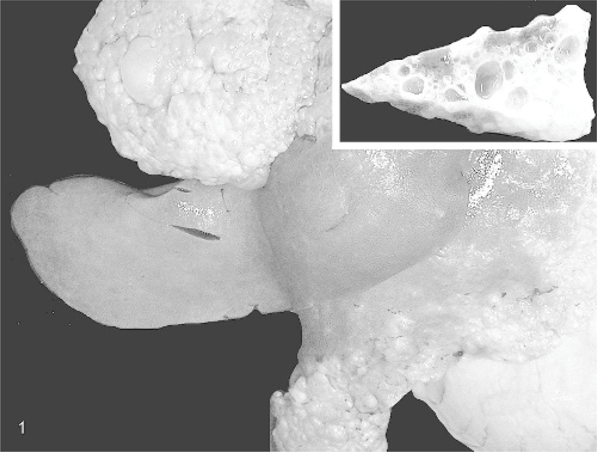
Cystic liver; pig. Numerous cysts are seen in all lobes. The cut surface reveals variable-sized cysts not only beneath the serous membrane but many in the parenchyma as well (inset).
Microscopically, cysts in the liver were located mainly in the large portal tracts and in the connective tissue of the portal tracts (Fig. 2). Near the cysts, there was a large bile duct with noncystic peribiliary glands (Fig. 3). Within serial sections, these cysts were solitary or multilocular, and they were separated from the bile duct. The cysts were lined by a single layer of columnar, cuboidal, and flattened epithelial cells (Fig. 4), and goblet cells were observed occasionally. Outside the epithelium lining there was loose connective tissue accompanied by a mild chronic inflammatory infiltrate in some areas. The hepatic lobules surrounded by cysts were deformed and remained like islets. In the other lesions, chronic lymphocytic cholangitis with lymph follicle formation was observed in the periportal connective tissues. The epithelial cells of cysts were stained with PAS/AB and HID/AB, indicating the presence of neutral mucin, sialomucin, and sulfomucin. Grimelius' method revealed the presence of endocrine cells in the cysts lining the epithelium. The Hall method did not show bile pigment in the cysts.
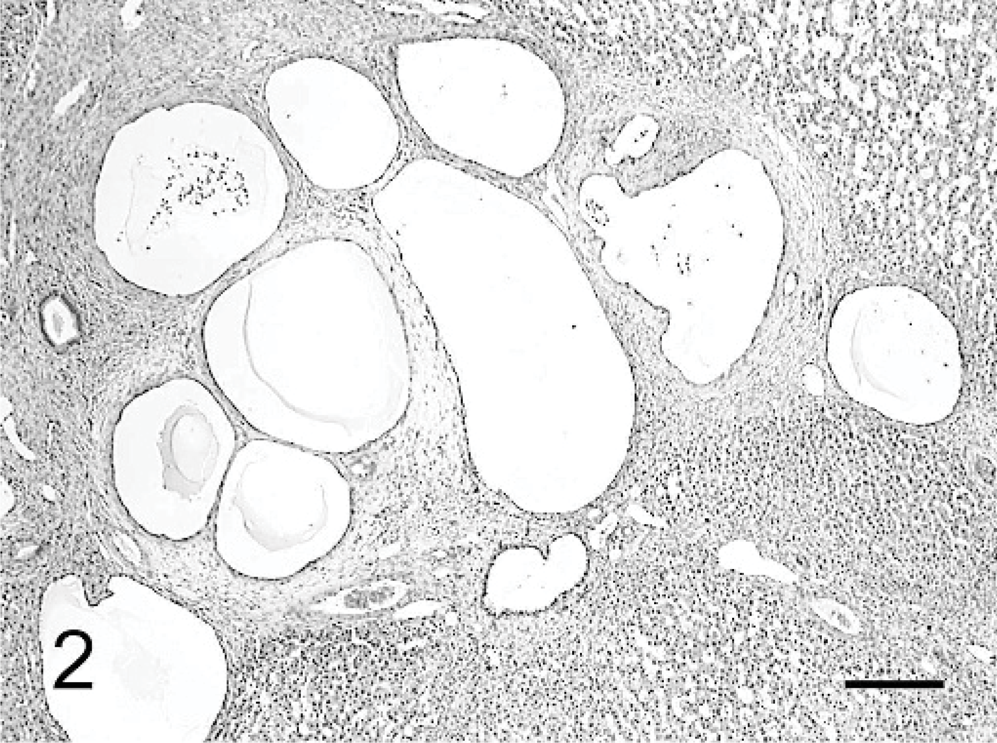
Cystic liver; pig. Variable-sized cysts are seen in the connective tissue of the large portal tracts. HE. Bar = 300 μm.
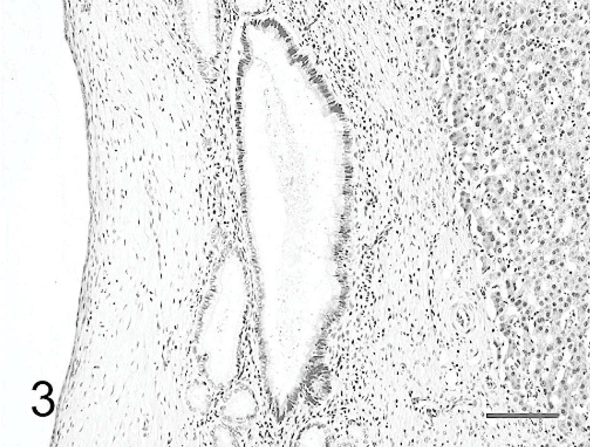
Cystic liver; pig. Large bile duct with noncystic peribiliary glands is seen near the cysts. HE. Bar = 100 μm.
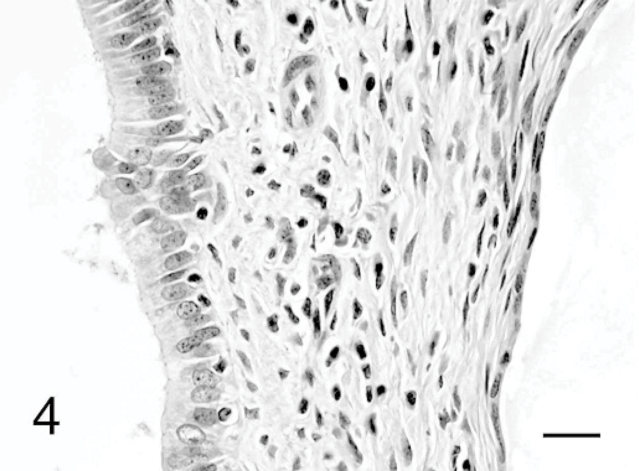
Cystic liver; pig. Cysts line a single layer of several types of epithelium. On the left, cysts are lined by columnar epithelial cells, and on the right they are lined by squamous epithelial cells. HE. Bar = 20 μm.
The multiple liver cysts in the present study were diagnosed as peribiliary cysts by histopathologic examination. Peribiliary cysts were first described by Nakanuma et al. in a human patient, since the cysts derived from peribiliary glands located in the hilum of the liver and larger portal tracts. 5 Although peribiliary cysts have been considered to be clinically asymptomatic, the cystic dilatation appeared to have been responsible for the progression of obstructive jaundice. 3, 7 Previous reports in humans showed that peribiliary cysts are usually detectable in preexisting hepatobiliary diseases, such as liver cirrhosis, hepatocellular carcinoma, idiopathic portal hypertension, intrahepatic cholangitis, systemic infection or septicemia, and autosomal dominant polycystic kidney disease. 3, 4 Therefore, two etiologic mechanisms have been considered to be involved in the formation of peribiliary cysts: one associated with inflammation or circulatory disturbance, leading to glandular obstruction, and the other with hereditary factors. 2, 3, 5, 8
Histologically, peribiliary cysts vary in size, are lined by a single layer of columnar or flattened epithelial cells without atypia, and do not communicate with the lumen of bile ducts. They are located within the connective tissue of the hepatic hilus and also within the larger portal tracts, whereas congenital cysts derived from bile ducts locate within parenchyma. 7
In the pig, peribiliary glands are formed around collecting bile ducts (Fig. 5) and secrete neutral mucin, sialomucin, and sulfomucin (data not shown). There are few reports about hepatic cysts in the pig. Webster et al. reported multiple liver cysts in a pig as a part of congenital polycystic syndrome of the kidneys. 9 The cysts were filled with bile and were considered to have been derived from bile ducts.
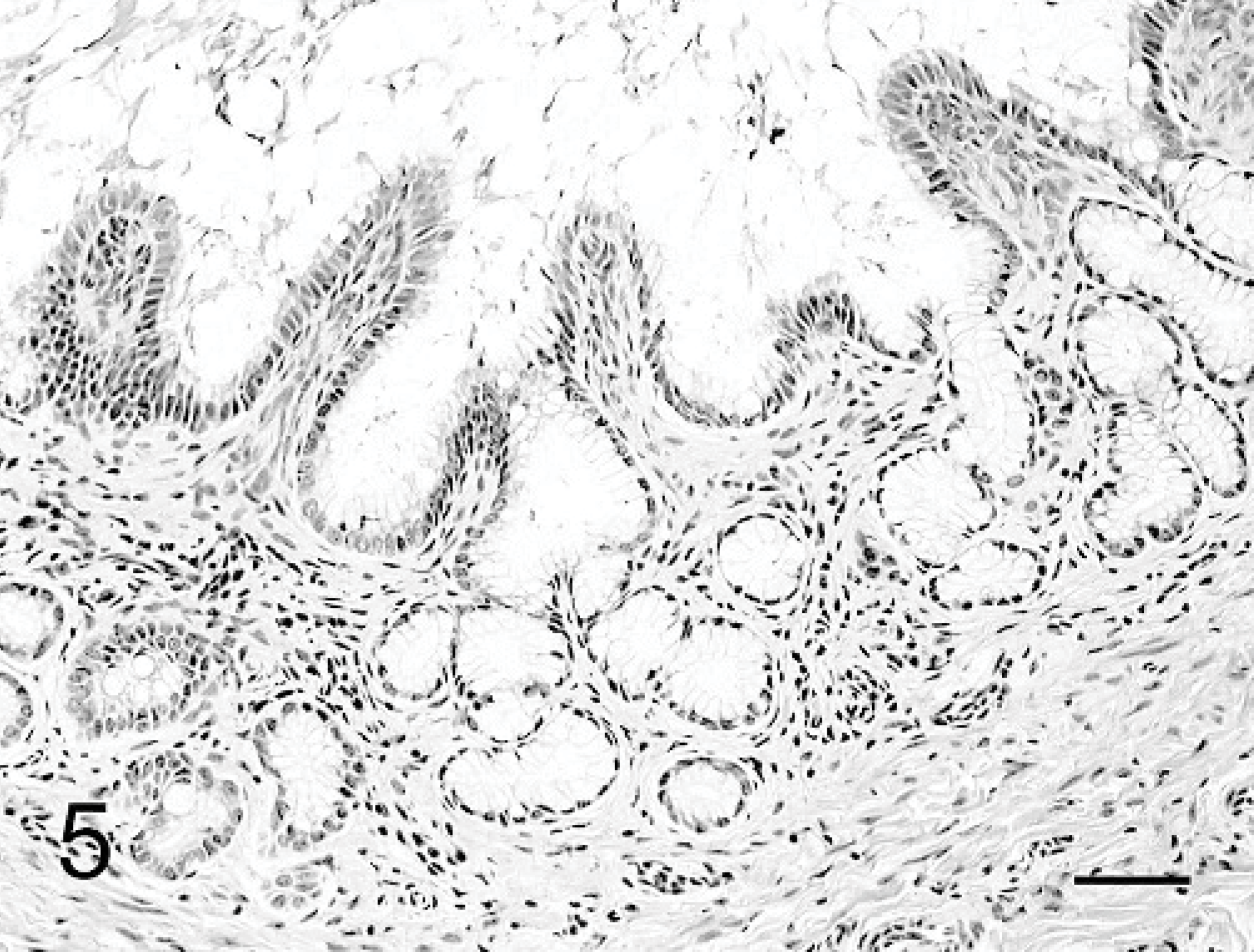
Normal liver; pig. Peribiliary glands are formed in lamina propria of collecting bile ducts. HE. Bar = 50 μm.
The cysts in the present case contained serous fluid, unlike those in the previous report. 9 On the other hand, they were like peribiliary cysts in humans, containing serous fluid and showing similarities in sizes, locations in the liver, and the characteristics of cells lining the cysts. In the present case, however, the cysts were observed not only in the hilum but also in the periphery of the portal tracts, although peribiliary cysts in humans have been observed in the hilum of the liver and larger portal tracts. We considered that this can be explained by the anatomical differences between human and pigs. Unlike humans, the pig has numerous peribiliary glands in the smaller bile ducts. 1, 10
These results support the conclusion that the cysts in this case were derived from peribiliary glands. We considered that these peribiliary cysts were causally related to dysplasia of the peribiliary glands, which are physiologically distributed along the large bile ducts.
Cysts of the liver occur in all species and are probably derived from bile ducts. But peribiliary glands are observed in various kinds of animals, not only in the pig but also in the cow, monkey, and cat. 6, 10 In conclusion, we showed in this study that cysts derived from peribiliary glands in a pig, suggesting that peribiliary cysts may occur even in other animals that have peribiliary glands.
Footnotes
Acknowledgements
This work was supported by a grant-in-aid to the High Technological Research Center (Rakuno Gakuen University) from the Ministry of Education, Culture, Sports, Science and Technology of Japan.