
Abstract
Growing rabbits from two rabbitries, fed with commercial concentrates and hay, developed painful thickenings of the extremities. Four rabbits from each farm were clinically examined and necropsied. All animals showed multiple moderate to severe osseous proliferations of extremities and mandibles and a mild to severe proliferative gastroduodenopathy. Histologically, periosteal and endosteal hyperostosis and a mild to severe proliferation of the gastric and duodenal mucosa were noted. Bone analyses revealed 12,700 and 15,000 ug fluoride per gram of bone ash in affected rabbits, compared with 550 ug fluoride in a control animal. A highly elevated fluoride content was found in concentrates. Vitamin A levels were moderately increased only in one concentrate, and copper levels were normal. Results indicate that alimentary fluoride intoxication caused prominent bony proliferations in the examined rabbits. Whether the proliferative gastroduodenopathy is related to the elevated fluoride intake or represents an incidentally occurring secondary disease remains to be determined.
Chronic fluorosis is a worldwide endemic disease in animals and humans, occurring especially in parts of India, Australia, Africa, and Turkey. 5, 10 It is usually caused by consumption of water or food abnormally high in fluoride and can often be observed in animals grazing near industrial plants that emit fluoride-bearing fumes and dusts. 5, 10 Most affected mammals are sheep, cattle, goats, and horses. 10
Major clinical manifestations of chronic fluoride intoxication are dental fluorosis and osteofluorosis. 6, 10 Dental fluorosis is characterized by enamel mottling and hypoplasia, and osteofluorosis by periosteal hyperostosis displaying a roughened irregular surface. 10 In Germany, chronic fluorosis represents a rare disease, occurring only infrequently in wild animals, especially in red deer (Cervus elaphus). 9, 14 In these animals, dental and bone lesions are found. 9, 14 There is also a report about fluoride accumulaton and toxicity in wild small mammals, including field voles (Microtus agrestis L.) and bank voles (Clethrionomys glareolus L.), in England. 3 Spontaneous alimentary fluoride intoxication of rabbits has so far only been described in one report from Mexico. 13 Affected rabbits exhibited bone deformation due to a high fluoride content in the rations. 13 The present report describes cases of spontaneously occurring alimentary fluoride intoxication in rabbits from two farms in Germany. Interestingly, osteofluorosis was associated with a proliferative gastroduodenopathy.
After feeding of a new lot of concentrated feed in a rabbitry (rabbitry 1) 30% of the 6- to 10-week-old cross-bred offspring developed apathy and reluctance to move and painful and swollen extremities. The animals were kept in wooden boxes on straw and were fed with hay and commercial pellets ad libitum. The ingestion period of the new concentrate is not known in detail. According to the owner, the feeding period varied between a few days and 40 days for the youngest and the oldest animals, respectively. Several litters were affected, but not each animal developed clinical signs. Three female rabbits and one male rabbit (Nos. 1–4), as well as a sample of the new lot of the pelleted commercial diet (concentrate 1) were sent to the University of Veterinary Medicine in Hannover, Germany, for a diagnostic work-up. Simultaneously, similar signs were reported in a rabbit breeding farm (rabbitry 2). Another 2 male and 2 female rabbits (Nos. 5–8) of this second rabbitry and a sample of the concentrate from this second farm (concentrate 2) were also submitted for further investigations.
Rabbits of both sources revealed identical clinical findings. They showed painful hard thickenings of the distal extremities and a reduced body condition. Moderate to high numbers of coccidia were detected in the feces of 4 animals (Nos. 2, 5, 6, and 7). Hematologic and plasma biochemical analyses were done on 4 rabbits (Nos. 3, 4, 5, and 6). An increased alkaline phosphatase value of 607 IU/l (maximal level for young rabbits: 397 IU/l) in rabbit No. 5 was the only abnormal finding (other data not shown). 8
Radiologically, the long bones of 2 investigated rabbits (Nos. 3 and 4) showed multifocal periosteal and endosteal hyperostoses.
Necropsy was performed on all 8 submitted rabbits. The hyperostoses were most prominent on radius, ulna, metacarpals, tibia, fibula, and metatarsals (Fig. 1). In addition, the mandibles of all animals revealed bulging osseous proliferations on their caudal ventral surfaces. Furthermore, 6 rabbits (Nos. 1, 2, 3, 4, 6, and 7) displayed a varying degree of multifocal mucosal proliferation in the fundic and pyloric glandular region of the stomach and in the duodenum. In the most severe cases, the gastric mucosa formed buttonlike projections (Fig. 2). In addition, rabbit No. 4 showed a suppurative lymphadenitis of the bronchial lymph node. Teeth of all animals were macroscopically normal and lacked brownish discoloration.
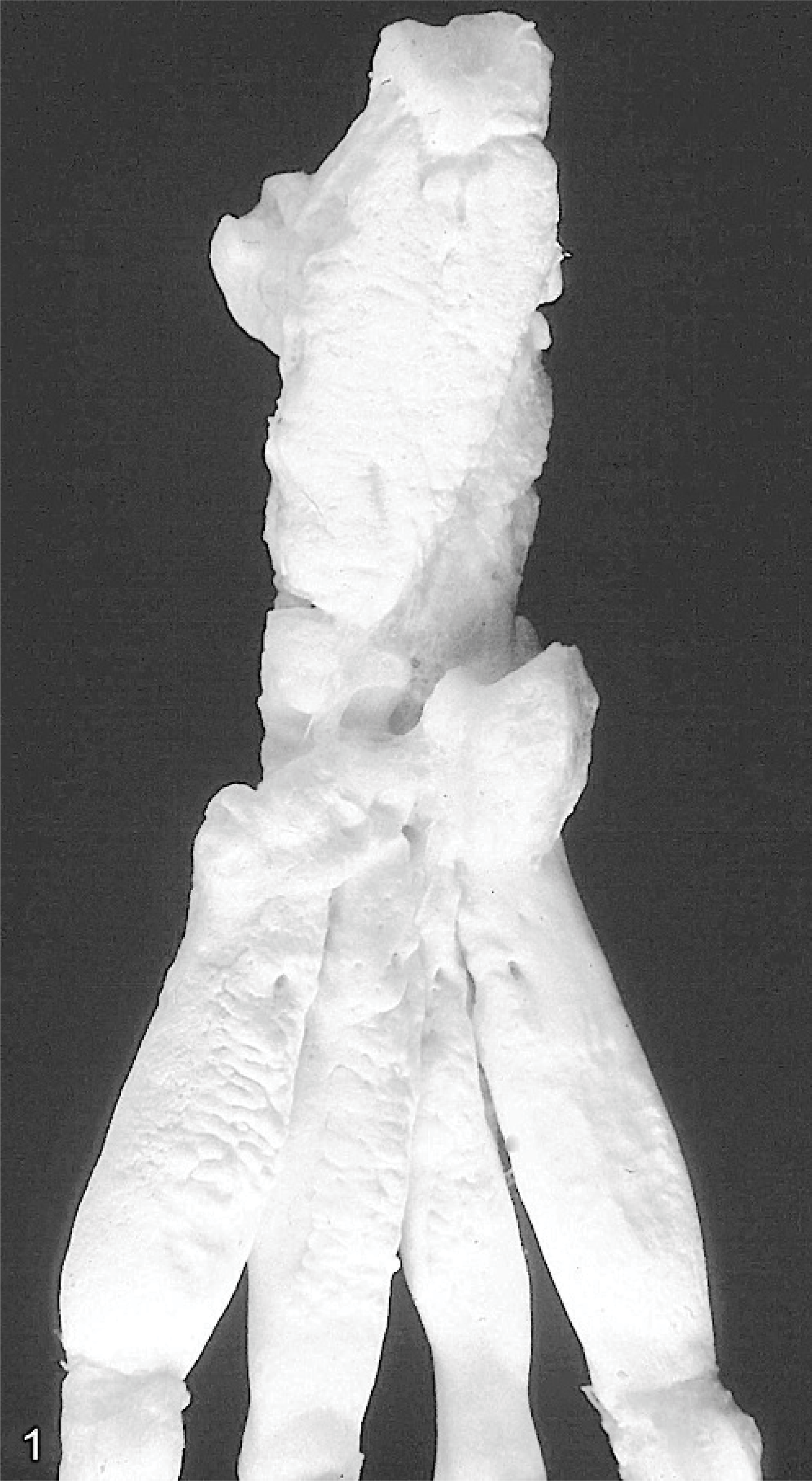
Tarsus and metatarsus; rabbit No. 3. Roughened bone surface due to severe periosteal hyperostosis.
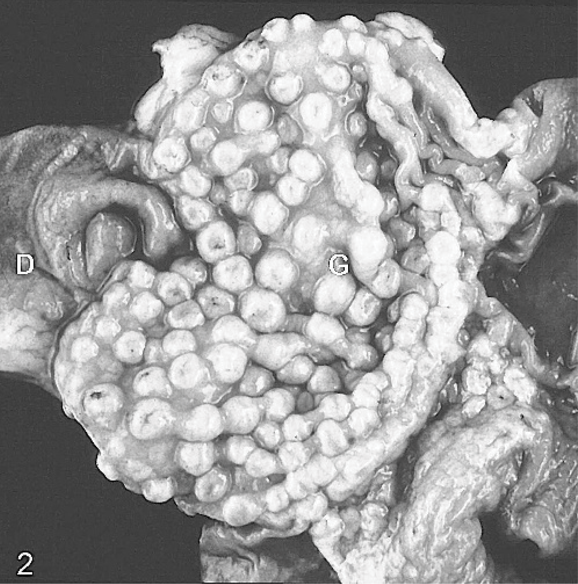
Stomach; rabbit No. 1. Buttonlike appearance of the gastric mucosa. D indicates duodenal mucosa; G, gastric mucosa.
For histologic investigation, the tissues were formalin fixed and embedded in paraffin wax. Bony tissue was also decalcified in 5% nitric acid at 37°C. The hyperostoses of the long bones were found predominantly periosteally and consisted of a prominent proliferation of woven bone and periosteal fibroblasts and a loss of preexisting lamellar bone (Fig. 3). Furthermore, moderate multifocal endosteal hyperostoses were noted, and a mild myelofibrosis was present. The stomach of all animals (including Nos. 5 and 8, which were not affected macroscopically) showed a thickened mucosa characterized by a mild to severe adenomatous proliferation consisting of branching villous projections covered by a hyperplastic columnar mucus-producing surface epithelium (Fig. 4). Similarily, the duodenal mucosa of all rabbits revealed varying degrees of adenomatous proliferations and a mild goblet cell hyperplasia. In addition, a mild to moderate multifocal to diffuse infiltration of lymphocytes, macrophages, and plasma cells was present in the gastric and duodenal lamina propria.
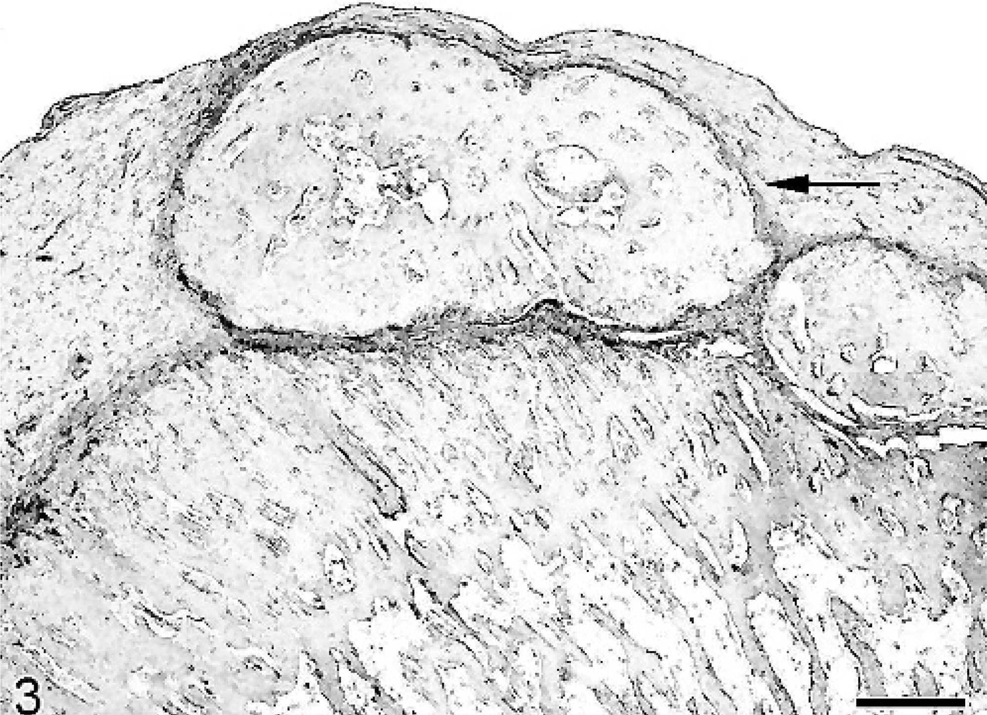
Bone; rabbit No. 3. Severe periosteal proliferation of woven bone (arrow) and proliferation of periosteal fibroblasts. HE after decalcification. Bar = 400 μm.
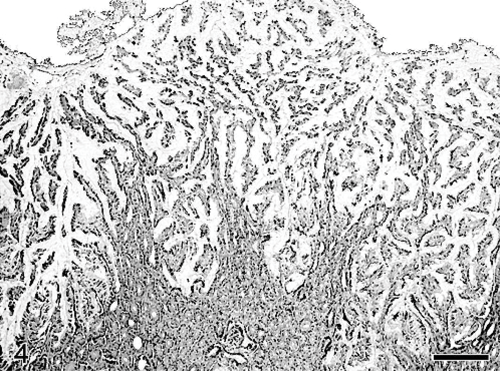
Stomach; rabbit No. 6. Thickened mucosa characterized by severe adenomatous proliferation consisting of branching villous projections. HE. Bar = 200 μm.
Using Warthin-Starry stain no Helicobacter-like or intracellular bacteria could be detected in the stomach of the animals. Bacteriologic investigation of the gastric mucosa of 2 rabbits (Nos. 3 and 4) revealed unspecific findings. Pasteurella multocida was isolated from the bronchial lymph node of rabbit No. 4.
The copper content of the liver of 3 rabbits (Nos. 1, 3, and 4) ranged within the marginal to adequate level (4.5, 4.9, and 6.5 mg/kg; marginal to adequate level: 4–50 mg/kg). 11 The liver vitamin A content of rabbit Nos. 1–4 was within the normal range, except for a minimal elevation in animal No. 3 (290, 213, 319, and 272 μg/g; normal range: 50–300 μg/g). 7
The fluoride content of the bones (tibia/fibula) of 2 affected rabbits (Nos. 4 and 5) was measured by ion-selective potentiometry with a fluoride-selective electrode in bone ash. A size- and age-matched control rabbit revealed a content of 550 μg fluoride per gram of bone ash. In contrast, 12,700 and 15,000 μg fluoride per gram of bone ash were detected in rabbit No. 4 (rabbitry 1) and rabbit No. 5 (rabbitry 2), respectively.
The results of the chemical feed analyses are given inTable 1. They show a severely increased fluoride level in both concentrates and a moderately increased vitamin A level in concentrate 1.
Analyses of concentrates fed in two rabbitries.
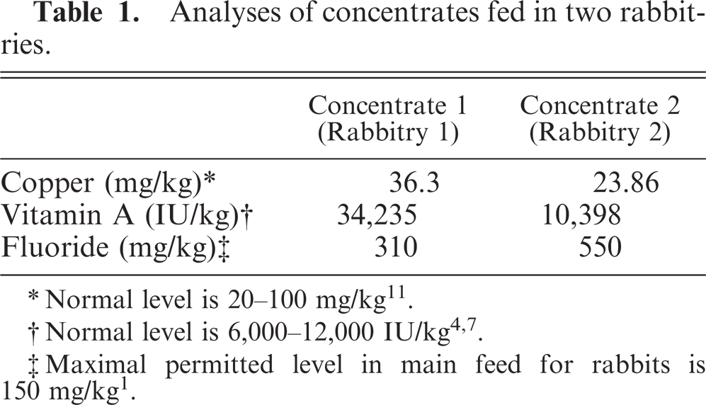
∗ Normal level is 20–100 mg/kg 11 .
‡ Maximal permitted level in main feed for rabbits is 150 mg/kg 1 .
Growing rabbits from two different rabbitries revealed a multifocal severe osteopathy associated with a highly elevated fluoride content in bones and feed. The bone fluoride content in the affected animals exceeded the control values by 23- and 27-fold. The source of the fluoride intoxication could be traced back to the concentrated rations used in both rabbitries, which were, according to the owners of the rabbits, produced by different mills. For cattle, sheep, goats, pigs, poultry, and chicken maximal permitted values of fluoride in main feed are defined. 1 For other animals, including rabbits, only a collective maximal value of 150 mg per kg diet has been determined. 1 The fluoride content of the pelleted commercial diets in the present case exceeded this maximal value by far. Based on the assumption of a calculated food intake of 60 g/kg body weight per day, the animals received between 18.6 and 33 mg fluoride per kilogram body weight per day. The toxic dose of fluoride in rabbits is 5.0 mg natrium fluoride per kilogram body weight per day, given over a period of 100 days. 11 The 6- to 10-week-old rabbits of the present case developed severe clinical signs in spite of a shorter period of fluoride ingestion, which was most likely due to the much higher daily fluoride intake. On a farm in Mexico, rabbits developed bone lesions after consumption of a diet with fluoride contents between 455 and 526 mg/kg. 13 Experimentally, West and Malcolm 17 induced osteofluorosis in rabbits by the ingestion of 500 mg/kg fluoride in the drinking water over a period of 30 days. 17 These amounts are comparable to the fluoride content of the diet in the present case, especially to the content of 550 mg/kg in concentrate 2. The reason for the elevated fluoride content in concentrates 1 and 2 could not be determined. The sources of the premixes were different in both mills, and a contamination of the rabbit concentrates with other mineral supplements, such as those produced for the more fluoride-tolerating poultry, was excluded by the mills. Fluoride contents of hay or drinking water were not measured, but industrial fluoride pollution and elevated fluoride contents in drinking water are not reported for the geographical region of the farms. A possible vitamin A intoxication induced by the relatively high vitamin A level in concentrate 1 was excluded, because the amount of vitamin A in the liver was within normal limits, except for a minimal elevation in animal No. 3. Copper contents were measured in the liver of 3 animals, because copper enhances osteosclerotic changes caused by prolonged fluoride toxicity. 11 However, no elevated copper levels could be detected. After changing the rations in both rabbitries, no further animals developed clinical signs of osteofluorosis.
A remarkable and unexpected finding of all 8 rabbits with osteofluorosis was the proliferative chronic gastroduodenopathy. Mucosal abnormalities in the stomach and duodenum are common findings in human osteofluorosis but, in contrast to the present observations, lesions in humans consisted of chronic atrophic gastritis and duodenitis without proliferative changes. 6 A possible effect of fluoride on the proliferation of the gastric mucosa of rabbits has not been reported so far. In an ultrastructural study on the effect of fluoride on the duodenal mucosa of rabbits, a “cracked-clay” appearance of the surface of microvilli and surface abrasions were observed, resembling findings in human atrophic gastritis caused by osteofluorosis. 6, 15 However, proliferative changes were not mentioned. 15 As a general mechanism, chronic atrophic gastritis in humans can lead to focal foveolar hyperplasia, resembling the histologic lesions of the present case. 12 In dogs, especially in the Basenji, a chronic hypertrophic gastritis of unknown origin is described. 2 This lesion displays similarities to Menetrier's disease of humans, which is characterized by foveolar hyperplasia. 2 Proliferative lesions resembling the findings of the present cases have hitherto not been reported in rabbits or other animals in association with fluoride toxicosis. A parasitic gastritis caused by Obeliscoides cuniculi or other parasites was excluded after histologic investigation. In primates, a mucinous hypertrophic gastropathy caused by polychlorinated biphenyls (PCBs) is described, whereas toxicity studies in rabbits revealed only megalocytosis in the liver. 16, 18 However, the animals of the present case lacked liver lesions; therefore, PCB toxicosis seems unlikely. Whether chronic fluoride intoxication has to be considered as a cause or trigger for the mentioned mucosal changes or whether these alterations represent an unrelated incidental finding remains to be determined. In conclusion, chronic fluoride intoxication leads to periosteal hyperostosis in rabbits and should be considered as a possible cause of a proliferative gastroduodenopathy in this species.