
Abstract
Clostridium septicum is the causative agent of histotoxic infections, including malignant edema and braxy (necrotizing abomasitis) in several animal species. The carcass of a 2-year–old, female New Zealand white rabbit with a history of acute depression and obtundation followed by death was received at the California Animal Health and Food Safety Laboratory System (San Bernardino, California) for necropsy and diagnostic workup. No gross lesions were detected at necropsy. Microscopically, there was moderate to severe, multifocal fibrinonecrotizing, transmural gastritis with numerous intralesional Gram-positive, sporulated rods, and disseminated thrombosis of the brain, lungs, heart, and liver, with occasional intravascular rods. The rods observed within the gastric wall and thrombi in the stomach and lung were positive for C. septicum by immunohistochemical staining. However, this microorganism was not isolated from stomach content. Clostridium septicum should be included in the list of possible etiologies of gastritis in rabbits.
Clostridium septicum is a Gram-positive, motile, spore-forming, highly virulent but poorly characterized anaerobic pathogen. 13 Clostridium septicum is the primary etiological agent of braxy, a suppurative and necrotizing abomasitis common in sheep5,7 and occasionally seen in calves. 20 The microorganism is also responsible for malignant edema in ruminants,8,14 traumatic myonecrosis in human beings,12,21 dogs, 17 horses, and gangrenous dermatitis and cellulitis in broiler chickens and turkeys. 22 Clostridium septicum was also associated with emphysematous gastritis in a horse. 4 This microorganism produces several extracellular toxins that include deoxyribonuclease, hyaluronidase, neuraminidase, and alpha-toxin. 11 The latter is considered to be the main virulence factor attributed to C. septicum. Alpha-toxin is a lethal, necrotizing, pore-forming cytolysin that is similar to the large lobe of aerolysin from Aeromonas hydrophila. 1 The C. septicum alpha-toxin is secreted as a water-soluble protoxin and cleaved at an RGKR motif by host cell proteases, such as furin, to form active monomers and a 45 amino acid C-terminal peptide.9,11 Toxin monomers bind to glycosylphosphatidylinositol-anchored proteins on the cell surface,10,11 oligomerize into hexameric complexes, and insert into the cell membrane to form a 1.3–1.6-nm pore.19,23 Therefore, the activity of the C. septicum alpha-toxin in a cell is associated with the presence of a receptor and an activating protease. 11 The C. septicum alpha-toxin has a range of effects over target cells, including lytic and vacuolating properties. 24
Gastritis has been described in most animal species and has been associated with a wide range of etiologies, including bacteria, viruses, fungi, and parasites, as well as chemical and nonchemical trauma. Gastritis has rarely been described in rabbits, with known etiologies including trichobezoars, trichothecene (T-2) mycotoxin, and Obeliscoides cuniculi larvae.15,18 In the current study, a case of necrotizing gastritis associated with C. septicum in rabbits is described.
The carcass of a 2-year-old, 5.40-kg, female New Zealand white rabbit (Oryctolagus cuniculus) from a university research rabbit colony in southern California, was received at the California Animal Health and Food Safety (CAHFS) Laboratory System (San Bernardino, California) for necropsy and diagnostic workup. The rabbit had a history of acute depression and obtundation followed by death. A full necropsy was performed within 6 hr of death, and samples were collected for multiple tests as described below. Unless specified, all tests were performed according to CAHFS standard operating protocols. Samples of small intestine, liver, lung, and colon were aseptically collected and inoculated onto 5% sheep blood Columbia agar plates a and incubated in 5–10% CO2 at 37°C for 48 hr. In addition, lung and stomach contents were inoculated onto Brucella, phenylethanol, and egg yolk agar plates a and incubated anaerobically at 37°C for 5 days. The DNA extracts of liver and colon samples were subjected to a real-time polymerase chain reaction (PCR) assay with primers designed to detect a fragment of the Salmonella-specific invA gene as previously described. 3 Salmonella culture was performed on the liver and colon. Samples of stomach, brain, trachea, lung, heart, liver, kidney, spleen, small and large intestine, vagina, and skeletal muscle were fixed by immersion in 10% buffered formalin (pH 7.2) for 24–72 hr. All tissues were processed by standard histological techniques for the production of 4-µm-thick sections that were stained by hematoxylin and eosin (HE). Selected sections of the stomach, lung, and brain were Gram stained.
Sections of stomach and lung were stained by an immunoperoxidase technique for C. septicum, Clostridium perfringens, Clostridium chauvoei, Clostridium novyi, and Clostridium sordellii using a commercial kit according to the manufacturer’s instructions. b Rabbit polyclonal anti–C. perfringens antibodies c and anti–C. chauvoei, C. novyi, C. septicum, and C. sordellii antibodies d were used as primary antibodies.
The liver was analyzed for heavy metals (lead, manganese, iron, mercury, arsenic, molybdenum, zinc, copper, and cadmium) by inductively coupled argon–plasma emission spectrometry. Selenium concentration in the liver was determined by inductively coupled plasma spectrometry using hydride generation. The feces were analyzed by fecal flotation exam.
At necropsy, the carcass was in a good state of postmortem preservation. The rabbit was well fleshed with excessive perirenal, retroperitoneal, and mesenteric depot fat. The stomach was filled with abundant fur and mucoid green ingesta, and the cecum and proximal colon contained semi-fluid green feces. The tracheal mucosa was dark red, and the lungs were mottled dark pink to red, and wet. There was severe, locally extensive congestion of the perivaginal tissue.
Microscopically, the glandular stomach showed moderate to severe, multifocal, fibrinonecrotizing gastritis characterized by scattered foci of superficial to deep mucosal necrosis (Fig. 1), with superficial clumps of fibrinohemorrhagic exudate, necrotic cellular debris, degenerate and viable heterophils, occasional dilated glands, and frequent capillary thrombosis. The submucosa was moderately distended with clear to acidophilic edema fluid (Fig. 2), and there was heterophilic inflammation and necrosis of the tunica muscularis (Fig. 3) and serosa.
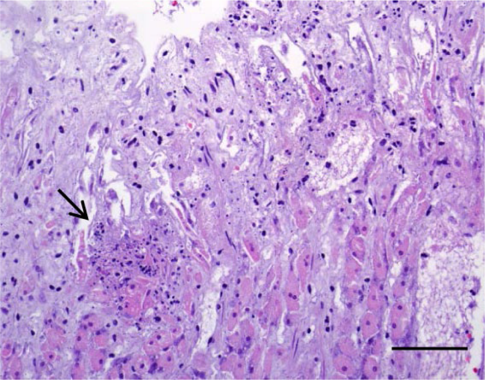
Section of the gastric glandular mucosa with multifocal areas of necrosis (arrow). Hematoxylin and eosin. Bar = 50 µm.
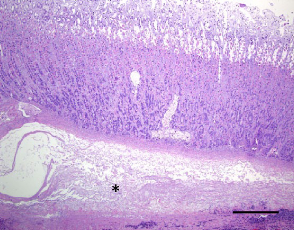
Section of gastric wall showing the submucosa moderately distended by clear to acidophilic edema (asterisk). Hematoxylin and eosin. Bar = 250 µm.
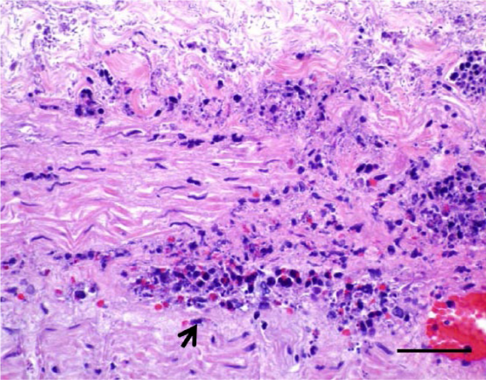
Multifocal heterophilic infiltration affecting the tunica muscularis of the stomach (arrow). Hematoxylin and eosin. Bar = 50 µm.
Myriad, approximately 1-µm wide by 5–10-µm long, Gram-positive bacilli with terminal or subterminal spores were multifocally to diffusely distributed throughout the mucosa, submucosa, muscularis, and serosa. Within the lung, heart, liver, brain, and cerebral meninges, numerous small- to medium-sized arteries and arterioles contained thrombi composed of fibrin, platelets, heterophils, lymphocytes, plasma cells, macrophages, nuclear debris (Fig. 4), and occasional individual or clustered bacilli were observed. There was marked congestion and moderate edema of the tracheal submucosa, and the perivaginal sinuses were severely congested with occasional fibrinocellular thrombi. The Gram-positive bacilli observed in the mucosa, submucosa, muscularis, and serosa of the stomach, lungs, heart, liver, brain, and meninges were consistently positive for C. septicum by immunohistochemical staining (IHC), and negative for C. perfringens, C. chauvoei, C. novyi, and C. sordellii. In addition, the occasional intravascular clostridial-like bacilli were also positive for C. septicum by IHC.
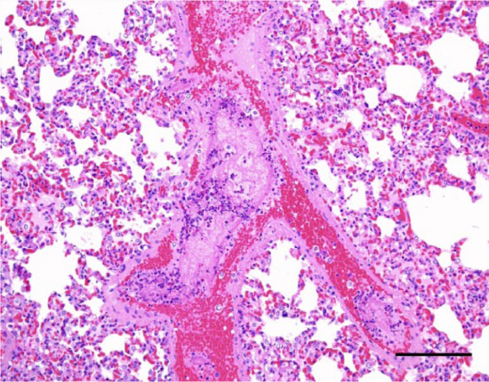
Lung; rabbit. Medium-sized vein with fibrinocellular thrombi composed of fibrin, platelets, heterophils, mononuclear cells, and nuclear debris. Hematoxylin and eosin. Bar = 200 µm.
Bacterial aerobic culture of small intestine, liver, lung, and colon yielded mixed flora, while bacterial anaerobic culture of stomach contents was positive for C. sordellii. Salmonella was not detected by culture or PCR from liver or colon. Hepatic copper and selenium concentrations were slightly low (4.9 and 0.89 ppm, respectively; normal range: 8–50 and 1–2 ppm, respectively). 16 The hepatic levels of other heavy metals were within the normal range for rabbits. No parasites were detected in feces by fecal flotation exam.
The severe transmural gastritis, accompanied by the presence of large numbers of Gram-positive bacilli throughout all gastric layers shown to be C. septicum by IHC (Fig. 5), strongly suggests that this bacterium played a significant role in the development of the disease. Furthermore, the disseminated thrombosis observed in the lungs, heart, liver, brain, and meninges are indicative of secondary septicemia and possibly also toxemia. Immunohistochemical staining for C. perfringens, C. chauvoei, C. novyi, and C. sordellii was consistently negative in several sections of the stomach, ruling out the possibility of a combined infection by these clostridial microorganisms. In addition, no other bacterial forms were observed in both HE- and Gram-stained sections of the stomach.
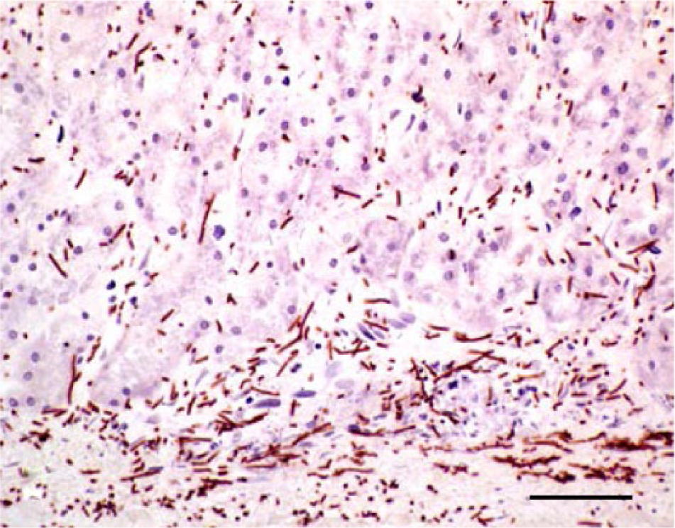
Clostridium septicum immunohistochemical staining of the glandular mucosa of the stomach showing myriad positive clostridia bacilli. Bar = 30 µm.
Surprisingly, cases of gastritis in rabbits are very scant in the literature. Reported causes include sublethal concentration of T-2 mycotoxin in the feed, which produces catarrhal gastritis, 6 and infestation of third-stage O. cuniculi larvae, which produces gross and histologic lesions similar to those of bovine ostertagiosis. 18 However, both of these conditions were experimentally induced, and neither caused lesions similar to the ones described herein.
Overall, the findings in this case are similar to those described for the necrotizing abomasitis and fatal bacteremia produced by C. septicum (braxy) in calves and lambs (Schamber GJ, Berg IE: 1986, Braxy or bradsot-like abomastitis caused by Clostridium septicum in a calf [letter to the editor]. Can Vet J 27:194). 2 Although the ingestion of cold or frozen feed has been suggested to be the main predisposing factor in cases of braxy, 8 this has never been proved. This was certainly not the case with the rabbit described herein, which was raised under controlled conditions for research purposes. Gastric bacterial infections occur infrequently in most animal species, possibly because of the acid intraluminal pH and mucosal barrier. However, traumatic micro-injuries to the gastric mucosa, or compromised blood flow to the stomach resulting in areas of ischemia, may be predisposing factors leading to clostridial invasion, proliferation, and toxin production. Although the presence of abundant fur in the stomach is not uncommon in rabbits, it may have facilitated gastric mucosal microinjuries in the present case, facilitating clostridial proliferation, possibly in combination with other stress factors. The rabbit was being fed a commercial rabbit ration that had, among other ingredients, an undisclosed amount of roughage. Although it is possible that some ingredients of the roughage caused gastric injuries, this is unlikely as this was the only rabbit in the colony to present the problem. The caretakers were warned, however, to review the diet of the rabbits in an attempt to avoid feeding the animals diets with components that might damage the gastric mucosa.
Clostridium septicum was not isolated from the stomach content, although it was demonstrated by IHC within the gastric wall. It is possible, albeit unlikely, that C. septicum was only present within the gastric wall but not in the content of this organ. Alternatively, it is also possible that C. sordellii was present in larger quantities in this content, which could have facilitated overgrowth of this microorganism over C. septicum. While C. sordellii was isolated from the stomach content, the negative IHC results for C. sordellii in multiple sections of stomach suggests that this organism was not associated with the lesions seen and was most likely an incidental finding. Indeed, C. sordellii is a common inhabitant of the intestine and may be isolated from the normal intestinal tract of domestic animals. 20 In the current case, it was possible that C. sordellii may have invaded the stomach from the small intestine due to duodenal reflux or was merely a sample contaminant. As in sheep, calves, and horses, C. septicum infection should be included in the differential diagnosis of necrotizing gastritis in rabbits.
Footnotes
a.
Hardy Diagnostics, Santa Maria, CA.
b.
Dako North America Inc., Carpinteria, CA.
c.
GenWay Biotech Inc., San Diego, CA.
d.
VMRD Inc., Pullman, WA.
Declaration of conflicting interests
The author(s) declared no potential conflicts of interest with respect to the research, authorship, and/or publication of this article.
Funding
Funding for the current report was provided by the California Animal Health and Food Safety Laboratory, School of Veterinary Medicine, University of California, Davis.