
Abstract
A 6-year-old male cross-breed rabbit (Oryctolagus cuniculus) was presented with lameness and severe swelling from the right shoulder to brachium. On 16-detector helical computed tomography images of the amputated right forelimb after being fixed in formalin, evident proliferative and destructive lesions of bone were observed. On histologic examination, the tumor was composed of proliferating neoplastic cells that resembled histiocytes, with abundant osteoid production. A large number of multinucleated giant cells were found throughout. This case was diagnosed as osteosarcoma by clinical, radiographic, and histologic findings. This is a rare case report of osteosarcoma in a rabbit consistent with canine predilection sites.
Osteosarcomas are primary malignant bone tumors characterized by the production of osteoid and/or bone by neoplastic osteoblasts. 11, 13 Osteosarcomas account for more than 80% of malignant bone tumors in dogs and about 70% in cats. 2, 15 Uterine adenocarcinoma is the most common spontaneously occurring tumor in rabbits. 5 Lymphomas are the second most common tumor of rabbits and the most common tumor of juvenile rabbits. Spontaneously occurring osteosarcomas in domestic rabbits appear to be rare, but a few cases have been reported. 1, 6, 8– 10 Osteosarcomas in laboratory rabbits have also been reported. 16, 17 This paper describes the pathologic features of an osteosarcoma in a domestic rabbit.
A 6-year-old male crossbreed rabbit that weighed 2.6 kg (Oryctolagus cuniculus) was presented with fever, lameness, and severe swelling from the right shoulder to the brachium. The rabbit had been purchased at a pet store and was reared as a domestic animal. Swelling of the right forelimb was assessed to be a malignant tumor on cytologic examination when using fine needle aspiration. Destructive lesions of bone from the distal scapula to the diaphysis of the humerus were observed radiographically, but no evidence of metastasis to the lungs was recognized. Serum alkaline phosphatase (ALP) activity was 77 U/l. Because the rabbit was suspected to have osteosarcoma, the right forelimb was surgically amputated (Fig. 1) and was submitted to the Nihon University for examination. The rabbit suddenly fell into cardiopulmonary arrest during the surgical operation and died without being revived. No postmortem examination was performed because of the owner's request. The amputated right forelimb was fixed in 10% neutral buffered formalin and was evaluated with 16-detector helical computed tomography (CT) (Aquilion16, Toshiba, Tokyo, Japan). The detector collimation (16-row unit) was 8 mm. The imaging parameters included a section thickness of 0.5 mm, kilovolt peak of 120, 200 mA, matrix of 512 × 512, and tube rotation time of 0.5 seconds. The images were reconstructed with a Virtual Place Advance PLUS (AZE, Tokyo, Japan). On helical CT images, evident proliferative and destructive lesions of bone from the distal scapula to the diaphysis of the humerus involving a glenohumeral joint space were observed (Fig. 2). For pathologic examination, several areas of the amputated right forelimb were embedded in paraffin wax and sectioned at 5 μm. The sections were stained with hematoxylin and eosin (HE) and Masson trichrome (M-T) stain. Immunohistochemistry was performed by the Streptavidin-biotin-peroxidase method. Primary antibodies used were rabbit anti-human lysozyme (diluted 1 : 1,000, DakoCytomation, Kyoto, Japan) and rabbit anti-multiple-antigen peptide osteocalcin (diluted 1 : 500, Cosmo Bio LSL, Tokyo, Japan).Normal rabbit lymph nodes and bones were used as controls to validate the specificity. According to the manufacturer's instructions, the anti-osteocalcin antibody cross-reacts with rat, bovine, human, and mouse osteocalcin. On histologic examination, the tumor of the right forelimb was composed of sheets of round-to-polyhedral neoplastic cells that resembled histiocytes, with round-to-oval nuclei (Fig. 3). The tumor was highly cellular, and pleomorphism and atypical nuclei and cytoplasm were prominent. There were about 12 mitoses per 10 high power fields. A large number of multinucleated giant cells were found throughout. Neoplastic cells produced abundant eosinophilic and amorphous neoplastic osteoid. Osteoid was stained blue with M-T stain (Fig. 4). Broad necrosis areas were also observed. On immunohistochemical examination, the cytoplasm showed positive immunostaining for osteocalcin in most of the neoplastic cells, but the cell membrane was also positive in some neoplastic cells (Fig. 5). Osteoid was negative for osteocalcin. The cytoplasm of osteoblasts on normal rabbit bone was positive for osteocalcin. Neoplastic cells were negative for lysozyme. Phagocytes in lymph nodes distinctly reacted to lysozyme, however, nonepithelial tissues were also partially and weakly positive.
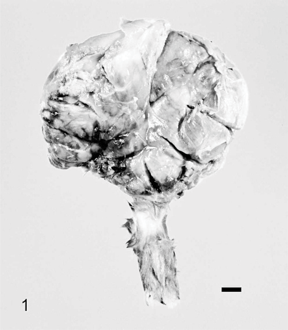
The amputated right forelimb; Rabbit. Severe swelling from the right shoulder to brachium. Bar = 1 cm.
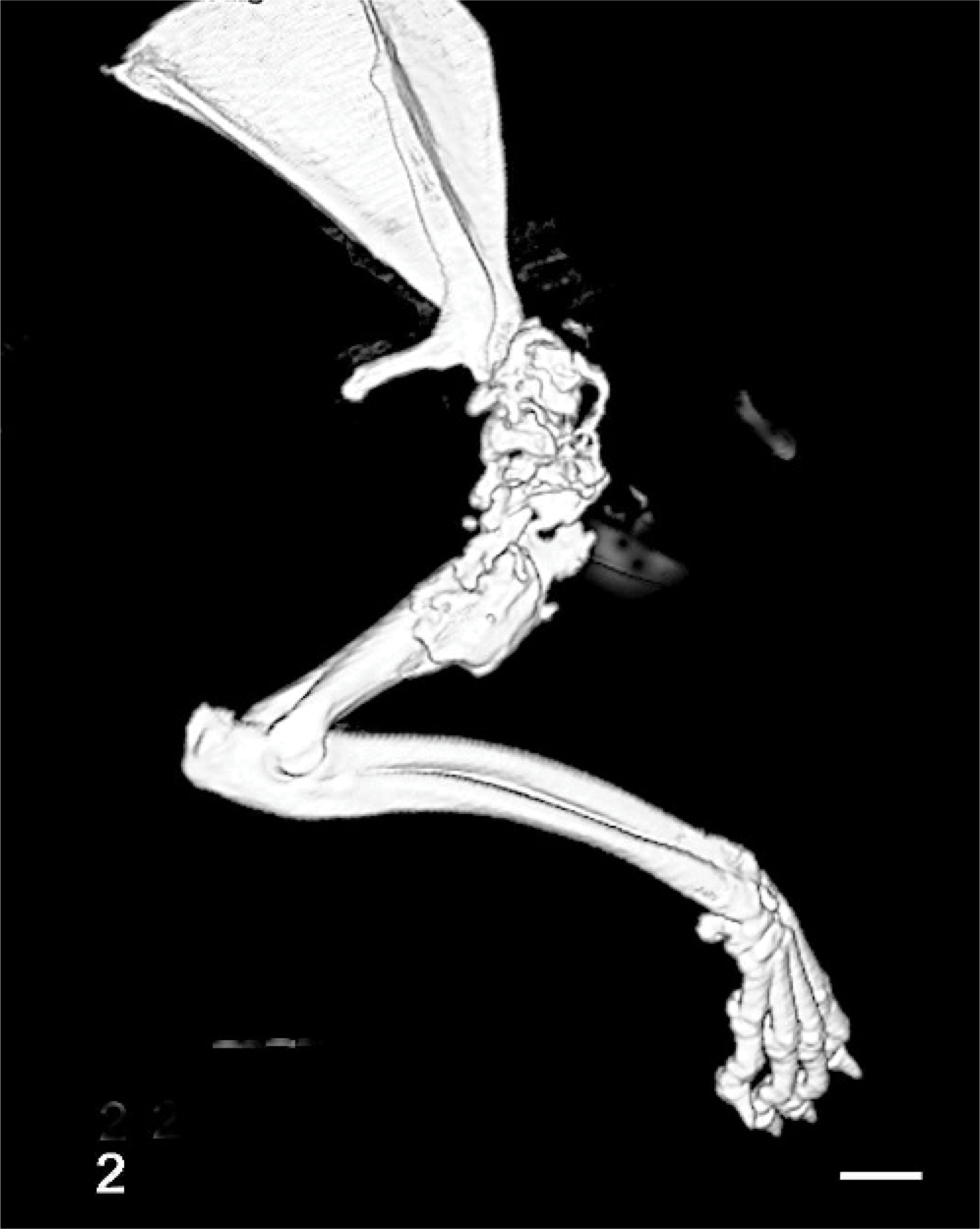
16-detector helical computed tomography images of the amputated right forelimb; Rabbit. Evident proliferative and destructive lesions of bone from the distal scapula to the diaphysis of the humerus. Bar = 1 cm.
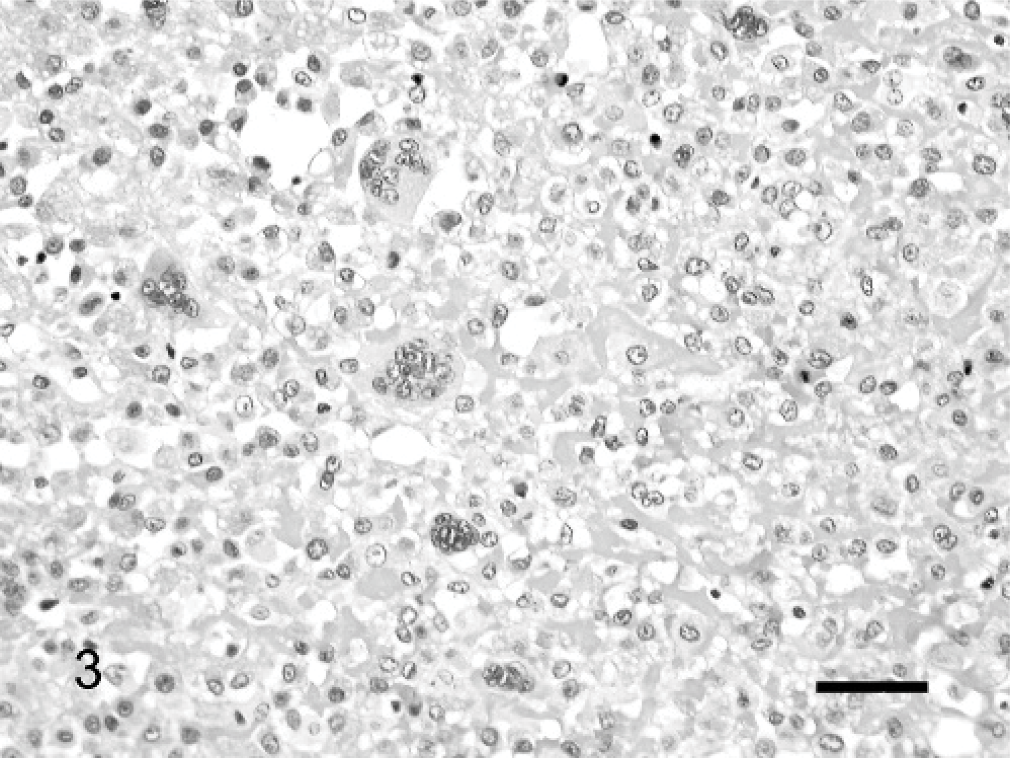
Osteosarcoma; Rabbit. Proliferation of neoplastic osteoblasts with osteoid production and multinucleated giant cells. HE. Bar = 50 μm.
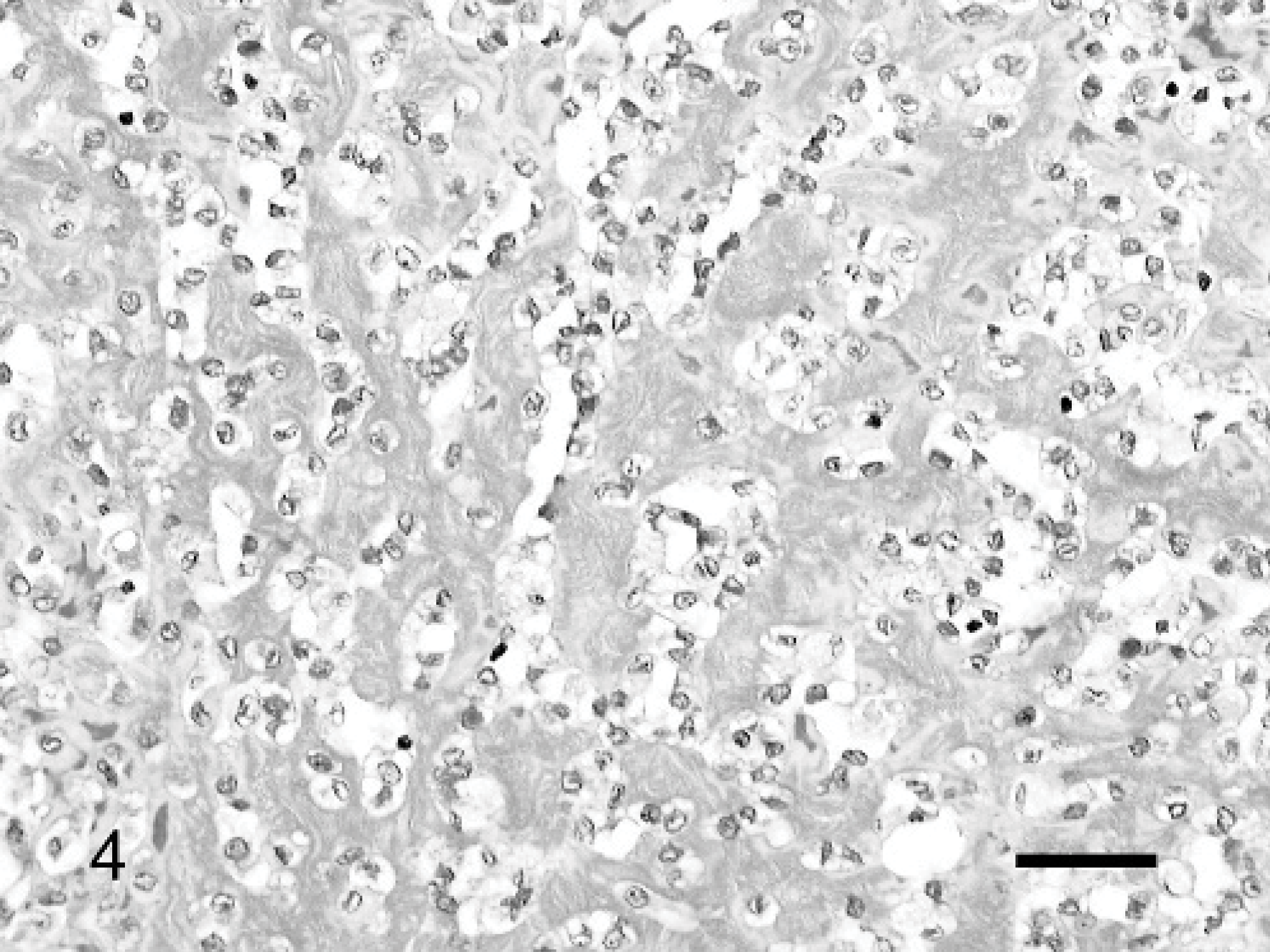
Osteosarcoma; Rabbit. Osteoid was stained blue. Masson trichrome stain. Bar = 50 μm.
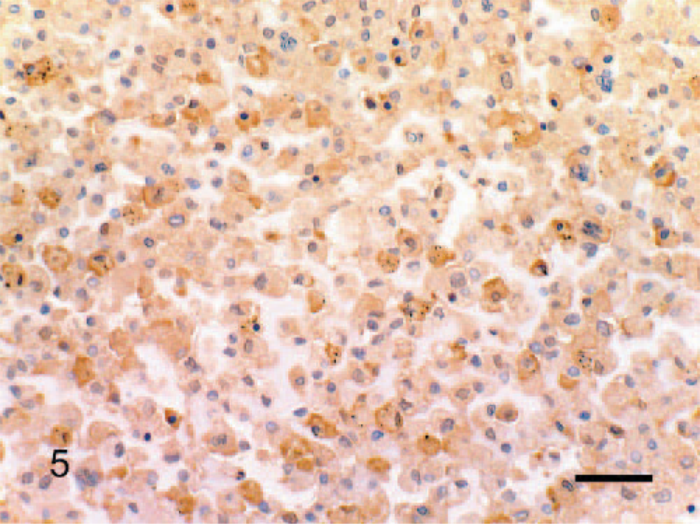
Osteosarcoma; Rabbit. The cytoplasm showed positive immunostaining for osteocalcin in most of neoplastic cells, but cell membrane was also positive in some neoplastic cells. Streptavidin-biotin-peroxidase method. Counterstaining with Mayer's hematoxylin. Bar = 50 μm.
On histologic examination, malignant osteoblasts varied from pleomorphic, spindle to plump, oval, or rounded cells. 13 In our case, the tumor was composed of proliferating neoplastic cells that resembled histiocytes, with abundant osteoid production. Neoplastic cells were positive immunostaining for osteocalcin but were negative for lysozyme. Osteocalcin is a specific protein marker for osteoblasts. In one study, osteoblasts staining for osteocalcin showed 70% sensitivity and 100% specificity. 4 In our study, the specificity for osteocalcin was confirmed in normal rabbit osteoblasts. Although the specificity of the anti-human lysozyme antibody was ambiguous, it was sufficient to exclude a possibility of histiocytic origin. By morphology, neoplastic cells that resembled histiocytes had an osteoblastic origin. By histology, osteosarcomas are classified into the following variants in dogs: poorly differentiated, osteoblastic, chondroblastic, fibroblastic, telangiectatic, and giant-cell types. 11, 13 In our case, multinucleated giant cells were prominently observed. Although no classification of osteosarcomas has been established in the rabbit, we suggest that our case could be classified as a giant-cell-type osteosarcoma by using the canine classification. It is important to distinguish the giant-cell-type osteosarcoma from a giant-cell tumor of bone. Giant-cell tumors are characterized by the presence of a large number of multinucleated giant cells that resemble osteoclasts, but osteoid is either absent or only weakly produced. 11 The diagnosis of a giant-cell tumor of bone was excluded, because the marked production of osteoid was noted. Although fibroblastic osteosarcoma has been reported in the rabbit, 9 there has been no report of giant-cell-type osteosarcoma.
Osteosarcomas are common malignant bone tumors in dogs and cats but are rare in other domestic animals. 13 The appendicular skeleton is affected 3 to 4 times as often as the axial skeleton, and the forelimbs are affected approximately twice as often as the hindlimbs. 2 This ratio closely corresponds to the ratio of weight distribution. In one study of site incidence in 314 canine osteosarcomas, 76% of the tumors occurred in 4 long bones: radius (23%), proximal humerus (19%), tibia (14%), and femur (12%). 18 Osteosarcomas of the ribs, skull, maxilla, upper lip, and left thigh have been reported in domestic rabbits, and osteosarcomas occurring in the mandible have been reported in laboratory rabbits. 1, 6, 8– 10, 16, 17 However, the predilection sites for osteosarcoma in rabbits are unknown. The predilection sites for osteosarcoma in rabbits may not correlate with those in dogs. The majority of dogs with appendicular osteosarcoma show metastasis to the lungs. 12 There are some reports of osteosarcomas with metastasis in rabbits. 1, 6, 8, 10 However, osteosarcomas seldom involve a joint space and spread through adjacent bone. 13 In our case, the tumor occurred from the distal scapula to the diaphysis of the humerus and involved a glenohumeral joint space. This was considered a rare case that involved a joint space, although the occurrence of our case was consistent with a canine predilection site. No evidence of metastasis to the lungs was recognized radiographically, but micrometastasis might have been present. The risk of osteosarcomas among large (40–80 pounds) and giant (more than 80 pounds) breeds of dogs are 7.9 and 60.9 times the risk of small breeds (less than 20 pounds), respectively. 14 The rabbit was normal sized. There are large breeds of rabbits (e.g., Flemish Giant), but a correlation between body size and the risk of osteosarcoma is unclear.
Serum ALP activity is often increased in dogs with osteosarcoma. 13 ALP is an enzyme found in many tissues, including the liver, intestine, kidney, placenta, and bone. Bone-specific ALP (BALP) is a more specific indicator of osteoblastic activity than ALP and, therefore, may be more useful as an indicator of bone-producing tumors. It has been reported that activities of serum ALP and BALP are important prognostic factors for appendicular osteosarcoma in dogs. 3 High preoperative activities of ALP and BALP were associated with a shorter survival interval. In one study, ALP activity in a laboratory rabbit with an osteosarcoma was elevated in comparison with normal values. 17 The normal reference range of serum ALP in rabbits is 4–16 U/l. 7 The BALP activity was unknown, but ALP activity was elevated to 77 U/l in our case. Although the rabbit was euthanatized in our case, we considered that evaluation of ALP is useful as a diagnostic and prognostic factor in rabbits with osteosarcoma.
It is important that the diagnosis of osteosarcoma is based not only on histologic findings but also on clinical, radiographic, and gross findings. Helical CT examinations have also been proven to be useful for the diagnosis of osteosarcoma in our study.