
Abstract
An 8-year-old chipmunk (Tamias sibiricus) showed a mass on the tail root. Histologically, the excised mass showed proliferation of spindle-to-polyhedral cells and abundant multinucleated giant cells, with the production of neoplastic osteoid. Immunohistochemically, neoplastic cells were positive for vimentin and weakly positive for osteocalcin. Osteoid was also positive for osteocalcin. This tumor was diagnosed as a giant cell-type osteosarcoma. This is the first case report of osteosarcoma in a chipmunk.
Osteosarcoma is a malignant tumor of osteoblasts, which is always accompanied by the production of osteoid and bone. This tumor accounts for approximately 80% of malignant bone tumors in dogs but is rare in other domestic animals.1,5 The chipmunk (Tamias sibiricus) is a species peculiar to Japan. There are no reports of neoplastic disease in the chipmunk. This is, therefore, the first report of an osteosarcoma in a chipmunk.
The chipmunk was an 8-year-old female weighing 80 g, which had been kept as a pet. The chipmunk had a history of a mass on the tail root being observed by the owner. On X-ray examination, no metastasis to other regions was observed. The tail was surgically excised with the mass. The excised lesion was fixed in 10% neutral buffered formalin, embedded in paraffin wax, and sectioned at 3 μm. The sections were stained with HE and Masson trichrome (M-T). Immunohistochemistry was performed by the streptavidin-biotin method. Primary antibodies used were mouse antipig vimentin (DakoCytomation, Kyoto, Japan; diluted 1:100), rabbit antihuman lysozyme (DakoCytomation, Kyoto, Japan; diluted 1:800), rabbit antibovine S-100 protein (DakoCytomation, Kyoto, Japan; diluted 1:500), and rabbit antirat osteocalcin (Cosmo Bio, Tokyo, Japan; diluted 1:500). Pieces of bone included in the same sections were used as positive controls to ascertain cross-reactivity with the chipmunk's tissue for osteocalcin,
The excised mass was 11 × 10 × 9 mm in diameter on gross inspection. The surface of the mass was dark red. Histologically, this mass showed proliferation of spindle-to-polyhedral cells and multinucleated giant cells, with the production of neoplastic osteoid and mineralization (Figs. 1, 2). Nuclei were spindle-to-plump shaped, and nucleoli were clear. Pleomorphism and atypia of cytoplasm and nuclei were prominent. Neoplastic cells extended into the dermis. There were 33 mitoses per 10 high-power fields, and bizarre mitoses were also observed. Multinucleated giant cells were scattered among neoplastic cells and had many nuclei, with a range from 2 to 20. Osteoid was stained deep blue by M-T stain (Fig. 3). There were hemorrhage and necrosis in some locations. The neoplastic cells showed positive immunostaining for vimentin and were weakly positive for osteocalcin. Osteoid and osteoblasts in pieces of non-neoplastic bone were also positive for osteocalcin (Fig. 4). Connective tissue in the same sections was weakly positive. The neoplastic cells were negative on immunostaining for lysozyme and S-100 protein.
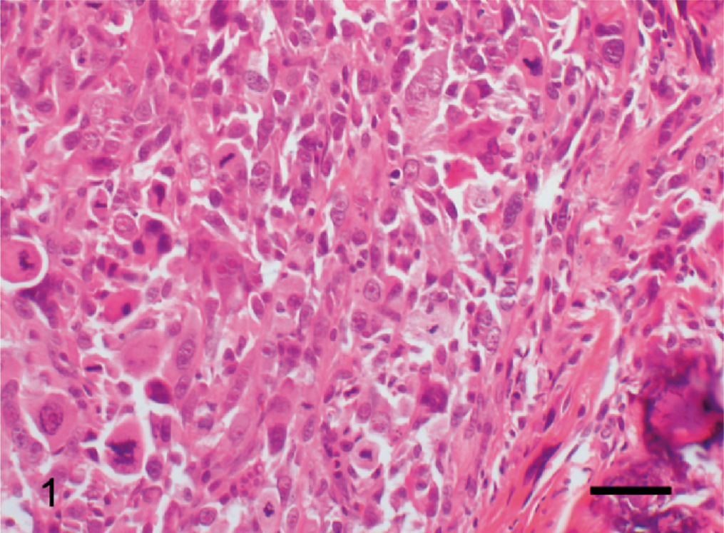
Osteosarcoma; chipmunk. Proliferation of spindle-to-polyhedral cells is seen. Numerous mitoses are observed. HE. Bar = 50 μm.
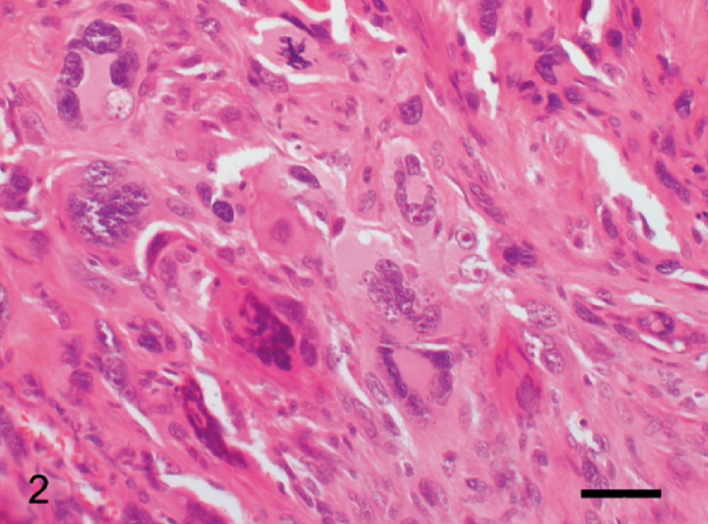
Osteosarcoma; chipmunk. Multinucleated giant cells show production of osteoid. HE. Bar = 50 μm.
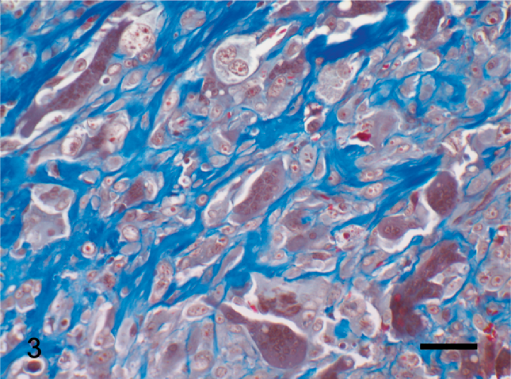
Osteosarcoma; chipmunk. Osteoid is stained deep blue. Masson trichrome. Bar = 50 μm.
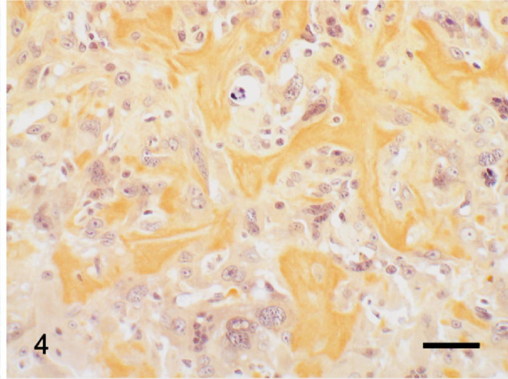
Osteosarcoma; chipmunk. The neoplastic cells and osteoid show positive immunostaining for osteocalcin. Streptavidin-biotin method, counterstaining with Mayer's hematoxylin. Bar = 50 μm.
A definitive diagnosis of osteosarcoma in all cases is based on the production of osteoid and/or bone by malignant mesenchymal cells.8 In our chipmunk, the production of osteoid by spindle-to-polyhedral cells was clearly observed. Osteosarcoma has various patterns histologically and is classified into the following types: poorly differentiated, osteoblastic (nonproductive and productive), chondroblastic, fibroblastic, telangiectatic, and giant cell.1 Cytoplasm and nuclei of osteoblast-like neoplastic cells in our chipmunk showed pleomorphism and atypia, and multinucleated giant cells were remarkable. Therefore, the tumor in this case was diagnosed as giant cell–type osteosarcoma. These observations were in accord with histologic features in osteosarcomas in dogs.8 Immunohistochemical findings were also useful for a diagnosis of osteosarcoma, being consistent with reports of osteosarcomas in dog and human subjects.2–4,9 Cytoplasmic and osteoid staining with osteocalcin showed a high sensitivity and specificity.2 Because neoplastic cells and osteoid were positively immunostained for osteocalcin, we conjectured that these cells were derived from osteoblasts. However, we considered from our results that cross-reactivity for osteocalcin was recognized and specificity was considered low in the chipmunk.
In dogs, the mean age for the development of osteosarcoma is around 7.5–8 years1,6,10 but remains unknown in chipmunks. It is said that a chipmunk's life span is from 5 to 10 years. So, the chipmunk in this case was of an advanced age. The risks of primary osteosarcoma among giant and large breeds of dogs are higher than those for small breeds.7 In dogs, the appendicular skeleton is affected 3–4 times as often as the axial skeleton, and the forelimbs, approximately twice as often as the hindlimbs.1,10 In domestic animals, it is suggested that site preference is closely related to weight balance and weight stress.8 The chipmunk's tail is as long as its body and very characteristic. The role of the tail is to communicate with other chipmunks and maintain balance in trees. As this is the first reported case of osteosarcoma in the chipmunk, definitive sites of predilection are uncertain in this species.