
Abstract
A 2-year-old intact female Golden Retriever presented due to rapidly progressing depression, ascites, dysuria, abdominal pain, and severe vaginal bleeding. At necropsy, the retroperitoneal space was expanded by multiple coalescing neoplastic nodules and the uterine wall was thickened with poorly defined neoplastic infiltrates. The urinary bladder was markedly thickened due to botryoid nodules exhibiting exophytic growth into the lumen. Metastases to lung, liver, kidney, and abdominal and thoracic lymph nodes were also noted. Microscopically, the genital tract and retroperitoneal masses were consistent with the alveolar subtype of rhabdomysarcoma, while the urinary bladder mass had characteristics of the embryonal subtype. Immunohistochemically, the neoplastic cells in all these tissue sites were intensely positive for desmin, sacromeric actin, and vimentin, while they were uniformly negative for cytokeratin and smooth muscle actin. Phosphotungstic acid hematoxylin stain revealed cross-striations in the cytoplasm of scattered neoplastic cells. Based on the gross findings, histopathology, and immunohistochemistry, genitourinary rhabdomyosarcoma with multisystemic metastases was made.
Rhabdomyosarcoma is a neoplasm originating from the skeletal muscle, although it can also derive from organs that lack striated muscle, such as the urinary bladder, uterus, and cervix. 2 Rhabdomyosarcoma has been rarely reported in various domestic animals, including dogs, 6 a cat, 9 and a horse. 4 In humans, rhabdomyosarcomas are classified into embryonal, alveolar, and pleomorphic variants based on histologic features. 2, 11 The majority of human pleomorphic-type rhabdomyosarcoma occur in adults, whereas the embryonal and alveolar types of rhabdomyosarcoma occur in young individuals with a predilection for the head and neck, genitourinary tract, and extremities. 5, 8 A clear pattern of breed, sex, and site prevalence has not been clearly documented in domestic animals, except for a relatively high incidence of sarcoma botryoides of the urinary bladder in young dogs, particularly Saint Bernards. 2 Here, we report a case of malignant metastatic rhabdomyosarcoma with primary involvement of the genitourinary tract and retroperitoneum in a young Golden Retriever dog.
A 2-year-old intact female Golden Retriever was presented to the Veterinary Medical Teaching Hospital of Seoul National University, Korea, with a history of rapidly progressing depression, anorexia, dysuria, abdominal distention and pain, and severe vaginal bleeding. Routine physical examination revealed a large, firm, palpable mass in the retroperitoneal region. After exploratory laparatomy, euthanasia was elected due to multiple organ involvement, tumor size and location, and expected poor prognosis. Necropsy was performed immediately after euthanasia.
At necropsy, the retroperitoneal cavity was almost completely occupied by multiple coalescing, tan, firm nodules, 2.0 to 5.0 cm in diameter. The uterine body, urethra, and distal colon were incarcerated in the retroperitoneal masses. The uterine wall was thickened with ill-defined tumor tissue in the myometrium and serosa, and the cervix and vagina were extremely expanded. On opening the cervix and vagina, the lumina were almost obliterated due to an impinging mass that extended from the uterine wall. No remarkable changes were noted in the ovaries and oviducts. The wall of the urinary bladder was markedly thickened, and the mucosa contained numerous botryoid nodules, soft to firm in texture, about 0.5 to 1.0 cm in diameter (Fig. 1). Both ureters were dilated, and the urethra was stenotic. On cut sections, most masses in the genital tract and retroperitoneum consisted of white to tan, multinodular masses with areas of necrosis and hemorrhage. Well-circumscribed similar neoplastic nodules, about 0.5 to 1.5 cm in diameter, were also present in the lung, liver, and kidney. Numerous lymph nodes in the abdominal and thoracic cavities were enlarged to 2 to 5 times their normal size with effacement of normal architecture.
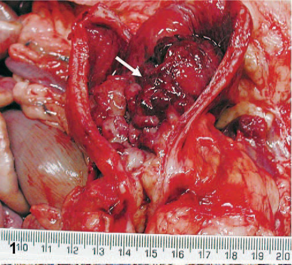
Mass from urinary bladder; 2-year-old Golden Retriever. A multilobulated mass resembling a cluster of grapes protrudes into the lumen from the mucosa. Also note necrosis and hemorrhagic of the mucosa (arrow).
For histopathology, tissue samples were fixed in 10% neutral phosphate-buffered formalin, processed in a routine manner, embedded in paraffin, and stained with HE. Replicate sections of the selected tumor samples were used for phosphotungstic acid hematoxylin staining. The standard avidin-biotin-peroxidase technique and commercially available antibodies were used to identify vimentin, desmin, cytokeratin, sarcomeric actin, smooth muscle actin, or S-100.
Microscopically, an unencapsulated, poorly circumscribed, and expansile neoplastic mass of the urinary bladder elevated the overlying epithelium and expanded the submucosa of the urinary bladder. The mass was composed of densely packed neoplastic cells, polygonal to spindloid in shape, in the deep submucosa, and strap-like spindle cells loosely embedded in myxomatous stroma at the periphery. The neoplastic cells had indistinct cell borders, scant to moderate eosinophilic cytoplasm, and round to ovoid, hyperchromatic nuclei with inconspicuous nucleoli. There were 1 to 3 mitotic figures per high power field (400 ×), as well as marked anisocytosis and anisokaryosis within the neoplastic foci. Multifocal mucosal ulceration and hemorrhage were noted. Aggregates of a few neutrophils, lymphocytes, and plasma cells were scattered through the neoplastic tissue.
Histologic features of neoplastic masses in the genital tract, retroperitoneum, and metastatic foci differed from those of the urinary bladder. The neoplastic tissues were unencapsulated, poorly circumscribed, and infiltrative and were primarily composed of relatively small to intermediate-sized neoplastic cells arranged in solid sheets. Occasional tumor cells were separated by thick fibrous connective tissue (Fig. 2). The neoplastic cells were round to polygonal with distinct cell borders and had variable amounts of eosinophilic cytoplasm and hyperchromatic, round to oval, occasionally pleomorphic nuclei with 1 or 2 prominent nucleoli. Mitoses were frequent (4–6 per 400 × field) with occasional aberrant mitotic figures. Tumor emboli were found frequently in both the blood vessels and lymphatics. Multifocal to coalescing areas of necrosis and hemorrhage were present in the tumor tissue.
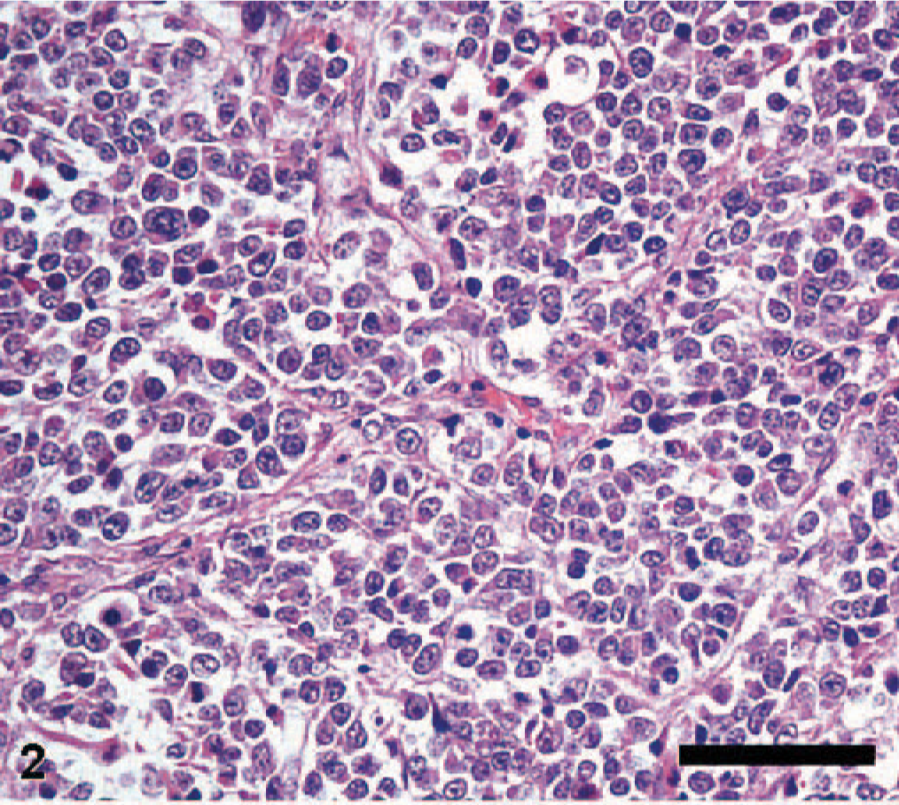
Uterine mass; 2-year-old Golden Retriever. Note round to polygonal neoplastic cells that are nested by fibrous septa resembling alveolar pattern. HE. Bar = 40 μm.
Immunohistochemically, the neoplastic cells were strongly and diffusely positive for desmin (Fig. 3), sarcomeric actin, and vimentin and multifocally positive for S-100. The neoplastic cells were, however, uniformly negative for cytokeratin and smooth muscle actin. Phosphotungstic acid hematoxylin staining revealed cross-striations in the cytoplasm of scattered neoplastic cells (Fig. 4).
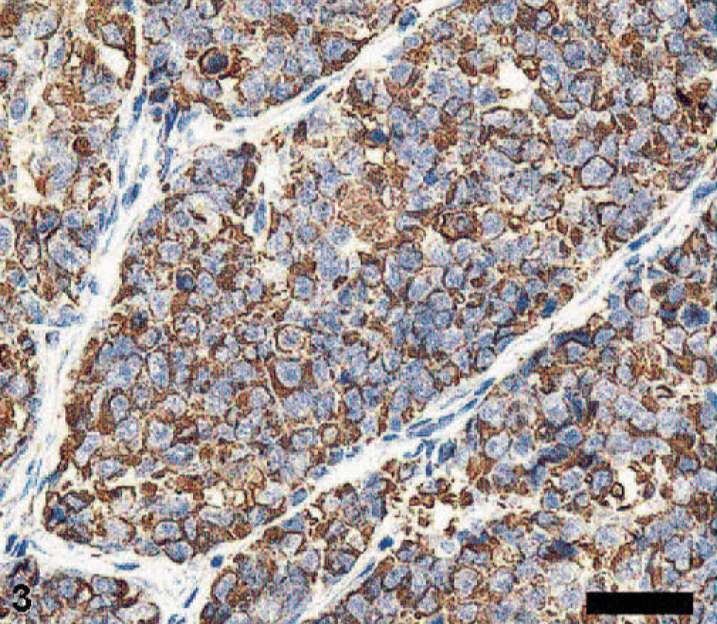
Uterine mass; 2-year-old Golden Retriever. The neoplastic cells are immunopositive to desmin. Avidin-biotin-peroxidase method, Mayer's hematoxylin counterstain. Bar = 20 μm.
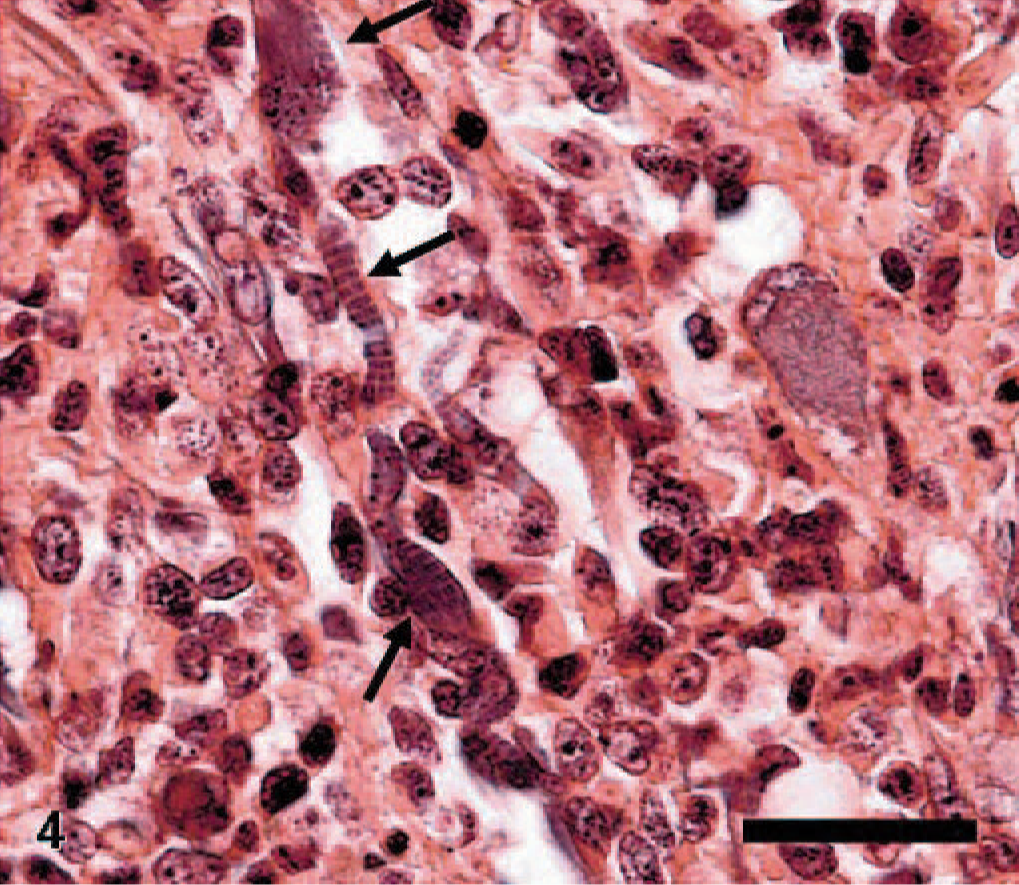
Urinary bladder mass; 2-year-old Golden Retriever. Note irregular and regular cross-striations in the cytoplasm of the neoplastic cells (arrows). Phosphotungstic acid hematoxylin stain. Bar = 40 μm.
After gross and histologic examination, leiomyosarcoma, carcinomatosis of uterine or ovarian carcinoma, dysgerminoma, and malignant mixed Müllerian tumor were considered for differential diagnoses. Dysgerminoma may mimic the histologic features observed but was ruled out because both ovaries were intact. Leiomyosarcoma was eliminated because there was no evidence of neoplastic spindle cells arranged in interlacing bundles. Carcinomatosis of uterine or ovarian origin and malignant mixed Müllerian tumor were excluded because the neoplastic cells were negative for cytokeratin but were positive for vimentin and a skeletal muscle-specific marker. Based on the age of the dog, gross and microscopic findings, and the immunohistochemical staining data, a diagnosis of rhabdomyosarcoma was made.
Histologically, rhabdomyosarcomas are classified into 3 variants. 2 The pleomorphic variant is extremely rare and contains cells of irregular size and shape with marked anisokaryosis. 2 The embryonal variant shows small spindle-shaped cells resembling primitive muscle cells in a copious myxoid matrix or round to polygonal deeply eosinophilic cells called rhabdomyoblasts. 2 The alveolar variant displays uniform, small, undifferentiated cells with round to oval hyperchromatic nuclei supported by dense fibrous septa. 2 In this dog, the genital tract and retroperitoneal masses were consistent with an alveolar variant of rhabdomyosarcoma, whereas the urinary bladder mass was sarcoma botryoides of the embryonal variant. All metastatic tumor tissues were rhabdomyosarcoma of the alveolar subtype. In humans, the histologic type of rhabdomyosarcoma is clinically important due to differences in prognosis. Alveolar and pleomorphic variants have an unfavorable prognosis, whereas the embryonal variant has a rather favorable prognosis. 3, 7 The biological outcomes in dogs of different histologic subtypes of rhabdomyosarcoma are not known.
The alveolar subtype of uterine or cervical rhabdomyosarcoma has rarely been reported in humans. These tumors are very aggressive, and hematogenous spread is a general outcome. 1, 3 Similarly, only a few cases of retroperitoneal rhabdomyosarcoma have been reported in humans. 10 We speculate that, in this dog, a primary genital tract tumor gave rise to widespread metastases, including spread to the retroperitoneum. We suggest that the urinary bladder is not the primary site because sarcoma botryoides in the urinary bladder of dogs has rarely been found to metastasize to other sites. 2 It is also possible that pleuripotential mesenchymal cells in the genital tract and urinary bladder are simultaneously transformed into 2 different subtypes of rhabdomyosarcoma in their respective locations. Although we suspect a genital tract origin, as argued above, the exact origin of this tumor has not been determined.
This report suggests that rhabdomyosarcoma should be considered as a differential diagnosis for tumors in the genital tract or retroperitoneum of juvenile animals. However, more cases need to be documented to evaluate the incidence, prevalence, and biologic behavior of such tumors.
Footnotes
Acknowledgements
This work was supported through the Brain Korea 21 Program for Veterinary Science and by Korean Research Foundation (KRF-2006-005-J02901). The authors thank Dr. Mac Law at North Carolina State University for critical review and comments and Sandra Horton (North Carolina State University) for expertise in staining.