
Abstract
An 11-year-old crossbred Pomeranian bitch displayed a large intramural, well-delineated uterine mass in one horn, near the junction with the uterine body. The mass was composed largely of mature adipose tissue, smooth muscle cells singly and in small clusters, anomalous medium-size and large arteries, and multifocal islands of cartilaginous and osseous tissues. Smooth muscle cells stained positively for desmin, and adipocytes and chondrocytes were positive for S-100 protein. This tumor has histologic and immunohistochemical features compatible with human uterine angiolipoleiomyoma, a rare tumor that has never been reported in the veterinary literature. This benign tumor is believed to be of a choristomatous nature.
Keywords
An 11-year-old crossbred Pomeranian bitch was brought to a veterinary clinic with a history of anorexia. At clinical examination, the animal had a fever, and abdominal palpation revealed the presence of a large mass. Thoracic radiographic examination showed no significant findings. On surgical exploration, a large mass located in the wall of a uterine horn was found, and an ovariohysterectomy was performed. Portions of the mass were immediately fixed in 10% neutral buffered formalin and submitted to the Département de Pathologie et Microbiologie of the Faculté de Médecine Vétérinaire de l'Université de Montréal for histologic examination. The remaining portion of the mass was kept frozen at −20 C. The resected uterus weighed 378 g. The mass was 7 × 9 × 11 cm, moderately well delineated, and located in one uterine horn, near the junction with the uterine body (Fig. 1). On gross examination, the tumor displayed a multinodular appearance with some areas that were soft and yellow and others that were firm and white. Islands of cartilaginous and osseous tissues were distributed randomly throughout the mass. Tissue sections were embedded in paraffin and stained with hematoxylin–eosin–saffron (HES) for light microscopic examination. Immunohistochemical analyses for the detection of S-100 protein and desmin were performed using commercial antibodies (anti–S-100 antibody, Dako, Mississauga, ON; anti-desmin antibody, BioGenex, San Ramon, CA). The avidin–biotin peroxidase method (Vectastain ABC kit, Vector Laboratories, Burlingame, CA) was used with 3-amino-9-ethylcarbamazole as the peroxidase substrate and Mayer's hematoxylin as the counterstain.
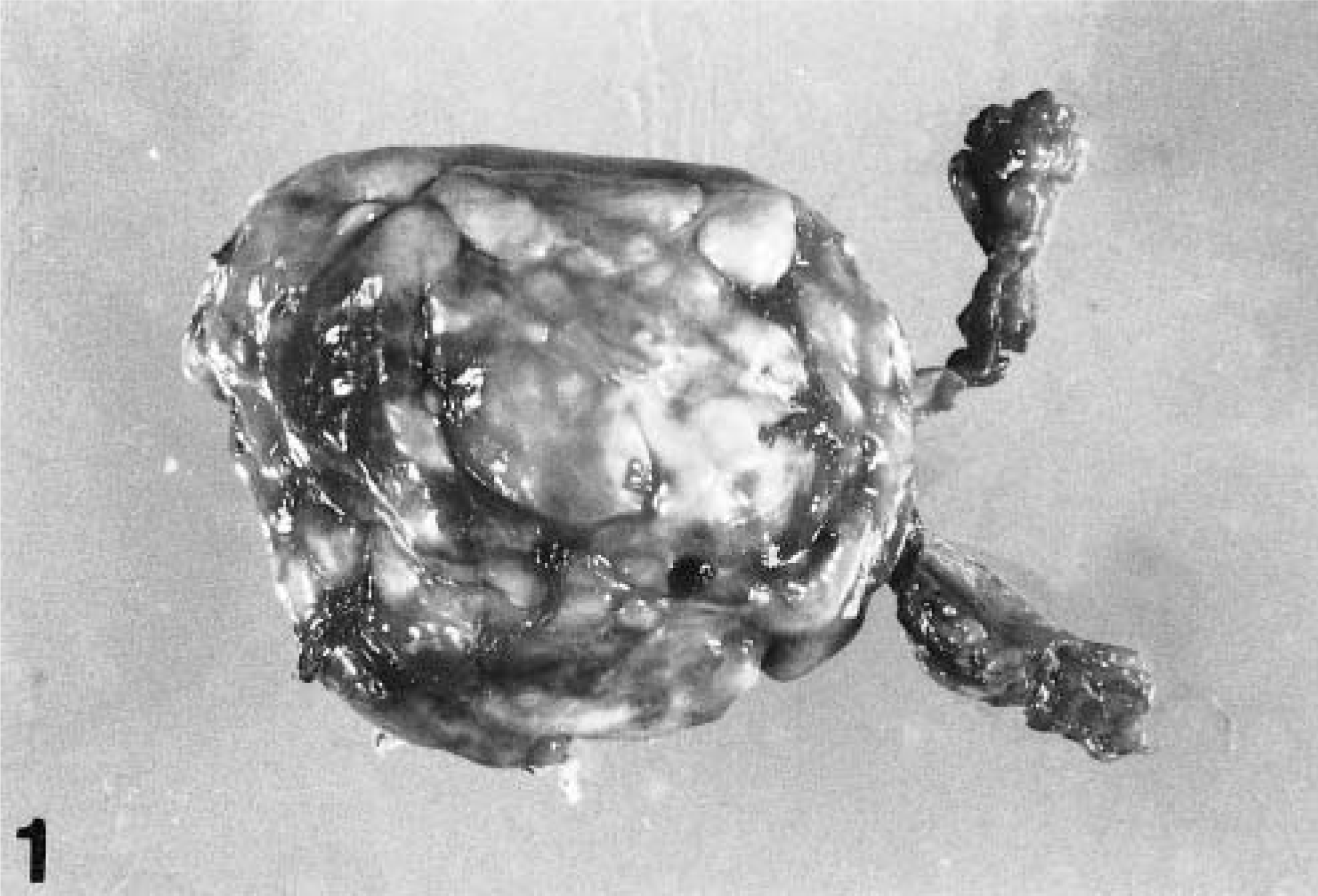
Uterine mass; dog. A large multinodular mass, 7 × 9 × 11 cm, located in the uterine horn near the junction with the uterine body.
Histologically, the mass was well circumscribed and unencapsulated and seemed to originate from the myometrium. The tumor consisted largely of mature adipose tissue divided into lobules by thin connective tissue septa (Fig. 2). Irregularly dispersed among the adipocytes were smooth muscle cells singly and in small clusters. In some areas, the smooth muscle component formed interlacing and whorled bands, typically seen in leiomyomas. Multifocally, hyalinized connective tissue with scattered fat cells and smooth muscle cells were present. Several islands of mature cartilage and bone, sometimes as large as 5 cm in diameter, were observed multifocally in the mass (Fig. 3). In addition, many anomalous, tortuous medium-size and large arteries were found throughout the tumor (Figs. 2, 4). The architecture of these vessels differed from that of normal blood vessels; walls were variably thickened, and lumina were irregular and/or reduced (Fig. 4). A small number of lymphocytes were seen throughout the mass. Immunohistochemical staining revealed that smooth muscle cells were strongly positive with the anti-desmin antibody (Fig. 5) and adipocytes (Fig. 6) and chondrocytes were immunoreactive with the anti-S100 protein antibody. These results confirmed the nature of the different mesenchymal components. In light of the histologic features, the immunohistochemical results, and the fact that this tumor did not correspond to any of the mesenchymal tumors (leiomyoma, fibroma, or lipoma) reported to occur in the canine uterus, 3 a uterine angiolipoleiomyoma was diagnosed. To our knowledge, this is the first case of uterine angiolipoleiomyoma reported in the veterinary literature.
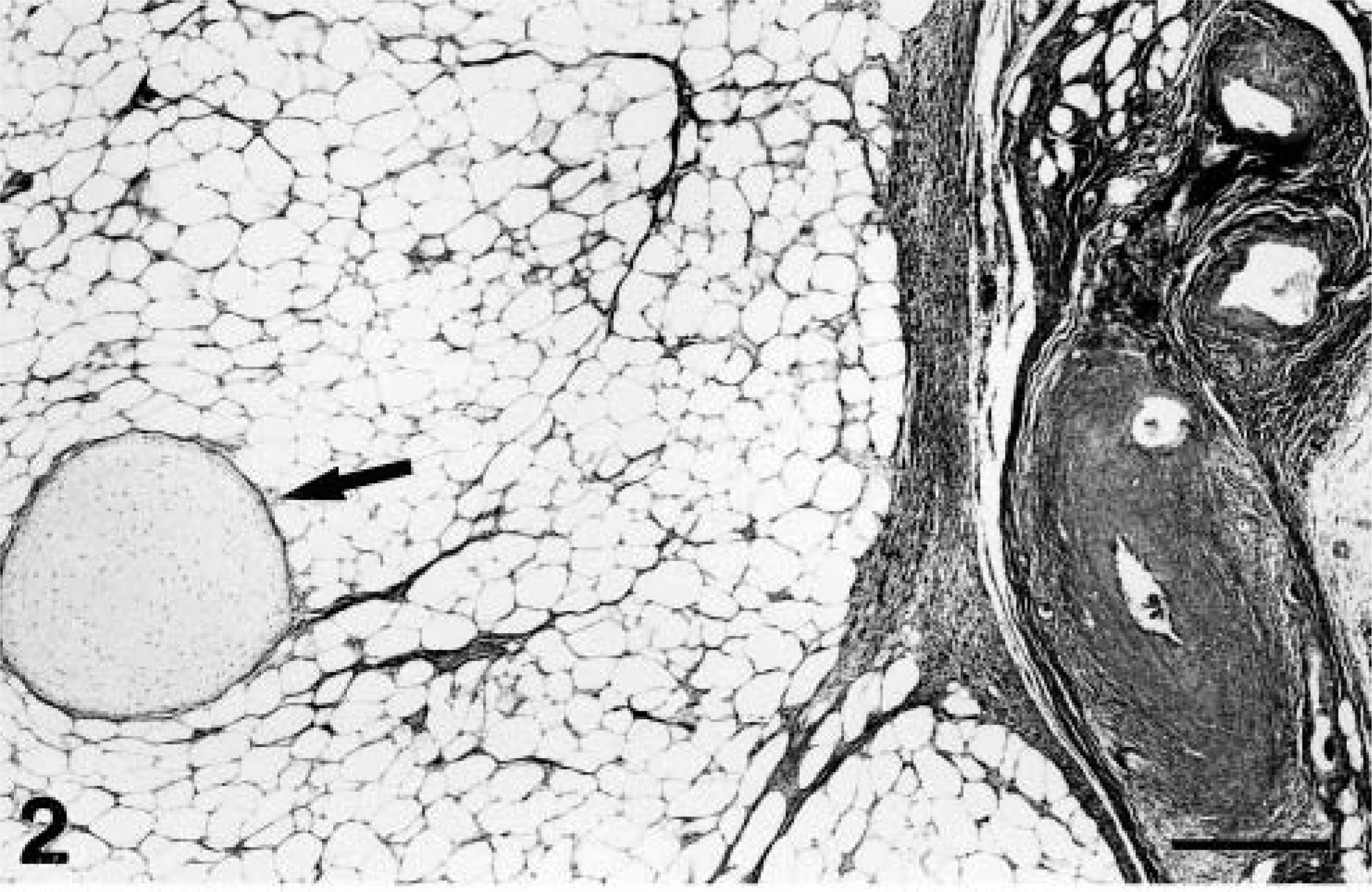
Uterine mass; dog. The tumor consists largely of mature adipose tissue supported by thin connective tissue septa, islands of cartilaginous tissue (arrow), and tortuous arteries of various sizes. HES. Bar = 300 µm.
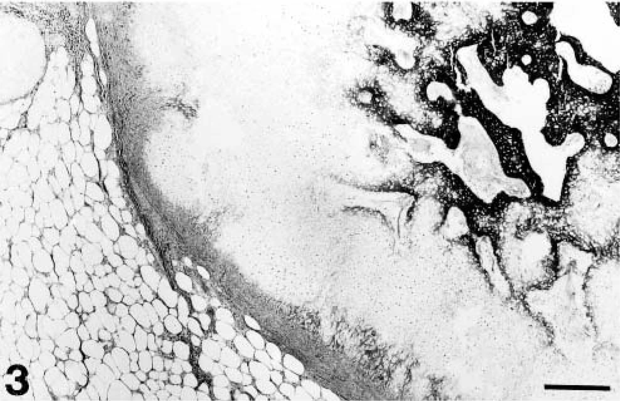
Uterine mass; dog. Large islands of mature cartilage and bone are present multifocally throughout the mass. HES. Bar = 300 µm.
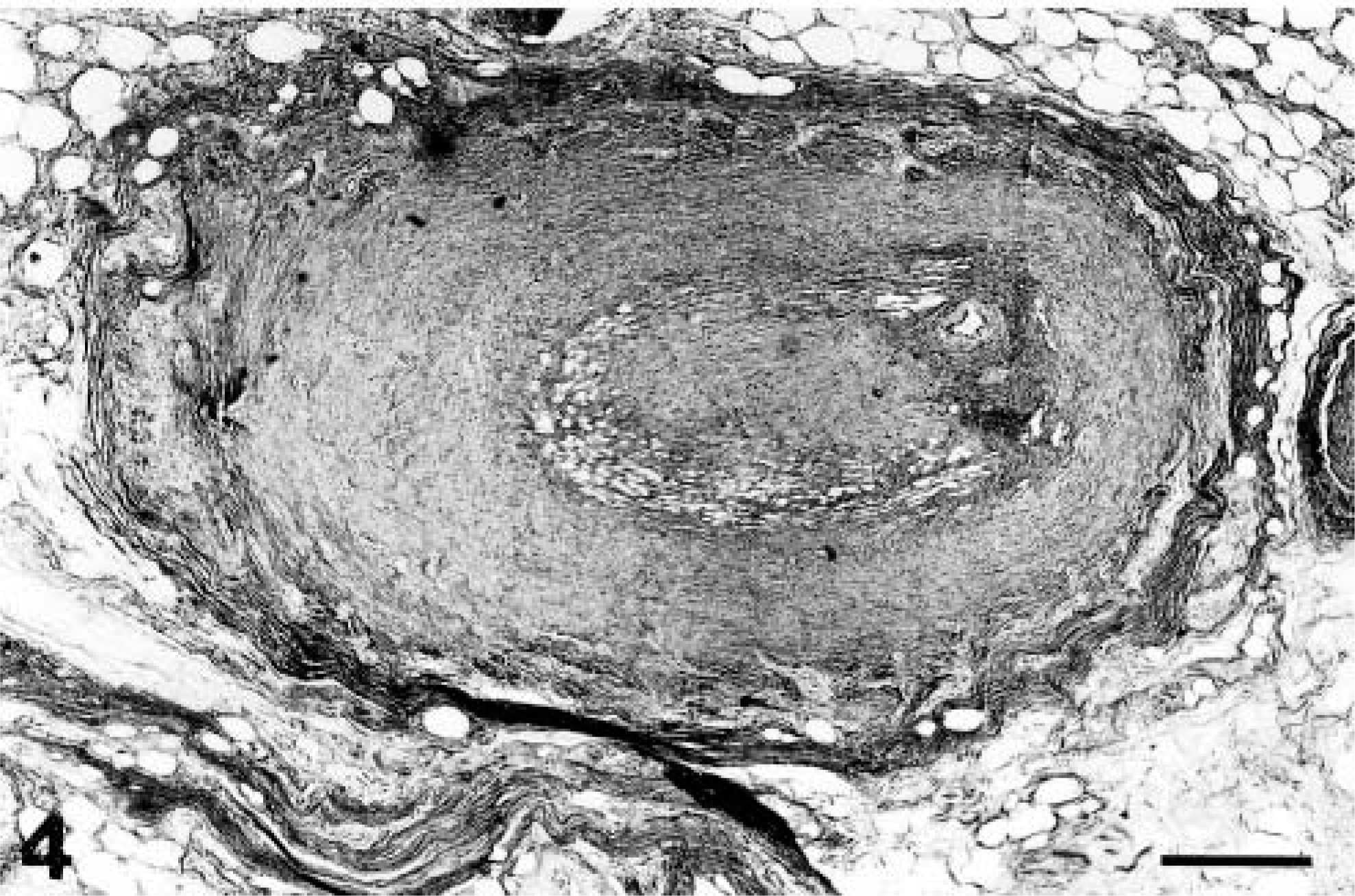
Uterine mass; dog. The walls of some anomalous, large arteries are thickened, and the lumina are severely reduced. HES. Bar = 300 µm.
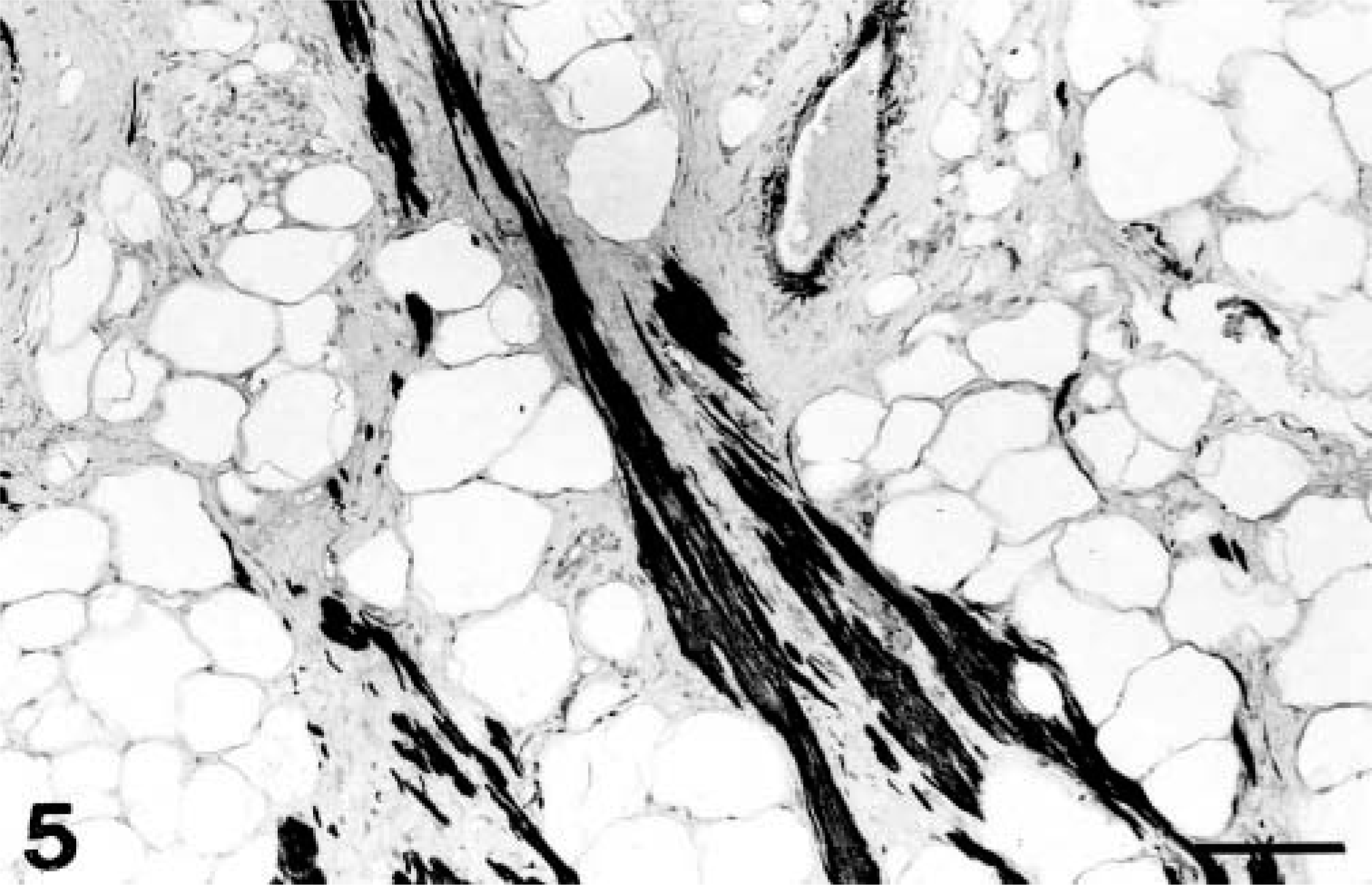
Uterine mass; dog. Smooth muscle cells stained positively for desmin. Immunohistochemistry on formalin-fixed sections using an anti-desmin monoclonal antibody. Avidin–biotin peroxidase complex method, 3-amino-9-ethylcarbamazole peroxidase substrate, Mayer's hematoxylin counterstain. Bar = 70 µm.
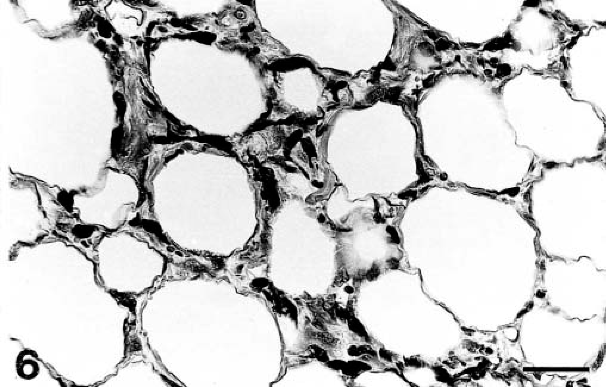
Uterine mass; dog. Adipocytes stained positively for S-100 protein. Immunohistochemistry on formalin-fixed sections using an anti–S-100 monoclonal antibody. Avidin–biotin peroxidase complex method, 3-amino-9-ethylcarbamazole peroxidase substrate, Mayer's hematoxylin counterstain. Bar = 30 µm.
Uterine lipoleiomyomatous tumors are rare and benign, usually occurring in the wall of the uterine body in postmenopausal women. 1,2,4–6,8 In the present case, 18 months after surgical resection of the uterine mass, there was no evidence of recurrence of the tumor. Gross aspects of these tumors differ as a consequence of the amount of lipomatous component. 4–6,8 The histogenesis of these neoplasms remains controversial. One researcher has speculated that uterine lipoleiomatous neoplasms are not a single histopathologic entity but, rather, consist of two distinct tumors: a lipoleiomyoma and an angiolipoleiomyoma. 6 These lipoleiomyomas, which are composed of variable amounts of adipose and smooth muscle tissue, might result from a lipomatous neometaplasia of preexisting uterine leiomyoma, whereas angiolipoleiomyomas, which comprise smooth muscle tissue, mature adipose tissue and anomalous arteries, would be of a choristomatous nature, similar to human angiomyolipomas of the kidney. 6 The presence of cartilaginous and osseous metaplasia, as seen in the present case, has been rarely reported. 7
Footnotes
Acknowledgements
We thank J. Deslandes for technical assistance and Dr. R. Pelletier, who referred the case.