
Abstract
This case report is the first description of a fibroepithelial hamartoma in a pig. The dysplasia, which covered half of the face of the newborn piglet, did not increase in relative size until the animal was euthanatized at 6 months of age. Histologic examination revealed a moderate orthokeratotic hyperkeratosis with variable degrees of epithelial proliferation. The main body of the dysplasia consisted of collagenous fibers. In addition, some proliferating small blood vessels as well as focally gathered dilated apocrine glands were evident. Given morphologic and clinical features, the diagnosis of a hamartoma seemed to be justified.
Tumors are an extremely rare event in pigs with the exception of melanomas in specific breeds.16 Neoplasms described in pigs include those of the hematopoietic system,2,7,14 genital leiomyomas, fibromas, and cyst-adenomas in high parity sows,1 as well as nephroblastomas and rhabdomyosarcomas in newborn or juvenile pigs.12 Hamartomas have to be differentiated from true neoplasms, as they represent an embryologic malformation resulting in overgrowth of one or more mature tissue types that are normally found in a given anatomic location. Fibropapillomas often show morphologic features of cutaneous hamartomas and are relatively common in small ruminants and cattle.11 Bovine papilloma viruses have been recognized as etiologic agents for fibropapillomas, which can affect various epidermal and mucosal surfaces and thus may cause severe illness depending on their mass and location.15 In pigs, viral papillomas and fibromas of the skin have not been described as yet.4 This case report describes a congenital tumor on a pig's face with histologic features of a fibroepithelial hamartoma rather than those of a neoplasm, although its gross morphology strongly suggested a soft-tissue sarcoma or fibropapilloma, respectively.
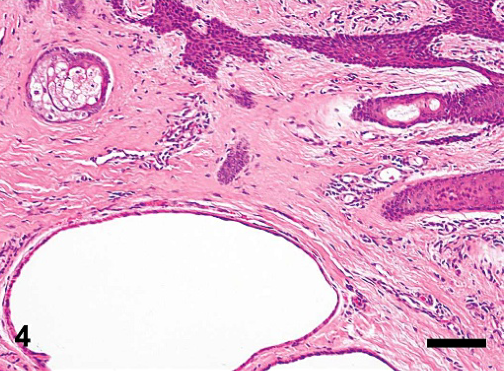
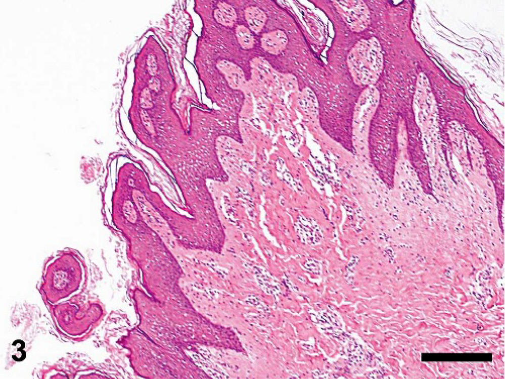
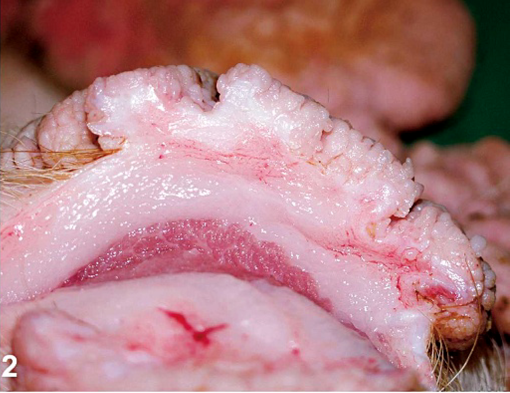
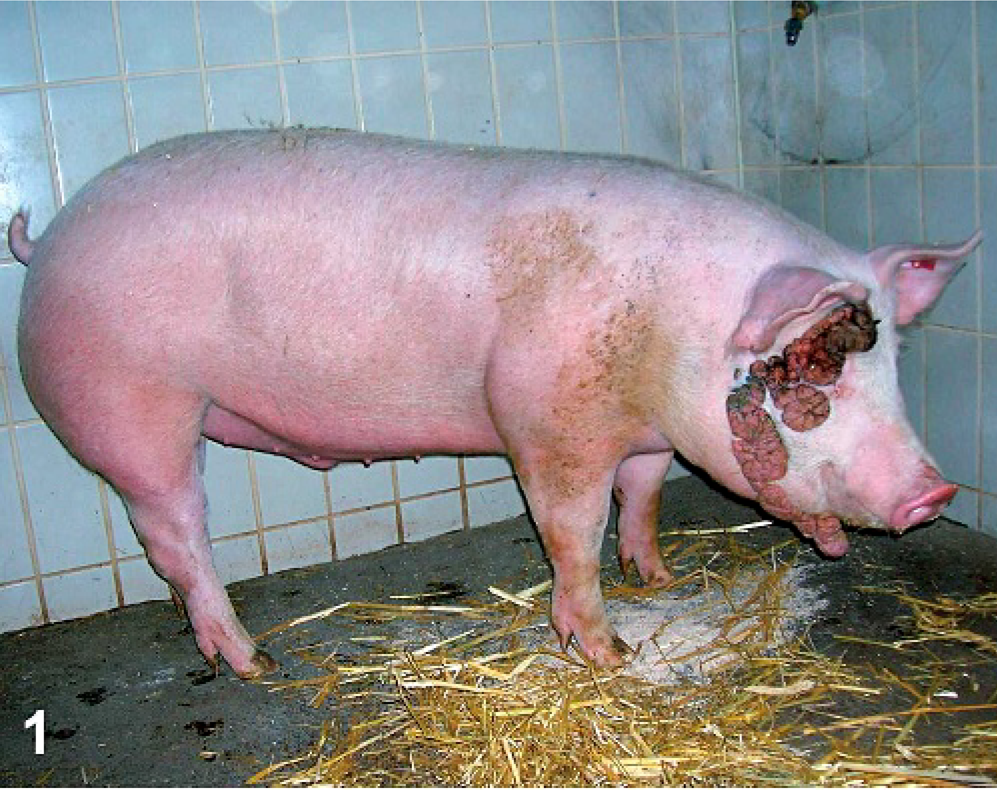
On a conventional closed-breeding and fattening farm, a newborn piglet showed a dysplasia (180 × 100 × 110 mm) covering the right side of the animal's head from the supraorbital region to the throat. The littermates were completely normal. The piglet suckled normally and grew satisfactorily after weaning. Nevertheless, the pig was submitted to the clinic for diagnostic reasons at about 4 months of age. At arrival, the pig was subjected to a physical examination. No clinical abnormalities were evident with exception of the dysplasia, which had not gained size during the piglet's life according to the farmer. The surface had a rugged contour and was of red-to-brown color. The consistency of the dysplasia was elastic-to-firm. Besides the main dysplasia, additional considerably smaller-sized formations of a similar phenotype were scattered on the pig's head. One such singular nodule was biopsied under ketamine-acepromazine anesthesia. Histologic examination revealed a papillomatous structure of the epidermis with mature fibrous tissue beneath. Therefore, a presumptive classification of fibropapilloma-like hamartoma was made. Over the following 2 months, the pig grew normally and was seemingly not hampered by the dysplasia, which did not change in relative size or structure over time (Fig. 1). After that period, the pig was euthanatized at a body weight of 123 kg. A thorough patho-anatomical examination did not yield any abnormal findings other than the facial mass. The surface of the cauliflower-like dysplasia had a deeply digitated or papillated configuration and was ulcerated focally, especially at the ventral (i.e., the mandibular) regions. The subcutis was not involved in the pathology (Fig. 2). Histologic examination revealed a mild-to-moderate acanthosis and orthokeratotic hyperkeratosis (Fig. 3). The bulk of the dysplasia was composed mainly of collagenous fibers with some small blood vessels as well as focally gathered dilated apocrine glands distributed throughout the body of the mass (Fig. 4). A mild-to-moderate perivascular infiltrate of predominantly histiocytes and plasma cells with scattered neutrophils was found in the collagenous tissue. Sebaceous glands and hair follicles had largely been destroyed, whereas some of the remaining hair follicles were heavily distended and filled with mature keratin. The adjacent healthy skin regions revealed normal hair structures. The regional lymph nodes (i.e., the mandibular and parotid lymph nodes) showed no signs of pathologic alterations.
Hamartoma; pig. Clusters of dilated apocrine glands and islets of sebaceous glands with degenerated sebaceous epithelial cells were distributed throughout the hamartoma. In addition, a mild perivascular infiltration with inflammatory cells is evident. HE stain. Bar = 100 μm.
Hamartoma; pig. The dysplasia was composed of a papilliferous hyperplastic epithelium, hyperkeratosis, and highly proliferative fibrous tissue. HE stain. Bar = 250 μm.
Hamartoma; pig. The hamartoma exhibited a cauliflower-like exophytic growth with no involvement of the subcutis beneath.
Hamartoma; pig. The relative size of the dysplasia did not change over time. The surface had a rugged contour of red-to-brown color.
Cutaneous hamartomas have not previously been reported in artiodactyls. Unlike in pigs, hamartomas are observed quite frequently in ruminants but involve other tissues or organs such as blood vessels, gonads, digestive tract, and lung.3,6,9,10,13,17,19–21 In contrast, a variety of epithelial and mesenchymal hamartomas of the skin have been identified in dogs and cats.8 Inflamed linear epidermal nevi are rare events in dogs and bear striking resemblance to inflamed linear verrucous epidermal nevus in humans. Despite some macromorphologic and histologic similarities to the presented pig, the acanthosis in inflamed linear epidermal nevi is more irregular and accompanied by severe parakeratosis, and the dermis has more prominent inflammation. Collagenous hamartomas are relatively common lesions in dogs and were first identified as fibromas, revealing the difficulties in discerning hamartomas from true neoplasms. These hamartomas are noncircumscribed nodules of redundant collagen within the dermis with a usually intact overlying epidermis and are thus well distinguishable from the lesion described in our pig.
Many different types of cutaneous hamartomas have been described in humans.18 Collagenous hamartomas in humans are very similar to those described in dogs, and sometimes a family history is present. In contrast to the presented pig, collagenous hamartomas do not have a papillated epidermis. Epidermal nevi are developmental malformations of the epidermis and associated with an excess of keratinocytes. In the majority of cases, an impressive hyperkeratosis, with papillomatosis of relatively broad and flat type, together with acanthosis is found. A distinct pathologic feature is the inflammatory linear verrucous epidermal nevus, which most often presents as a pruritic linear eruption on the lower extremities. However, an exact classification of the tumors is not always possible, as demonstrated by a case of a congenital subcutaneous lesion composed of a mixture of T and B cells, epithelial cells, and, finally, dendritic cells as the predominant cell type. This lesion seemed to be benign, as no recurrence could be observed after total removal.5
This case report describes for the first time a cutaneous hamartoma in a pig. The classification of the dysplasia as hamartoma was not only based on morphologic features, but also on the clinical history of congenital origin. Seemingly, the large size of the hamartoma did not disturb the animal. This finding was supported by the fact that, despite the mild-to-moderate inflammatory process, the regional lymph nodes did not show any reactions typical for inflammation in the tributary region. By comparison with dermatopathology literature covering other veterinary species and humans, an exact analogy with any known epithelial and mesenchymal hamartoma of the skin could not be found.
Footnotes
Acknowledgements
We thank Prof. Wilhelm Behringer, MD, from the Medical University of Vienna, for valuable discussion and Klaus Bittermann for excellent technical assistance.