
Abstract
An adult female California sea lion (Zalophus californianus) that stranded in central California was found to have a small glossal polypoid mass on gross necropsy. Histologically, the mass was consistent with a fibropapilloma, and intranuclear inclusions were found within endothelial cells lining small arterioles within the mass. Electron microscopy revealed 40-nm virions within endothelial intranuclear inclusions. Rolling circle amplification was used to obtain a partial viral genomic sequence. Sequence analysis identified the virus as a novel polyomavirus, tentatively named California sea lion polyomavirus 1. In addition, the sea lion had a severely thickened small intestine and swollen pale kidneys on gross examination. Severe renal amyloidosis with chronic interstitial nephritis was diagnosed histologically as well as T-cell intestinal lymphoma, which was confirmed via immunophenotyping and molecular clonality. The relationship, if any, between polyomavirus infection and the other disease processes in this sea lion is not known, but it is considered unlikely that the polyomavirus induced the lymphoma.
Members of the Polyomaviridae family are small DNA viruses that infect birds and mammals. Among mammals, infection has been identified in humans, nonhuman primates, rodents, bats, and cattle. 7,9,14,21 In non-immune-compromised endemic mammalian hosts, natural infections have not typically been linked with severe disease but may lead to subclinical, persistent, ubiquitous infections. 9 In humans, reactivation can occur in immune-suppressed individuals, leading to severe and sometimes fatal disease including hemorrhagic cystitis, progressive multifocal leukoenceph-alopathy, and Merkel cell carcinoma. 5,9,10,20 Some human polyomaviruses, including Simian virus 40 (SV40), BK polyomavirus, and JC polyomavirus, can cause cancer in animal models or neoplastic transformation of cell cultures. 15 Hamster polyomavirus is associated with skin tumors. 6,7 In contrast, polyomavirus infection in avian species can cause acute and chronic diseases such as budgerigar fledgling disease caused by Psittacine polyomavirus and hemorrhagic nephritis and enteritis of geese caused by Goose hemorrhagic polyomavirus. 10 The purpose of the current report is to describe the histopathologic and molecular features of polyomavirus infection and intestinal T-cell lymphoma in a free-ranging California sea lion (Zalophus californianus), both of which have not been previously reported in any marine mammal species.
An adult female sea lion was found stranded along the central California coast and was taken to The Marine Mammal Center Sausalito, California). On initial examination, the sea lion was lethargic and was unable to raise itself from sternal recumbency. Blood work abnormalities included elevated blood urea nitrogen, creatinine, sodium, and phosphorus levels, consistent with renal disease and hypoalbuminemia. Ultrasound examination revealed bilaterally enlarged kidneys, with multifocal cysts of varying size. Differential diagnoses included acute nephritis, amyloidosis, leptospirosis, and chronic renal disease. 8 Urogenital carcinoma was also considered as a differential diagnosis due to the sonographic finding of renal cysts, as ureter obstruction and hydronephrosis are common sequelae of metastatic urogenital carcinoma in sea lions. 4 Because of poor prognosis, the sea lion was humanely euthanized.
On gross examination, the dorsal surface of the tongue had a 0.7 cm in diameter, light tan papillary, nodular mass (Fig. 1). In addition, the proximal one third to one half of the small intestine, including the duodenum, was markedly thickened and plicated. On cut section, the intestinal mucosa was thickened and rough. There was no evidence of ureter obstruction or hydronephrosis. However, the kidneys were enlarged, swollen, and diffusely pale tan with loss of corticomedullary and renule differentiation. Multiple cysts were scattered throughout the cortex. Representative tissue sections were placed in 10% neutral buffered formalin and submitted to the Pathology Service, William R. Pritchard Veterinary Medical Teaching Hospital, University of California (Davis, California) for evaluation. Tissues were embedded in paraffin, processed routinely, sectioned at 5 μm, and stained with hematoxylin and eosin for routine light microscopy. Representative sections of tongue and kidney were stained with Congo red. Immunohistochemistry was completed on sections of tongue and small intestine. Sections of tongue were incubated with polyclonal antibodies to Bovine papillomavirus-1 (rabbit polyclonal antibody). a Sections of intestine were incubated with monoclonal antibodies to clusters of differentiation (CD)3 (CD3-12), b CD79a (HM57), c and CD20 (rabbit polyclonal antibody) d as previously described. 16
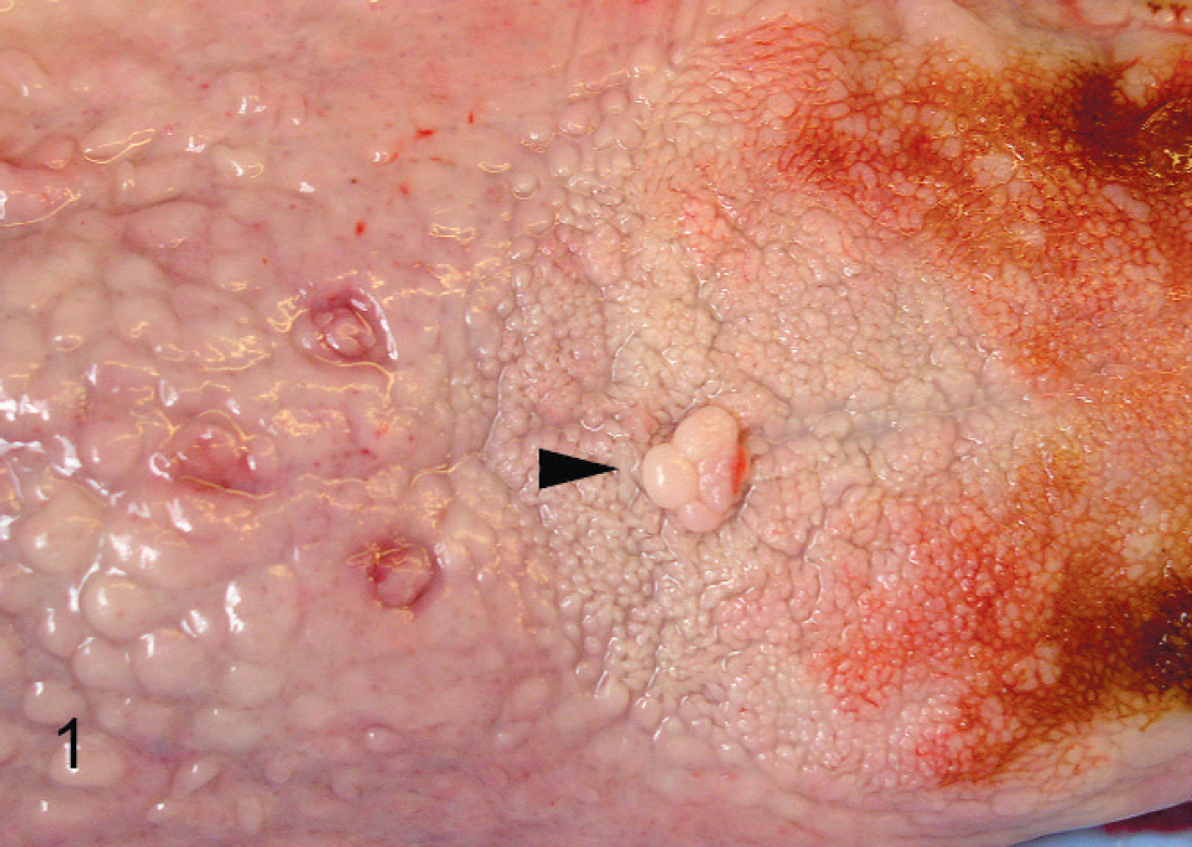
Tongue from a California sea lion (Zalophus californianus) with a small focal polypoid mass (arrow) on the dorsal mucosal surface.
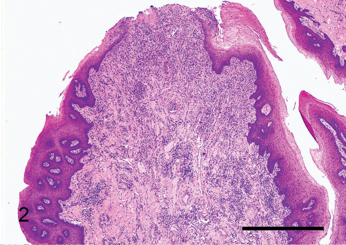
Tongue mass; California sea lion (Zalophus californianus). The hyperplastic epithelium overlies a thick stalk of dense fibrovascular connective tissue. There is mild superficial erosion and segmental ballooning degeneration in the epithelium. Hematoxylin and eosin. Bar = 1 mm.
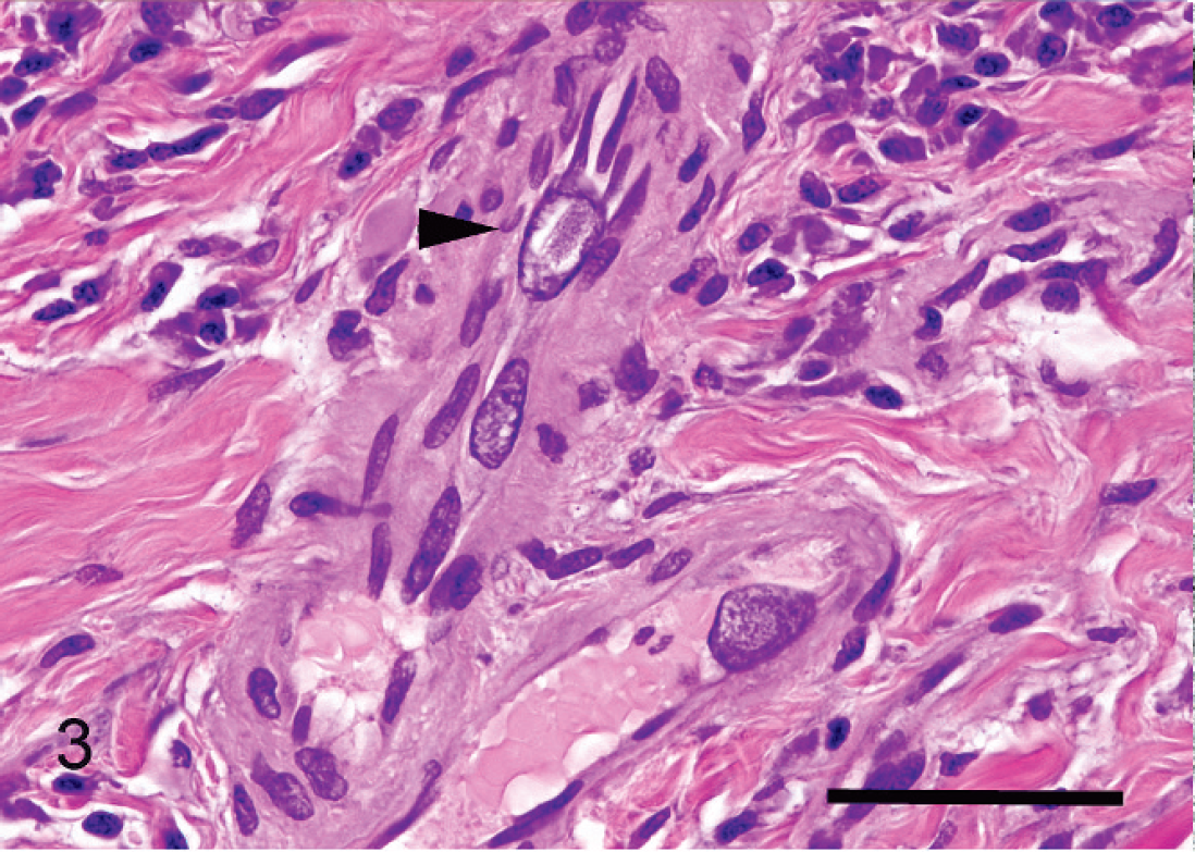
Tongue mass; California sea lion (Zalophus californianus). Blood vessels within the fibrous connective tissue contain amyloid deposits and occasionally are lined by endothelial cells with enlarged nuclei, peripheral chromatin, and pale basophilic inclusions (arrow). Hematoxylin and eosin. Bar = 50 μm.
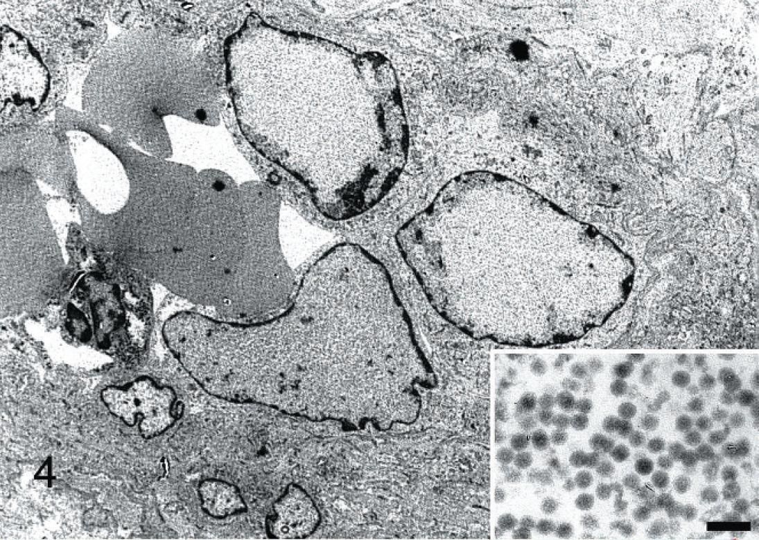
Tongue mass; California sea lion (Zalophus californianus). Transmission electron microscopy of a blood vessel with large endothelial nuclei and intranuclear inclusions containing numerous 40-nm virions (inset, higher power). Bar = 100 nm.
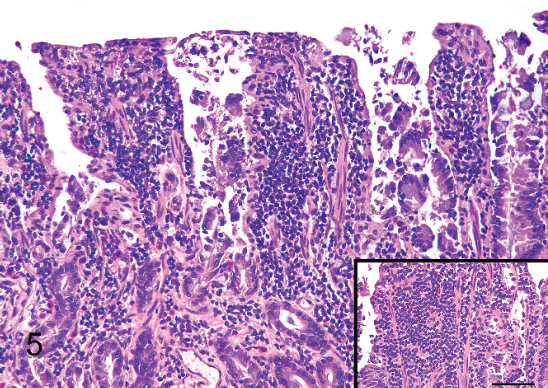
Small intestine, California sea lion (Zalophus californianus). The lamina propria contains a monomorphic population of densely packed neoplastic lymphocytes. Inset, Higher magnification of neoplastic lymphocytes. There is occasional epitheliotropism. Hematoxylin and eosin. Bar = 100 μm.
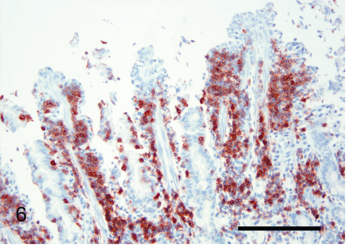
Small intestine, California sea lion (Zalophus californianus). Immunohistochemical staining for the T-cell marker cluster of differentiation (CD)3 illustrating diffuse immunoreactivity of the neoplastic lymphocytes. Bar = 200 μm.
For transmission electron microscopy (TEM), formalin-fixed sections of tongue were processed and embedded in epoxy resin e as previously described. 11,24 Cut thick sections were mounted on glass slides, stained with Toluidine blue O, and examined by light microscopy. Thin sections were mounted on 150-mesh copper grids, stained as previously described, and examined under a transmission electron microscope f at 60-kV accelerating voltage. 19
DNA was extracted from the fresh tongue lesion material and paraffin-embedded intestine tissue using a commercial kit, g and rolling circle amplification was performed with the DNA amplification kit h following the manufacturer's instructions. Products were digested using EcoRI enzyme, i cloned into pUC18, j expanded in One Shot Max Efficiency DH5alpha-T1R Competent Cells, k and sequenced using M13 primers. The resultant sequences were compared with those in GenBank (http://www.ncbi.nlm.nih.gov/Genbank/), European Molecular Biology Laboratory (http://www.embl.org/), and Data Bank of Japan (http://www.ddbj.nig.ac.jp/index-e.html) databases using the BLASTX algorithm. 2
Histopathologically, the mass on the tongue was composed of hyperplastic stratified squamous epithelium supported by a thick base of dense fibrovascular connective tissue (Fig. 2). There was segmental superficial erosion of the epithelium accompanied by mild neutrophilic inflammation. Segmen-tally, epithelial cells in the superficial layer exhibited mild ballooning degeneration; however, no inclusions were noted in the epithelium. In the underlying fibrous connective tissue, many small arterioles had thickened walls containing smudgy pale eosinophilic, acellular, congophilic material, consistent with amyloid. 3 Several of these vessels were lined by endothelial cells with markedly enlarged nuclei containing pale basophilic smudgy inclusions and peripheral chromatin (Fig. 3). Immunohistochemistry for papillomavirus was negative. Using TEM, numerous nonencapsulated virions averaging 40 nm in diameter were noted within endothelial cell nuclei (Fig. 4). The size and morphology of the virions were consistent with viruses of the families Papillomaviridae or Polyomaviridae. Rolling circle amplification of the tongue DNA resulted in a 904-base pair (bp) product. BLASTX results of the 904-bp product showed the highest identity score with Myotis polyomavirus major capsid protein (VP1; GenBank accession no. FJ188392.1; 68% amino acid identity) and Murine pneumotropic virus major capsid protein (GenBank accession no. EF186666.1; 67% amino acid identity), confirming the identity of the virus as a polyoma virus. The virus was tentatively named California sea lion virus 1, strain CSL6994. The sequence was submitted to GenBank (accession no. GQ331138). Sections of intestine were negative for polyomavirus DNA using the rolling circle amplification polymerase chain reaction (PCR) method and negative using the papillomavirus immunohistochemical stain.
In sections of the small intestine, the mid and superficial lamina propria was expanded and contained a monomorphic population of medium-sized immature lymphocytes (Fig. 5). Villous tips were often fused and disrupted, with the epithelium artifactually lifted away from the villi. The immature lymphocytes were densely packed and occasionally formed small aggregates within the mucosal epithelium. Cells had scant amounts of basophilic cytoplasm and round to indented nuclei with clumped chromatin (Fig. 5, inset). The immature lymphocytes were mixed with small to moderate numbers of small mature lymphocytes, plasma cells, globular leukocytes, and eosinophils, especially in the lower portion of the lamina propria. Scattered immature lymphocytes and plasma cells were noted in the submucosa and muscularis, often surrounding blood vessels. No inclusions were noted within the affected intestine or any other tissue examined.
The dense monomorphic, occasionally epitheliotrophic population of immature lymphocytes in the lamina propria were diffusely immunoreactive to CD3 (Fig. 6). Lymphocytes in the more heterogeneous population in the lower lamina propria occasionally expressed CD20. CD79a, a monoclonal antibody specific for B cells, did not cross-react with sea lion B cells. Based on these results, T-cell lymphoma was diagnosed.
Further confirmation of T-cell intestinal lymphoma was accomplished via molecular clonality analysis for T-cell receptor gamma (TCRG) gene rearrangements using PCR. DNA was extracted from 25-mm sections cut from paraffin blocks containing small intestine as previously described. 16 Amplification of the CDR3 region of TCRG by PCR was used to assess TCRG gene rearrangement. Consensus primers derived from the feline TCRG V and TCRG J segment were used as previously described for feline samples. 16 The positive clonal control was DNA extracted from formalin-fixed, paraffin-embedded sections of feline intestinal T-cell lymphoma, and the polyclonal control was DNA extracted from feline lymph node. Template DNA was omitted from the negative control reaction. The PCR reactions were run in triplicate to distinguish true clonal samples from pseudoclonal samples. The PCR analysis using feline-specific primers amplified DNA bands of 108 and 124 bp, consistent with a clonal T-cell population with biallelic TCRG rearrangement. Sequencing of amplified sea lion DNA confirmed that the products were 80-90% homologous to the feline TCRG.
In addition, the liver contained several small nodular aggregates of monomorphic lymphocytes similar to those noted in the affected small intestine. These cells were diffusely immunoreactive to CD3, suggestive of early metastasis of the intestinal T-cell lymphoma to the liver. Renal lesions included severe interstitial and glomerular amyloidosis with interstitial fibrosis and lymphoplasmacytic nephritis, similar to lesions previously described in sea lions with amyloidosis. 3 In addition, sections of vagina contained segmental areas of intraepithelial neoplasia consistent with sea lion urogenital carcinoma. 4
Although advanced renal disease was the primary cause of this sea lion's clinical signs, hematological abnormalities, and biochemical abnormalities, the intestinal T-cell lymphoma may have contributed to debilitation. The glossal mass was small and unlikely to have caused significant adverse effects. Although polyomavirus infection has been associated with skin tumors in hamsters and with Merkel cell carcinoma in humans, the relationship between polyoma-virus infection and the glossal fibropapilloma in this sea lion is not clear. 6,20 In Merkel cell carcinomas, viral DNA can be detected within tumor cells but not in nonneoplastic cells. 20 As viral particles were noted within only endothelial cells and not within epithelial or stromal cells within the glossal mass, it is possible that neoplasia and severe renal disease caused reactivation of latent viral infection.
The relationship, if any, between the polyomavirus infection and the T-cell lymphoma is unknown. Viral inclusions were noted only in the tongue lesion, and polyomaviral DNA was not detected in the intestine. T-cell lymphoma may cause immunosuppression, resulting in reactivation of latent virus. Polyomaviral disease has been reported in human T-cell lymphoma patients. 5 On the other hand, some polyomaviral T-antigens have been found to interfere with tumor suppressor genes including p53 and Rb, promoting oncogenesis. 1 Although DNA from polyomaviral-induced tumors has been shown to induce lymphomas when injected into hamsters, these lymphoid tumors did not contain detectable polyomaviral particles. 7 There is conflicting evidence regarding an association between SV40 and lymphomas, and even those studies finding an association have regarded B-cell lymphomas rather than T-cell lymphomas. 17
Lymphoma has rarely been reported in pinniped species, and there is a single documented case of lymphoma in a California sea lion. 12,13,22,23 Immunophenotyping has rarely been performed in reported cases. 12 The morphologic characteristics of the lymphoid infiltrate in the sea lion in the present case were very similar to those noted in early T-cell, mucosal-associated intestinal lymphomas in cats. In cats, mucosal T-cell lymphoma is characterized by dense lymphoid aggregates that often form a band-like pattern in the mid and superficial lamina propria. There is often epitheliotropism and only mild extension into the submucosa and muscularis. 16 In cats, it is thought that these lymphomas arise from the diffuse mucosal-associated lymphoid tissue (MALT) in the small intestine. This tissue consists of both an intraepithelial and lamina propria component and is composed predominantly of CD3+ T cells. 18 The MALT of sea lions has not been characterized; however, a similar pathogenesis may exist. In feline inflammatory bowel disease (IBD), there is an expansion of the MALT T-cell populations similar to what occurs in feline intestinal T-cell lymphoma. Molecular clonality analysis can help to distinguish lymphoma from IBD. In cases of IBD, a smear or ladder of bands covering a range of PCR product sizes would be expected, indicating a polyclonal, reactive lymphoid population within the sample. 16 In the sea lion in the current study, crossreactivity of the feline TCRG primers with the TCRG region of the California sea lion enabled confirmation of clonality within the intestinal lymphoid population.
Neither polyomavirus infection nor primary intestinal lymphoma has previously been described in marine mammals. Further information is needed to determine the pathogenicity and tissue tropism of the newly discovered sea lion polyomavirus. In addition, the present case illustrates the utility of molecular phenotyping in diagnostic pathology of nondomestic species.
Footnotes
a.
Dako North America Inc., Carpinteria, CA.
b.
Serotec Inc., Oxford, UK.
c.
Dako North America Inc., Carpinteria, CA.
d.
Lab Vision Corp., Fremont, CA.
e.
Eponate-12 epoxy resin, Ted Pella Inc., Redding, CA.
f.
Zeiss 906E, Zeiss Electron Microscopy, Thornwood, NY.
g.
DNeasy, Qiagen Inc., Valencia, CA.
h.
TempliPhi, Amersham Biosciences, Piscataway, NJ.
i.
Invitrogen Corp., Carlsbad, CA.
j.
Clontech Laboratories Inc., Mountain View, CA.
k.
Clontech Laboratories Inc., Mountain View, CA.