
Abstract
Testicular tumors are rarely reported in cats. We describe a case of interstitial cell tumor and Sertoli cell tumor in a cat that developed aggressive behavior and inappropriate urination 7 years after it was obtained from a shelter as an allegedly castrated 2 year old. At physical examination, the urine odor and the presence of penile papillae implied testosterone production. Testes were not palpable, but the left testis was found in the scrotum by surgical exploration and was mostly replaced by the 2 tumors. The interstitial cell tumor, but not the Sertoli cell tumor, was immunohistochemically positive for Melan-A, consistent with steroid production. Behavior improved after excision of the testis and penile papillae began to regress, but the cat was euthanatized 3 1/2 months after castration at the owner's request. Neither tumor had metastasized. The right testis was never found and was presumed to have been removed during the reported castration procedure.
Keywords
Testicular tumors are seemingly rare in cats; none were reported in surveys of more than 2200 feline tumors in the United States3,7,13,19,21 and the United Kingdom.5 McEntee encountered only 1 feline case, an interstitial cell tumor, among 749 testicular tumors from domestic mammals.10 The first published cases of feline testicular neoplasia may have been in Meier's 1956 report of Sertoli cell tumor in 2 cats.11 Additional reports document 1 or 2 seminomas,4,6 4 interstitial cell tumors,6,18,22 3 Sertoli cell tumors,1,2,20 and 2 teratomas8,12 in the testes of cats. The purpose of this report is to describe an interstitial cell tumor and a Sertoli cell tumor in the left testis of a 9-year-old Russian blue cat.
The cat was adopted from an animal shelter at the age of 2 years. The adoptive owners were informed that the cat had already been castrated. Seven years later, it developed aggressive behavior (attacking and biting the owner), inappropriate urination, and strong “tom cat” urine odor. At physical examination, spines (papillae) were noted on the glans penis. The left scrotal sac was thickened with what was thought to be scar tissue. Surgical exploration revealed a testis-like structure, which was removed by blunt dissection and submitted for histologic examination.
The formalin-fixed biopsy specimen was 9 mm × 9 mm × 18 mm and not identifiable as testis macroscopically. When it was bisected longitudinally, 2 masses were evident: a light-to-dark brown, roughly spherical, 8-mm diameter nodule, and a 9 mm × 7 mm, ovoid, pale-yellow nodule that was interlaced by bands of white fibrous tissue (Fig. 1). Each half of the bisected testis was processed routinely for histologic examination. Paraffin sections were stained with HE and by immunohistochemistry (IHC) for cytokeratins (mouse monoclonal antibody, clone AE1/AE3; M3515, Dako Corp., Carpinteria, CA), vimentin (rabbit monoclonal antibody, clone RM-9120; Neomarkers, Lab Vision Corp., Fremont, CA), calretinin (rabbit polyclonal 18-0211; Zymed, San Francisco, CA), Melan-A (mouse monoclonal antibody, clone A103, M7196; Dako Corp.), protein gene product (PGP) 9.5 (rabbit polyclonal Z5116; Dako Corp.), and neuron-specific enolase (mouse monoclonal antibody, M0873; Dako Corp.) according to standard laboratory procedures.15,17 Normal feline testis was used as control tissue for IHC.
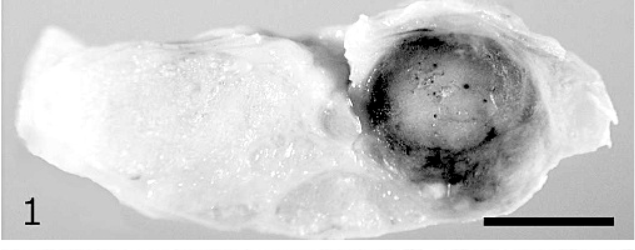
Left testis; cat. In sagittal section, the interstitial cell tumor is the darker spherical nodule that occupies about half the testicular parenchyma. Remaining testicular tissue is mostly comprised by a pale ovoid nodule of Sertoli cell tumor. Bar = 5 mm.
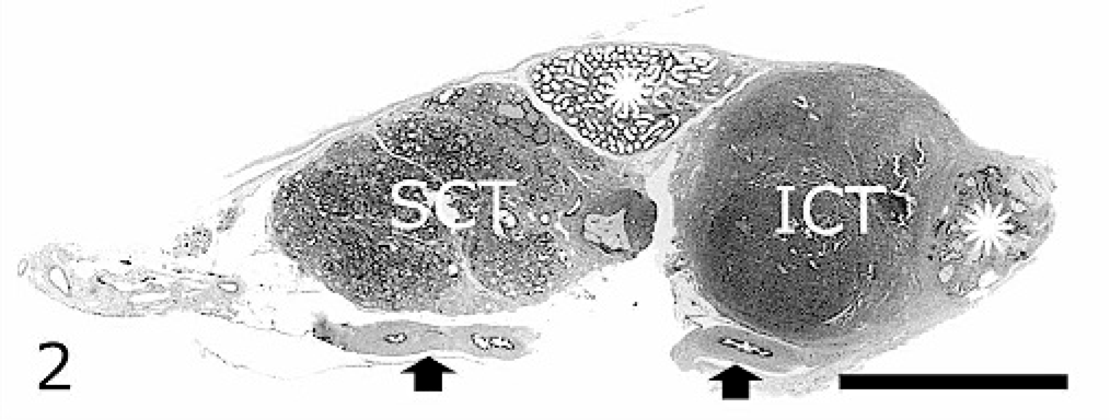
Histologic sections included testis, epididymis, ductus deferens, and spermatic cord (Fig. 2). The 2 macroscopic nodules comprised the bulk of testicular parenchyma. The brown spherical nodule was well circumscribed and composed of round-to-polyhedral cells with a round hypochromic nucleus, moderate variation in nuclear diameter, distinct nucleolus, no mitotic figures in ten 400× fields, and ample-to-abundant pale eosinophilic or amphophilic cytoplasm with faint cell borders (Fig. 3). Cytoplasm was granular or vacuolated and sometimes contained golden-brown pigment. Tumor cells were closely associated with scanty stroma. Histologic diagnosis was interstitial cell tumor.
Interstitial cell tumor, testis; cat. Lobules of round-to-polyhedral cells with vacuolated cytoplasm are separated by minimal stroma. HE stain. Bar = 250 μm.
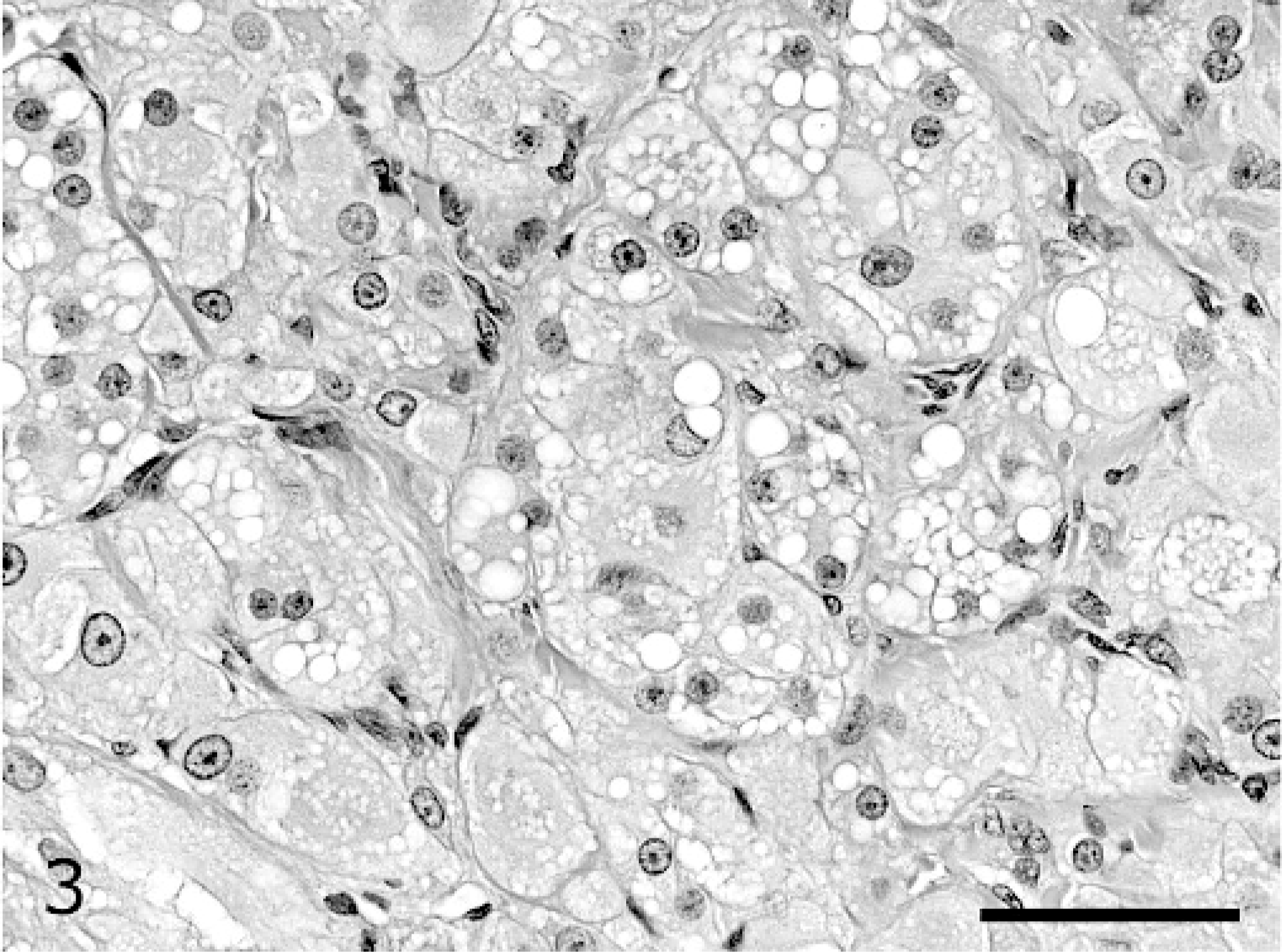
Left testis; cat. Sub-gross view of histologic section. Sertoli cell tumor (SCT) and interstitial cell tumor (ICT) comprise the bulk of testicular parenchyma. Also evident are the body and tail of the epididymis (star) and ductus deferens (arrows). HE stain. Bar = 5 mm.
The paler ovoid nodule was a Sertoli cell tumor. Most tumor cells were within seminiferous tubules, but individual cells or clusters of cells were observed multifocally in the excessive interstitial fibrous tissue. Neoplastic Sertoli cells were polyhedral or pyriform with their base at the tubular basement membrane zone and the long axis of the cell tapering toward the tubular lumen (Fig. 4). Tumor cells had a hypochromic oval nucleus, finely stippled chromatin, moderate variation in nuclear diameter, distinct central nucleolus, 2 mitotic figures in ten 400× fields, and ample-to-abundant faintly eosinophilic granular or coarsely vacuolated cytoplasm with indistinct cell borders.
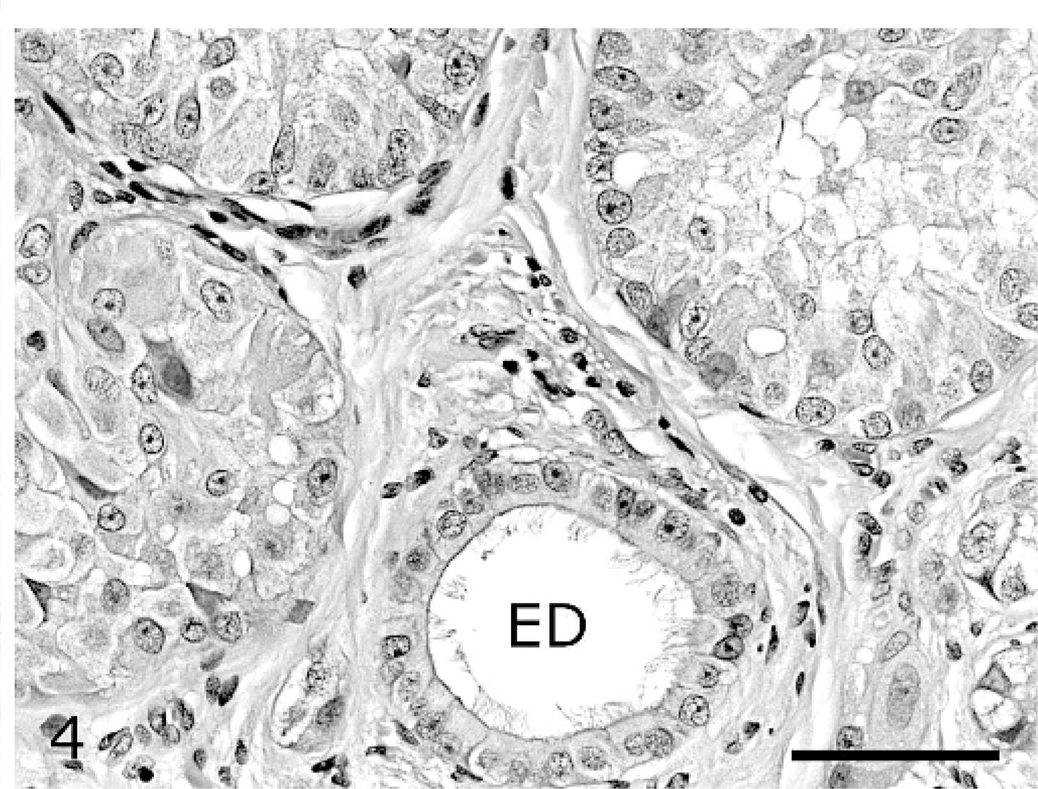
Sertoli cell tumor, testis; cat. Three seminiferous tubules are filled with polyhedral-to-pyriform cells and separated by fibrous stroma. An efferent ductule (ED) is lined by ciliated columnar epithelial cells. HE stain. Bar = 250 μm.
Normal seminiferous tubules were not observed, and spermatogenesis was not evident. Efferent ductules were focally dilated by faintly eosinophilic secretion. Epididymal tubules were devoid of spermatozoa. Tumor cells were not observed in structures of the spermatic cord or in peritesticular tissue.
Immunohistochemically, the epithelial cells of the efferent ductules and epididymis reacted strongly to antibody against cytokeratin; neoplastic interstitial and Sertoli cells were immunohistochemically negative for cytokeratin. The neoplastic interstitial cells had patchy moderate-to-strong staining for vimentin, whereas neoplastic Sertoli cells reacted weakly or not at all. Immunohistochemistry for calretinin resulted in focal staining of efferent ductular epithelium, but no staining of tumor cells. Cells of the interstitial cell tumor, but not those of the Sertoli cell tumor, were labeled by immunohistochemistry for Melan-A. Antibody to PGP 9.5 reacted intensely with nerve fibers; cells of the interstitial cell tumor were negative or weakly positive for PGP 9.5; the Sertoli cell tumor was negative for this marker. In normal testicular control tissue, PGP 9.5 antibody reacted variably with germ cells, weakly with interstitial cells, and not with Sertoli cells. Antibody to neuron-specific enolase also reacted intensely with nerve fibers, but only with a few cells of neoplastic or control interstitial or Sertoli cells.
Penile papillae persisted and behavioral problems had not resolved completely at 8 weeks after removal of the left testis, so the abdominal cavity, inguinal canals, and right scrotal sac were explored surgically. The right scrotal sac contained adipose and fibrous tissue that surrounded remnants of the spermatic cord and ductus deferens, but no testicular tissue was found grossly or microscopically in the scrotum, inguinal canals, or abdominal cavity.
Although its behavior improved, the owner requested euthanasia of the cat 3.5 months after surgery. By this time, penile papillae had partially regressed. No important lesions were detected at necropsy. No testicular tissue was found and neither testicular tumor had metastasized.
Feline testicular tumors are rare, so the presence of 2 tumors in 1 testis of a reportedly castrated cat was unexpected. The cells of the interstitial cell tumor were positive for Melan-A as they are in interstitial cell and other steroid-producing tumors of dogs.16 Though Melan-A is also detected in most canine Sertoli cell tumors,16 the neoplastic Sertoli cells in this cat were generally negative, which may indicate lack of steroid production. Feline neoplastic and nonneoplastic Sertoli cells are reported to be immunohistochemically positive for vimentin and neuron-specific enolase2,20; however, in our laboratory, although vimentin was detected in interstitial cells, neither marker was detected in more than a few neoplastic or normal Sertoli cells. Another neuronal marker, PGP 9.5, was strongly expressed in nerve fibers (as was neuron-specific enolase), but interstitial and Sertoli cells were generally negative. PGP 9.5 is considered a specific marker of spermatogonia in pigs9 and stained spermatogonia in the control feline testis in this study. Germ cells were not found, however, in the neoplastic testis. Calretinin is expressed in canine testicular Leydig and Sertoli cells and in their tumors as well as in canine seminoma,14 but in this cat, calretinin was appreciated only in efferent ductular epithelium. Only efferent ductular and epididymal epithelial cells were positive for cytokeratin. Interspecies variation may account for some of the variability in expression of these markers.
Fourteen cases of testicular neoplasia in domestic cats have been reported in the past 50 years; 7 were from the United States (Table 1). Interstitial cell tumor and Sertoli cell tumor each accounted for 5 cases. Although most canine testicular tumors are benign,10 2 of 5 reported feline Sertoli cell tumors,11,20 and 1 seminoma,4 metastasized. At least 2 of the 5 reported feline Sertoli cell tumors2,20 and 1 teratoma12 developed in a cryptorchid testis. Feminization has not been reported in cats with Sertoli cell tumor and was not evident in this case. On the other hand, although feline interstitial cell tumors have been incidental findings at necropsy,10 in this and in at least one other case,18 interstitial cell tumor was found in a supposedly castrated cat that developed masculine physical features and behavior. In the previous case, the tumor was found in ectopic testicular tissue attached to the spermatic cord remnant 11 years after castration. The cat had a round facial profile, muscular conformation, a 2-year history of urine spraying, and elevated serum testosterone concentration.18
Features of 14 reported testicular tumors in cats.∗
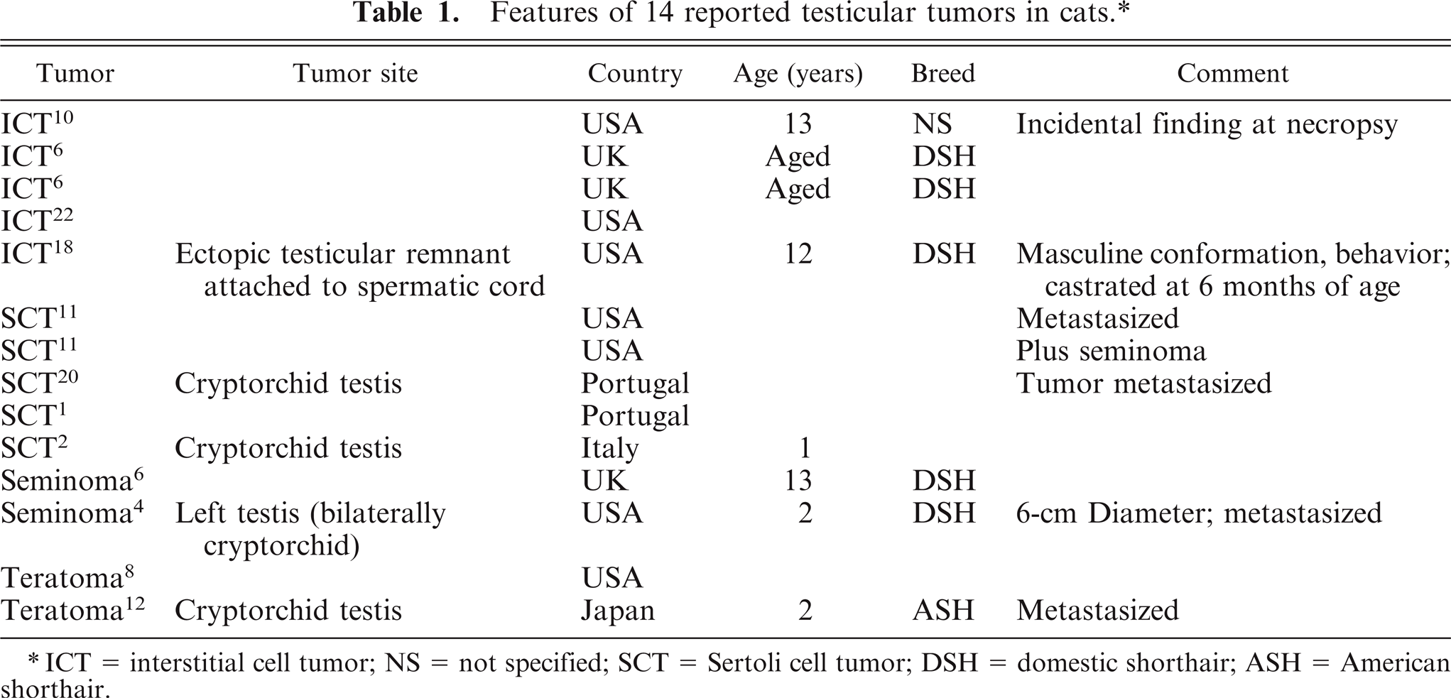
ICT = interstitial cell tumor; NS = not specified; SCT = Sertoli cell tumor; DSH = domestic shorthair; ASH = American shorthair.
We were unable to examine early medical records of the cat of this report and do not know why both testes were not removed during the initial castration procedure. However, even with 2 tumors, the remaining testis was smaller than a normal feline testis and no normal testicular parenchyma was observed. If hypoplastic or not fully descended, the testis may not have been found during the initial surgery. Hormone assays were not performed in this case, but the presence of penile papillae indicated elevated testosterone levels. Urine odor and behavioral problems were also attributed to testosterone influence. Positive immunohistochemistry for Melan-A in the interstitial cell tumor may indicate steroid production by the tumor cells. Postsurgical regression of penile papillae, lessening of inappropriate urination, and decreased aggression all suggest that the neoplastic testis was a source of testosterone.
Footnotes
Acknowledgements
We are grateful to the Midwest Association of Veterinary Pathologists for the opportunity to present portions of this study at their summer meeting, East Lansing, MI, August 17, 2006.