
Abstract
Conidiobolomycosis is reported in the state of Piauí, in the semiarid region of northeastern Brazil. Affected sheep had depression, weight loss, serous or mucohemorrhagic nasal discharge, and cranium-facial asymmetry from exophthalmos of 1 eye, generally with increased volume of the eyeball, keratitis, and corneal ulceration. At necropsy of 60 sheep, friable masses were observed in the posterior region of the nasal cavity, often destroying the ethmoturbinate bones. Frequently, the lesions invaded the nasal sinuses, cribiform plate, orbit, and brain. The masses were irregular, granular with moist surfaces, and soft and friable with white, yellow, or tan coloration. Dissemination of the lesion to lungs was observed in 27 sheep, to the brain in 26, to lymph nodes in 3, to the kidney in 2, and to the gallbladder and heart in 1. The microscopic examination showed granulomatous inflammation composed of central necrosis surrounded by lymphocytes, epithelioid and giant cells, and fibrous tissue. In all lesions, negatively stained structures representing hyphae were surrounded by Splendore-Hoeppli material. Coagulative necrosis, thrombosis, and vasculitis were also observed. Grocott methenamine silver stain showed 8–30-μm-thick hyphae, rarely septate or ramified, irregular in shape, and with black contoured wall, sometimes with bulbous dilatation in the extremities. On electron microscopy, the hyphae had a thick double wall surrounded by cellular remnants and an inflammatory exudate. Conidiobolus coronatus was isolated from the lesions of 6 sheep. Conidiobolomycosis is an important disease of sheep in the state of Piauí, and other regions of northeastern Brazil.
Conidiobolomycosis is a zygomycosis caused by fungi of the class Zygomycetes, order Entomophthorales, affecting humans and animals. The main Conidiobolus species involved are C. coronatus, C. incongruus, and C. lamprauges. Conidiobolus spp. are found mainly in soil, decaying vegetation, and insects of tropical and subtropical regions.22 In humans, C. coronatus is the most common agent isolated causing infections of the upper airways.2,22,29,35 In animals, conidiobolomycosis caused by C. coronatus, C. lamprauges, and C. incongruus are reported mainly as subcutaneous infections in horses,6,18 llamas,11,28 and dogs12,14 and rhinofacial, nasopharyngeal, or oral infections in horses3,13,16,21,24,26,27,36 and dogs.1,12 In sheep, the disease has been reported in Australia5,18 and Trinidad Tobago.30 In Brazil, nasal zygomycosis, caused probably by Conidiobolus species, has been reported in sheep in the state of Paraíba, but without identification of the causal agent.31 Sheep affected by C. incongruus had mucohemorrhagic nasal discharge, dyspnea, anorexia, enlargement of anterior or posterior nasal cavity, and exophthalmos.5,18,30 Gross lesions were characterized by granulomatous necrotic tissue in the ethmoidal region extending through the turbinate bones into the brain and also into soft tissues of the nose with dissemination to the lung and lymph nodes.5,18,30 Histologically, the presence of Splendore-Hoeppli material surrounding fungal hyphae is characteristic of the infection.5,18,30
Undiagnosed cases of nose lesions in sheep with high fatality rate are frequently observed by farmers and practitioners in the state of Piauí. In this paper, we report the pathology of this disease and the isolation of C. coronatus from the lesions. Because many practitioners used to diagnose the disease as enzootic ethmoidal tumor, electronic microscopy of the lesions was also performed.
Material and Methods
Sixty sheep with lesions suggestive of conidiobolomycosis from 25 farms in 12 municipalities of the north and northeastern regions of the state of Piauí were necropsied. After anesthetization with an intravenous injection of 25 mg/kg sodium thiopental, the animals were euthanatized with intravenous injection of 30% potassium chloride. All procedures were performed according to the Brazilian guide for care and use of animals, and all experimental protocols used were previously approved by the Ethics Committee of the Federal University of Piauí.
During necropsy, samples from affected tissues were fixed in 10% buffered formalin, embedded in paraffin, and sectioned at 6 μm. All sections were stained by HE, and selected sections were also stained by Gomorís methenamine silver and periodic acid–Schiff (PAS) stains for fungi.
For electron microscopy, samples of nasal and lung lesions of 10 sheep were fixed in 2% glutaraldehyde with 2% paraformaldehyde in 0.4 M cacodylate buffer (pH 7.4), postfixed in 1% osmium tetroxide buffered in 0.4 M sodium cacodylate (pH 7.4), and embedded in Araldite 502 (Polysciences, Warrington, PA). Semithin sections were stained with methylene blue. Ultrathin sections were stained with lead citrate and uranyl acetate and examined with an EM 10 Zeiss transmission microscope.
To isolate the fungus, fragments of the nasal lesions were placed on Sabouraud dextrose agar (SDA), with 90 g/liter of chloranphenicol, and potato dextrose agar (BDA). The cultures were incubated at 25°C–28°C for 7 days. The colonies were stained with lactophenol blue for morphologic evaluation. Cultures of the fungi isolated were sent for identification to the Mycology Laboratory at the Institute of Biological Sciences in the Federal University of Minas Gerais and to the Institute of Tropical Medicine in São Paulo. They were identified following the criteria mentioned by Dreschler,7 King,19 and King and Jong.20
Results
Clinical signs and epidemiology
From January 2002 to December 2004, the incidence of disease on the 25 farms varied from 0.1 to 14.3%, with a mean annual incidence of 2.8%. Farmers informed that fatality rate is 100%. Fifty-three out of 60 cases occurred from April to July. Most sheep were hair sheep of the Morada Nova and Santa Inês breeds or crossbreeds. The region in which the disease was studied was in the Brazilian semiarid, North of the state of Piauí, northeastern Brazil, situated between 2:44′ and 10:52′S and between 40:25′ and 45:59′W. Altitude is between 59.6 and 256 m. Temperature varies between 19 and 36°C, and relative humidity is between 46 and 69% in September during the dry season and 78 and 82% in February during the rainy season. Annual rainfall varies from 1,000 to 1,600 mm, with a rainy season from December to April and a dry season from May to November. The disease was observed only in sheep. Cattle and goats, usually raised together with sheep, were not affected. Most affected sheep were 1–5 years old. Some farmers were aware of the disease for as long as 30 years, but on most farms, the first cases appeared in the last 5 years.
Clinical signs were characterized by serous or mucohemorrhagic nasal discharge and dyspnea with noisy and difficult respiration. Cranium-facial asymmetry because of exophthalmos of 1 eye (Fig. 1) was observed in 55 (91.6%) sheep. In those cases, the animals had also increased volume of the eyeball, blindness, keratitis, and corneal ulceration. Marked depression, sometimes maintaining the head down, or head pressing were observed in some cases. Sheep lost weight, and the clinical manifestation period was from 1 to 5 weeks.
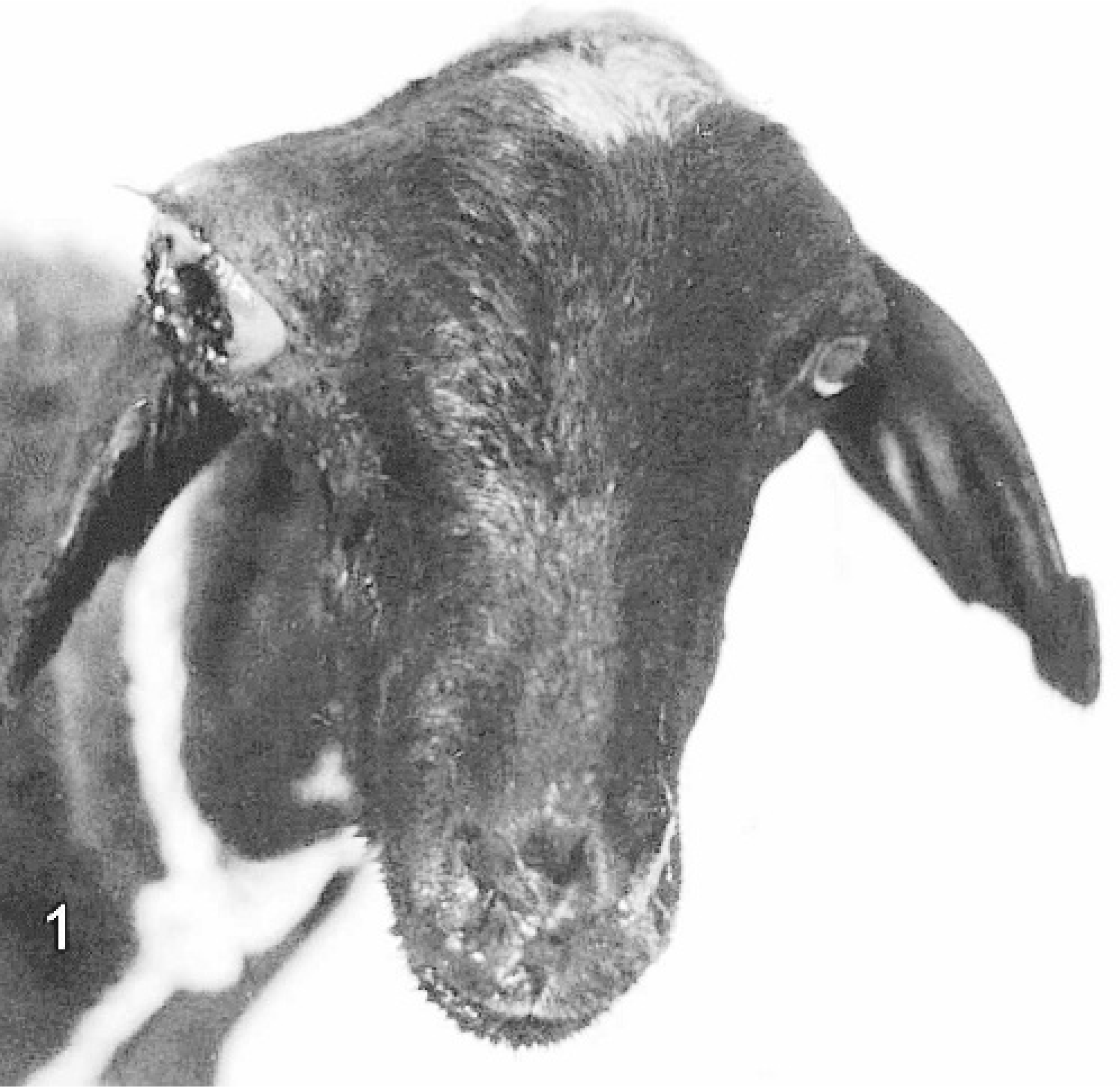
Conidiobolomycosis. Sheep with nasal discharge, cranium-facial asymmetry, and exophthalmos of the right eye. The eyeball is enlarged with keratitis and corneal ulceration.
Gross lesions
On sagittal section of the heads, a whitish or yellow friable mass with irregular and granular consistency was observed in the ethmoidal region (Fig. 2). The size of the lesions was 3–12 cm cranium-caudal and 2–8 cm dorsum-ventral. The mass infiltrated the ethmoid and turbinate bones. Most often, the orbit was also affected, causing cranium-facial asymmetry and exophthalmos. Occasionally, the cribiform plate and frontal sinuses were also affected. The nasal septum was distorted, and the pharynges and larynges had yellow, greenish, or sometimes black exudates with fetid smell. The turbinate spaces were sometimes filled by mucus.
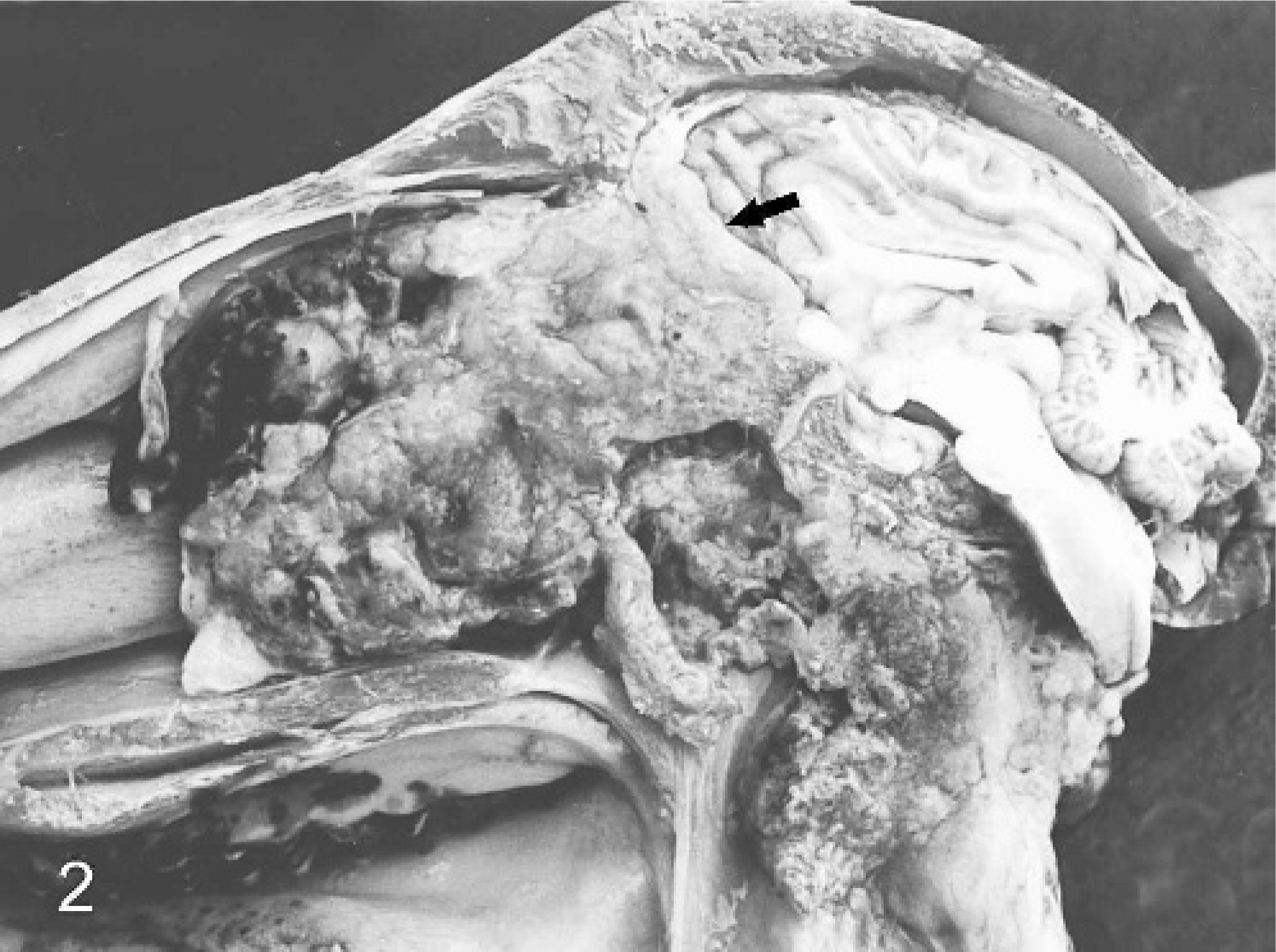
Sheep conidiobolomycosis. Sagittal section of the head. The ethmoid bone and most of the turbinate bones are destroyed and have been replaced by a yellow mass. The pharynges and larynges are full of exudates. The lesion extended to the frontal meninges, which is thickened (arrow).
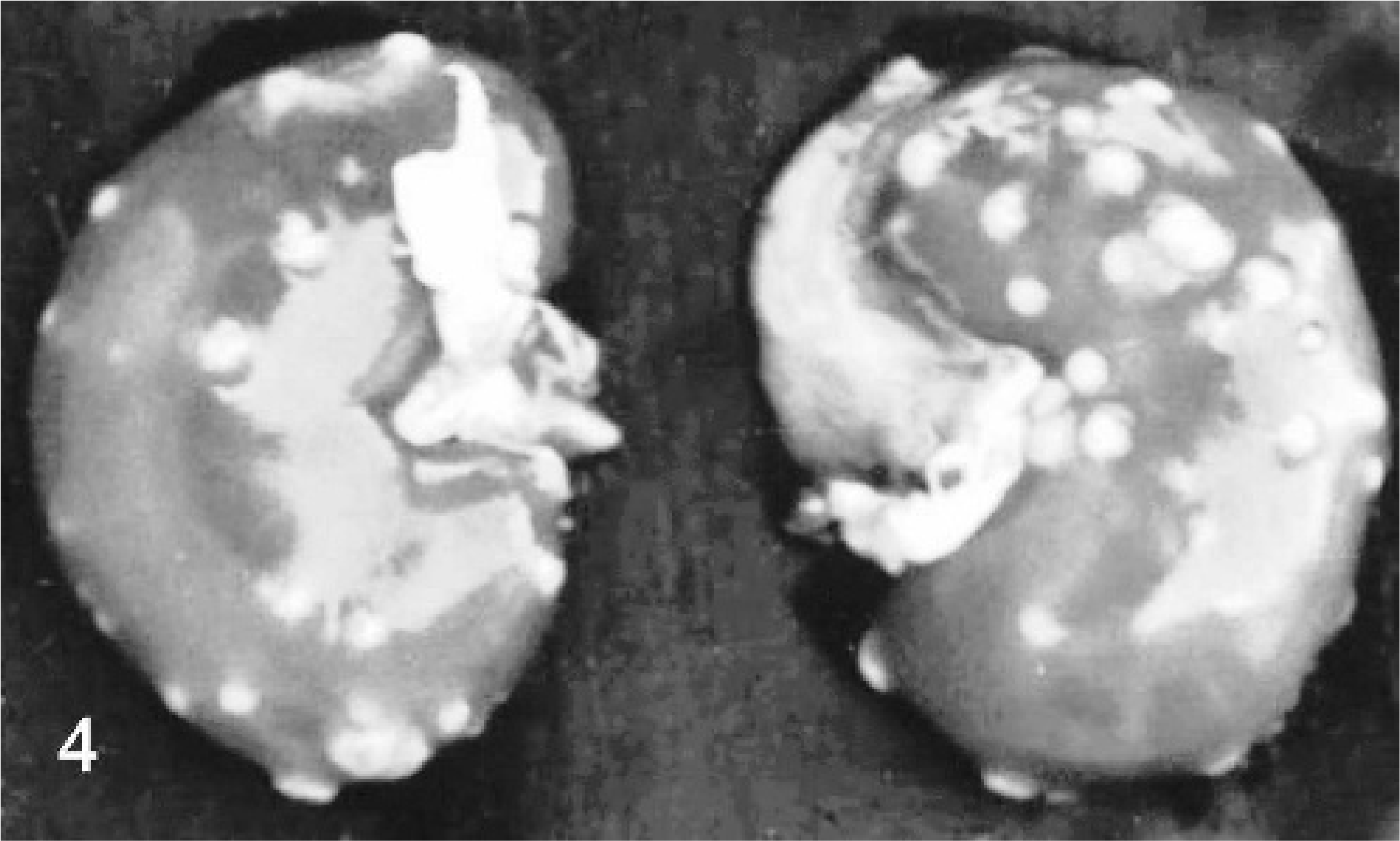
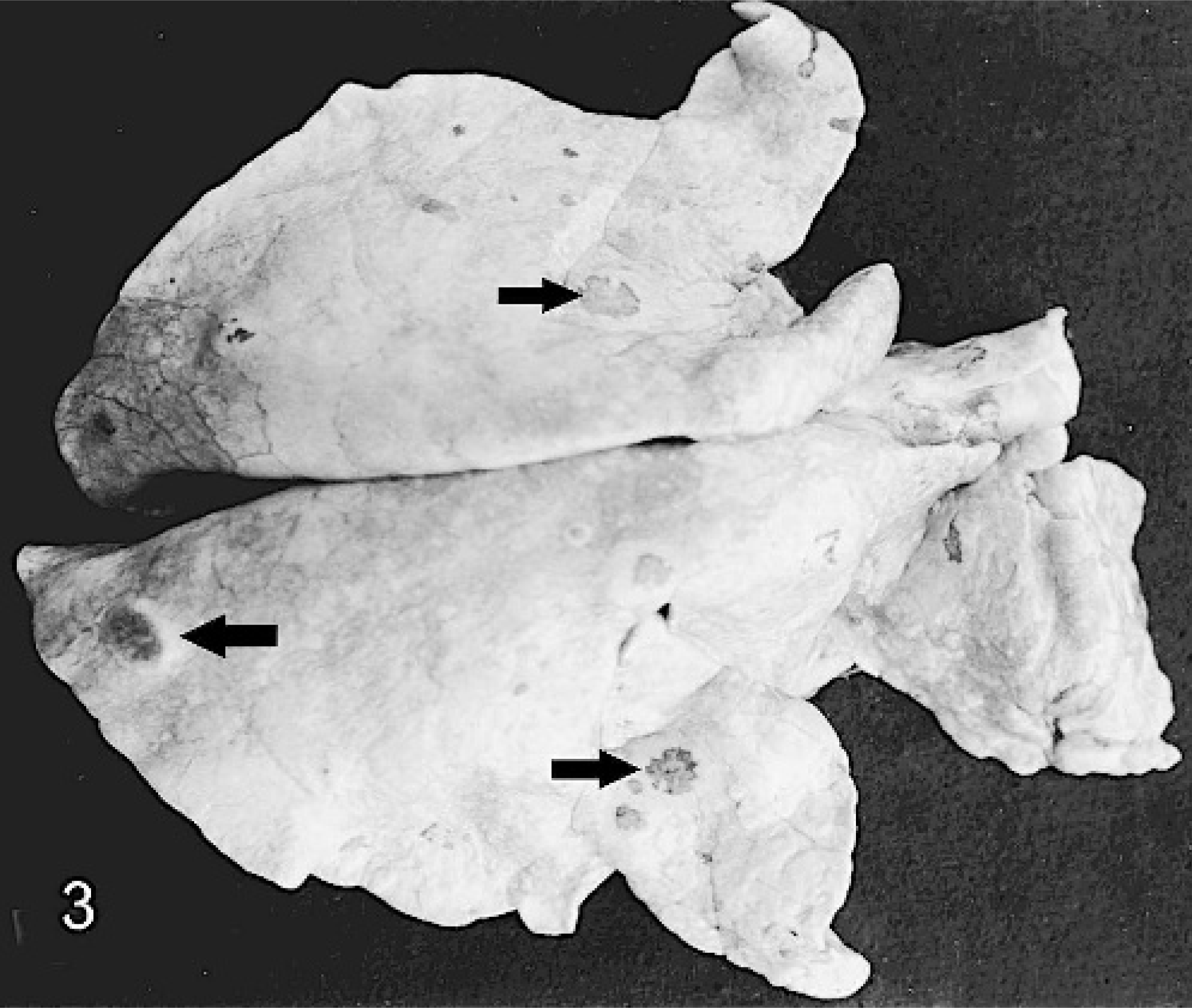
The ethmoid bone was affected in all sheep. In 59 (98.3%) sheep, the lesions extended rostrally to the turbinate bones. In 55 (91.6%) sheep, the orbit was affected, and in 36 (60%), the cribriform plate of the ethmoidal bone was affected. In 26 (43.3%) sheep, the lesion invaded the frontal brain and meninges, and in 7 (11.7%), the lesions were limited to the meninges. In 17 (28.3%) sheep, the frontal sinuses had yellow or whitish exudates, with reddish or edematous mucosal surface. In 52 (86.7%) sheep, the lesion extended to the pharynx, which was full of exudates. Twenty-seven (45%) sheep had lung lesions characterized by multifocal whitish nodules with central grey to red areas and a hard consistency measuring 0.5–3 cm (Fig. 3). Two (3.3%) sheep had similar lesions in the kidneys (Fig. 4), and in 1 of these, the heart and gall bladder were also affected. In 3 sheep, the submandibular lymph nodes were hard and enlarged, with multifocal yellowish or greenish areas. In other sheep, the submandibular and sometimes the mediastinal lymph nodes were enlarged with a whitish and wet cut surface. Areas of consolidation were observed in the apical lung lobes in 10 sheep.
Sheep conidiobolomycosis. Kidneys. Multifocal whitish nodules measuring 0.5–3 cm are observed.
Sheep conidiobolomycosis. Lung. Multifocal whitish nodules measuring 0.5–3 cm (arrows).
Microscopic lesions
Main lesions were multifocal, sometimes coalescent, granulomas with a central necrotic area with negatively stained structures representing fungal hyphae surrounded by eosinophilic Splendore-Hoeppli material (Fig. 5). Sometimes the hyphae had a slightly eosinophilic cytoplasm. Within the necrotic areas, deposits of eosinophilic granules, presumably the remnants of eosinophils, were also observed. The necrotic areas were surrounded by inflammatory cells, mainly eosinophils, neutrophils, mononuclear cells, and epithelioid and giant cells. Hyphalike structures were sometimes observed within giant cells. Bone trabeculae within the lesions were reabsorbed and substituted by fibrous tissue. Congestion and hemorrhages were occasionally observed. Thrombosis, vasculitis, and coagulation necrosis were observed in some blood vessels. Occasionally, the nasal epithelium was ulcerated, but in other cases, had no significant lesions. Most glands within the lesions were destroyed.
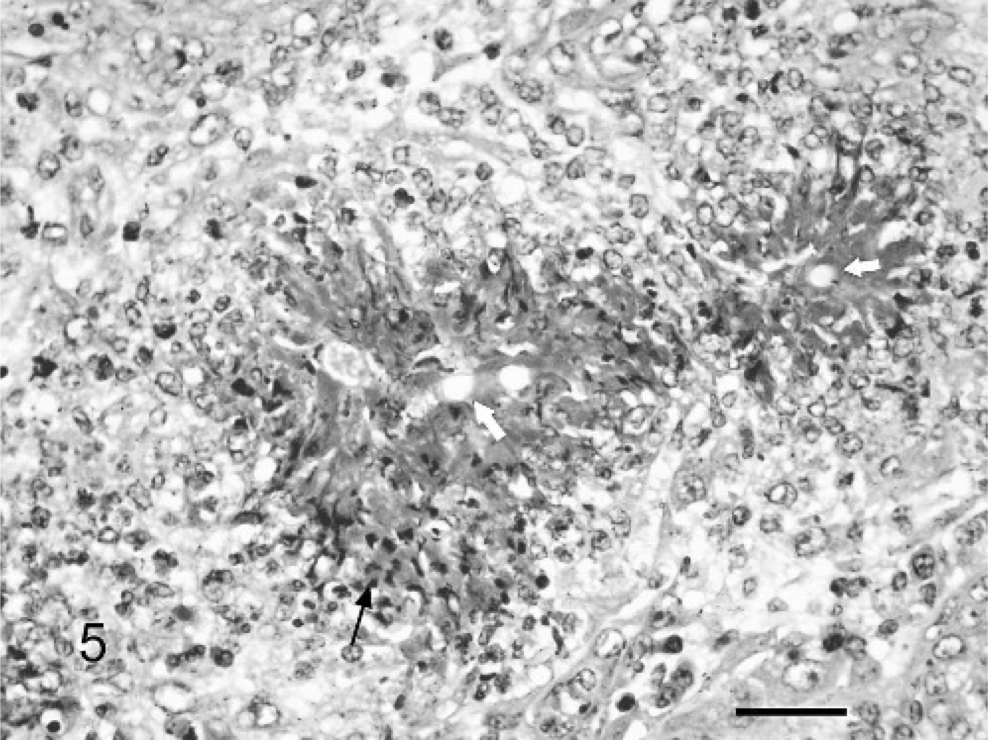
Sheep conidiobolomycosis. Nasopharyngeal tissues. A granuloma is observed containing hypha-like structures (white arrows) surrounded by eosinophilic Splendori-Hoeppli material and some necrotic eosinophils (black arrows) and neutrophils. The central area is surrounded by epithelioid macrophages. HE. Bar = 60 μm.
Multifocal to coalescent granulomas, similar to those observed in the nose, were present in the lung, meninges, kidneys, heart, gall bladder, and some lymph nodes. The bronchiolar epithelium was hyperplasic with proliferation of fibrous tissue and infiltration of lymphocytes and plasma cells. Mononuclear cells, neutrophils, and Splendore-Hoeppli material were observed within the bronchioles or in peribronchial tissues. The septa were thickened and infiltrated by mononuclear cells. Thrombosis of blood vessels was occasionally observed. The meninges were thickened by an inflammatory reaction of neutrophils, lymphocytes, plasma cells, and giant cells. Diffuse or focal gliosis, perivascular cuffing of mononuclear cells, and occasionally microabscesses were observed in the frontal lobe of the brain. In the kidneys, the granulomatous reaction destroyed the parenchyma, causing a granulomatous glomerulonephritis. Tubular degeneration, hyaline casts, tubular dilatation, and interstitial nephritis with infiltration of mononuclear cells and fibrosis were also observed. Hyalinization, vacuolation or necrosis of muscular fibers, and infiltration by mononuclear cells were observed in the heart. Most lymph nodes examined had follicular hyperplasia.
In the Gomorís methenamine silver stain of the nose, lung, kidney, heart, gallbladder, and meninges, numerous, 8–30-μm-thick hyphae were observed (Fig. 6). They were rarely septate or ramified, irregular in shape, and with black contoured wall, sometimes with bulbous dilatation in the extremities. With the PAS stain, the hyphae were slightly colored.
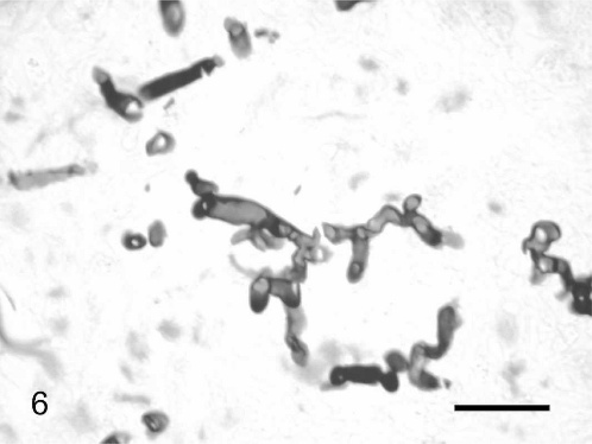
Sheep conidiobolomycosis. Nasopharyngeal tissues. Walled, occasionally septate or ramified hyphae, irregular in shape, sometimes with bulbous dilatation in the extremities are observed in the center of the granuloma. Gomori methenamine silver. Bar = 100 μm.
Electron microscopy
On electron microscopy, the hyphae had a thick double wall, 1 laminar and 1 undulated, surrounded by cellular remnants (Fig. 7). The cytoplasm had a dense granular matrix, liposomes, Golgi apparatus, and myelin figures. The lesion comprised macrophages, sometimes with hyphae within the cytoplasm, lymphocytes, fibroblasts, epithelioid and giant cells, collagen, and a large number of degenerated cells and cell remnants. No viral particles were observed in the nose or in the lung lesions.
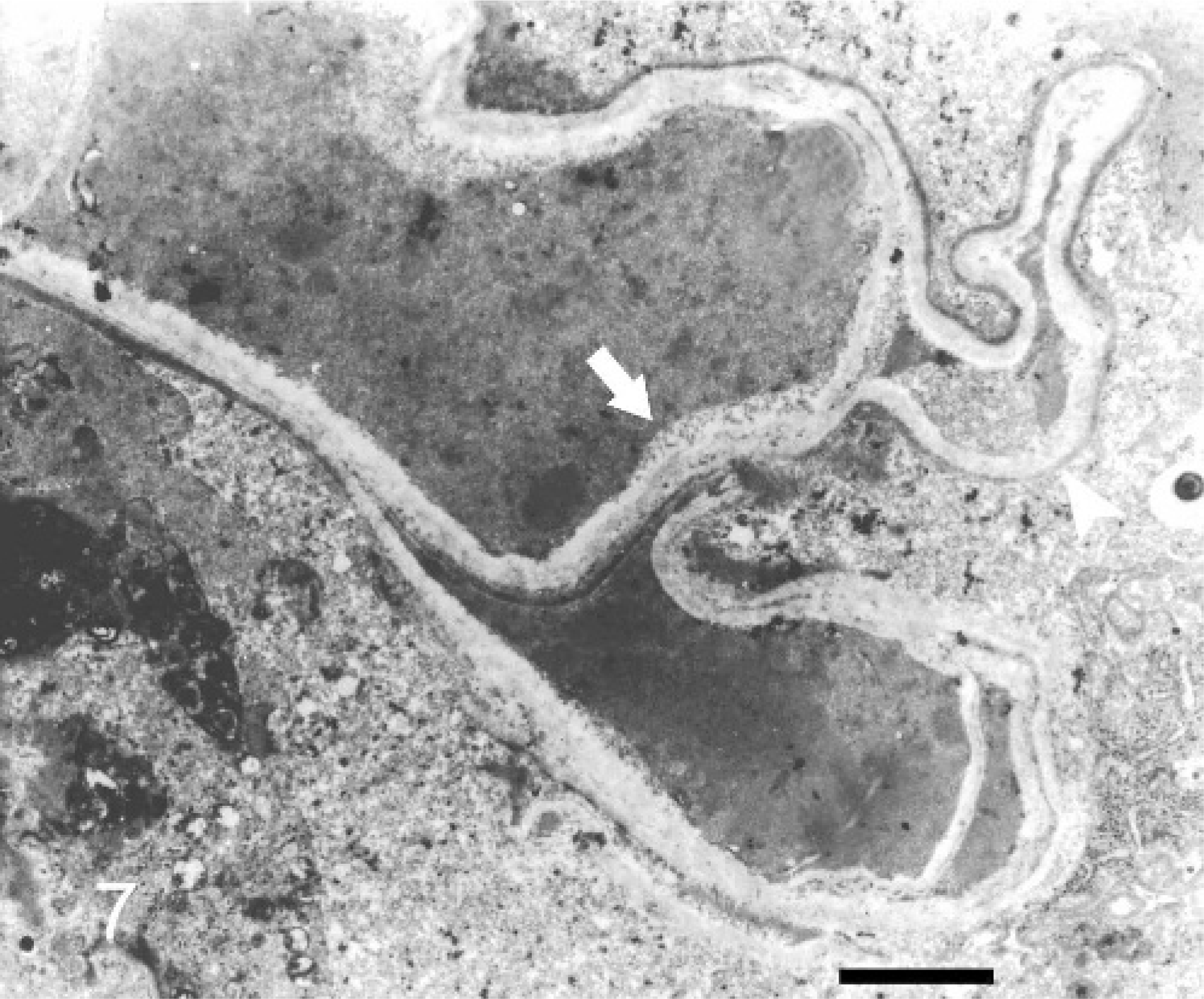
Electron microscopy micrograph. Sheep conidiobolomycosis. Nasopharyngeal tissue. Hyphae in the center of a granuloma with a thick double wall—the inner laminar (arrow) and the outer undulate (arrowhead)—surrounded by cellular remnants. Bar = 10 μm.
Isolation of the agent
On BDA, the colonies were smooth, with a cereous aspect. Satellite colonies of the same fungus were observed around the main colony. The colonies appeared after 24 hours incubation and, in 5 days, covered the whole plate. In SDA, the colonies were whitish or yellowish with a powdery and corrugated aspect; they also formed satellite colonies. Microscopically the fungus had wide hyphae, with few septa and irregular ramifications. The conidia were rounded, with a diameter of 14–22 μm, thin walls, and prominent papillae. Numerous rounded zygospores with thick or duplicated walls and diameters of 13–21 μm were also present. The fungus was identified as C. coronatus by the Mycology Laboratory at the Institute of Biological Sciences in the Federal University of Minas Gerais and at the Institute of Tropical Medicine in São Paulo.
Discussion
Conidiobolomycosis caused by C. coronatus reported in this paper is similar to conidiobolomycosis caused by C. incongruus reported in sheep in Australia5,18 and Trinidad Tobago.30 It is characterized by granulomatous and necrotic lesions in the ethmoidal and nasopharingeal regions extending to the turbinate bones and frequently invading the brain and the orbit. A very characteristic clinical sign of the disease is the exophthalmos and eye lesions because of the extension of the granuloma to the retrobulbar tissues, which was observed in 91.6% of the cases. Similar lesions are observed in human conidiobolomycosis causing opthalmoplegia and protrusion of the eye.9
An important aspect of the disease in this semiarid region of the state of Piauí is its enzootic character, with high frequency over a large geographic region. Probably this high frequency is a consequence of the climatic characteristic of the region: high temperatures, varying from 19 to 39°C with high rainfall of 1,000 to 1,600 mm during the rainy season from December to April. In other regions of the state of Piauí and other states in semiarid Brazil in which temperatures are also high, but with rainfall varying from 300 to 800 mm annually,23 the disease also occurs during the dry season, but is sporadic, with lower frequency.
In most cases reported here, the infection affected other organs, including brain, lymph nodes, lung, kidney, heart, and gallbladder. In Australia, the infection by C. incongruus in sheep affected also the lung, lymph nodes, and brain, but not other organs.5,18 In humans, horse, and llama, conidiobolomycosis caused by C. coronatus is restricted to the nasal cavity3,7,10,26,28 or neighboring regions.10,33 There is only 1 report of human conidiobolomycosis caused by C. coronatus affecting the lungs,17 but the infection by C. incongruus with dissemination to the lungs, lymph nodes, esophagus, and liver was fatal.4 Conidiobolomycosis (C. incongruus) in a deer affected organs of the thoracic and abdominal cavities, but without nasal lesions.34
The main histologic lesion was the presence of multifocal granulomas with fungal hyphae surrounded by Splendore-Hoeppli material, which is characteristic of infections by Entomophthorales.3,5,10,14,15,18,25,27,29,30,32,33,35 One important observation was the presence of vasculitis and thrombosis in the lesions of most sheep. Those lesions are rarely reported in infections by Entomophthorales, being more frequent in infection by Mucorales; such a characteristic has also been used to differentiate between mycosis caused by these 2 orders of fungi.22 Vascular lesions are occasionally reported in human infections by C. coronatus 4,17,36 and also in sheep and deer infected by C. incongruus.5,34 The presence of thrombi in the nasal lesions explains the hematogenous dissemination to other organs.
The ultrastructural examination of the nasal and pulmonary lesions confirmed the presence of a granulomatous inflammation and the absence of viral particles, also in areas of alveolar and bronchial proliferation. These finding are important, considering that many veterinarians in northeastern Brazil misdiagnosed the disease as enzootic ethmoidal tumor and that the bronchiolar and alveolar proliferative lesions were attributed to pulmonary adenomatosis, a disease rarely reported from Brazil.8
Conidiobolomycosis by C. coronatus is an endemic disease of sheep in the semiarid region of northeastern Brazil. In most cases, the infection disseminates from the nasal lesions to other organs, including brain, lymph nodes, lung, kidney, heart, and gallbladder. The disease is similar to those described previously in sheep and other species, but the large number of animals affected, the high fatality rate, and the endemic nature of the condition are responsible for severe losses. The high frequency of the disease in the semiarid state of Piauí is probably a consequence of the climatic characteristics of the region: high temperatures varying from 19 to 39°C with high rainfall of 1,000 to 1,600 mm during the rainy season from December to April.
Footnotes
Acknowledgements
The authors thank Elisabeth Maria Heins-Vacarri and Natalina Takahashi de Melo from the Mycology Laboratory of the Tropical Medicine Institute of Sao Paulo, and Leonardo Rodrigues from the Mycology Laboratory of Institut of Biological Sciences of the Federal University of Minas Gerais for identification of the fungi. The revision of the manuscript by Dr. Gordon Carter is kindly acknowledged.