
Abstract
A case of protothecosis is reported in an adult goat with inspiratory dyspnea and stertor. Dermatitis with prominent ulcerated nodules up to 3 cm in diameter was observed in the muzzle at the mucocutaneous junctions of nasal and lip skin, and in the border of the pinna. Histologic lesions were necrotizing pyogranulomatous dermatitis and rhinitis with myriads of walled sporangia, characteristic of Prototheca wickerhamii. This seems to be the first report of protothecosis in a goat.
Protothecosis is an uncommon disease of humans and animals caused by Prototheca spp., aclorophylic algae-like unicellular organisms, which belong to the family Chlorellaceae. 3, 10, 12 Prototheca are ubiquitous organisms, which can be isolated from plants, soil, sewage, mud, feces of wild and domestic animals, and many water sources, including lakes and sewage-laden areas. 8, 11 There are 2 pathogenic species, Prototheca zopfii and Prototheca wickerhamii. Most human cases of protothecosis are caused by P. wickerhamii; 11 P. zopfii mainly affects animals. 2, 3, 5, 7, 10
In humans, 3 forms of protothecosis are reported: cutaneous, olecranon bursitis, and disseminated. 9, 11 Most infections occur in individuals who are immunologically compromised by immunosuppressive therapy, acquired immune deficiency syndrome, malnutrition, renal or hepatic disease, cancer, or autoimmune disorders. In contrast, individuals with olecranon bursitis are usually not immunocompromised. Cutaneous protothecosis is usually secondary to trauma and restricted to the site of inoculation. Disseminated disease occurs in individuals who are severely immunocompromised. 9, 11
In dogs and cats, protothecosis is most often caused by P. zopfii, 3, 7, 10 which produces systemic or cutaneous infections, but infections by P. wickerhamii also occur. 13 Systemic infections affect a number of organs, including the eye, kidney, central nervous system, liver, heart, large intestine, skeletal muscle, myocardium, lymph nodes, thyroid, pancreas, and peritoneum. Clinical signs depend on the organs affected. 3, 10, 13 Bovine protothecosis is characterized by mastitis caused by P. zopfii, 1, 2, 4, 5 but a case of disseminated protothecosis was also reported. 12
In Brazil, Prototheca spp. were isolated from cases of mastitis in cows in the states of Mato Grosso do Sul, São Paulo, Minas Gerais, Paraná, Rio Grande do Sul, Santa Catarina, Pernambuco and Goiás. 2– 5 Human cases were also reported. 14 The objective of this article is to report a case of protothecosis in a goat in northeastern Brazil.
Protothecosis was diagnosed in a 3-year-old, nonlactating, nonpregnant, female Nubian crossbred goat, with nasal discharge, inspiratory dyspnea and stertor, and weight loss. Dermatitis with prominent ulcerated nodules up to 3 cm in diameter involved both sides of the muzzle at mucocutaneous junctions of the nostrils and lips (Fig. 1). An ulcerated nodule was also observed at the edge of the right pinna. Clinical signs were first observed in January 2006, during the rainy season, 9 months before the goat was presented to the Veterinary Hospital. The owner stated that the goat was in good body condition before the appearance of nasal lesions and respiratory signs. Treatment with tetracycline was not effective. On the same farm, about 200 Nubian and crossbred goats bred for meat production were not affected. This farm was in the municipality of São José do Bomfim, in the semiarid rangeland of the state of Paraíba, northeastern Brazil. The flock was in a pasture around a large pond for long periods. Because the affected goat was cachectic and finally recumbent, it was euthanatized.
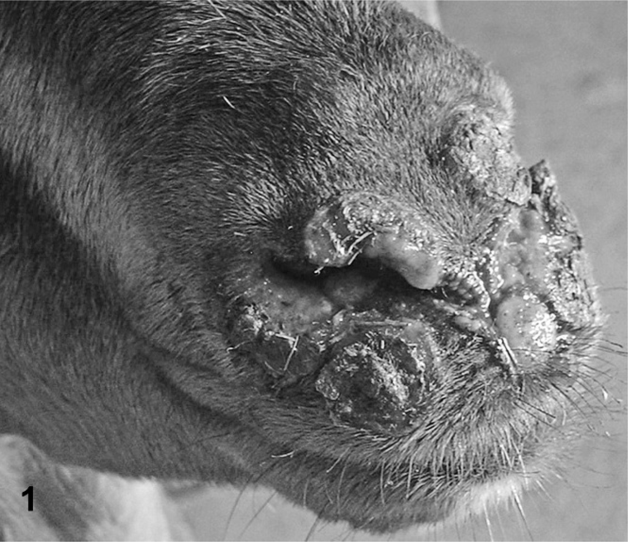
Muzzle; goat. Coalescing ulcerated granulomas are concentrated in the skin around the external nares.
At necropsy, coalescing yellowish nodules, up to 3 cm in diameter, were observed in the subcutis over the nasal bone. The mucosa of the nasal vestibule had an irregular, ulcerated, and fibrinonecrotic surface that extended from the external nares to the nasal conchae (Fig. 2). The mucosa of the rostral aspect of the ventral turbinates and nasal septum was also ulcerated. No gross lesions were observed in regional lymph nodes, lungs, liver, digestive tract, kidneys, spleen, heart, central nervous system, thyroid gland, adrenal glands, pancreas, or skeletal muscles.
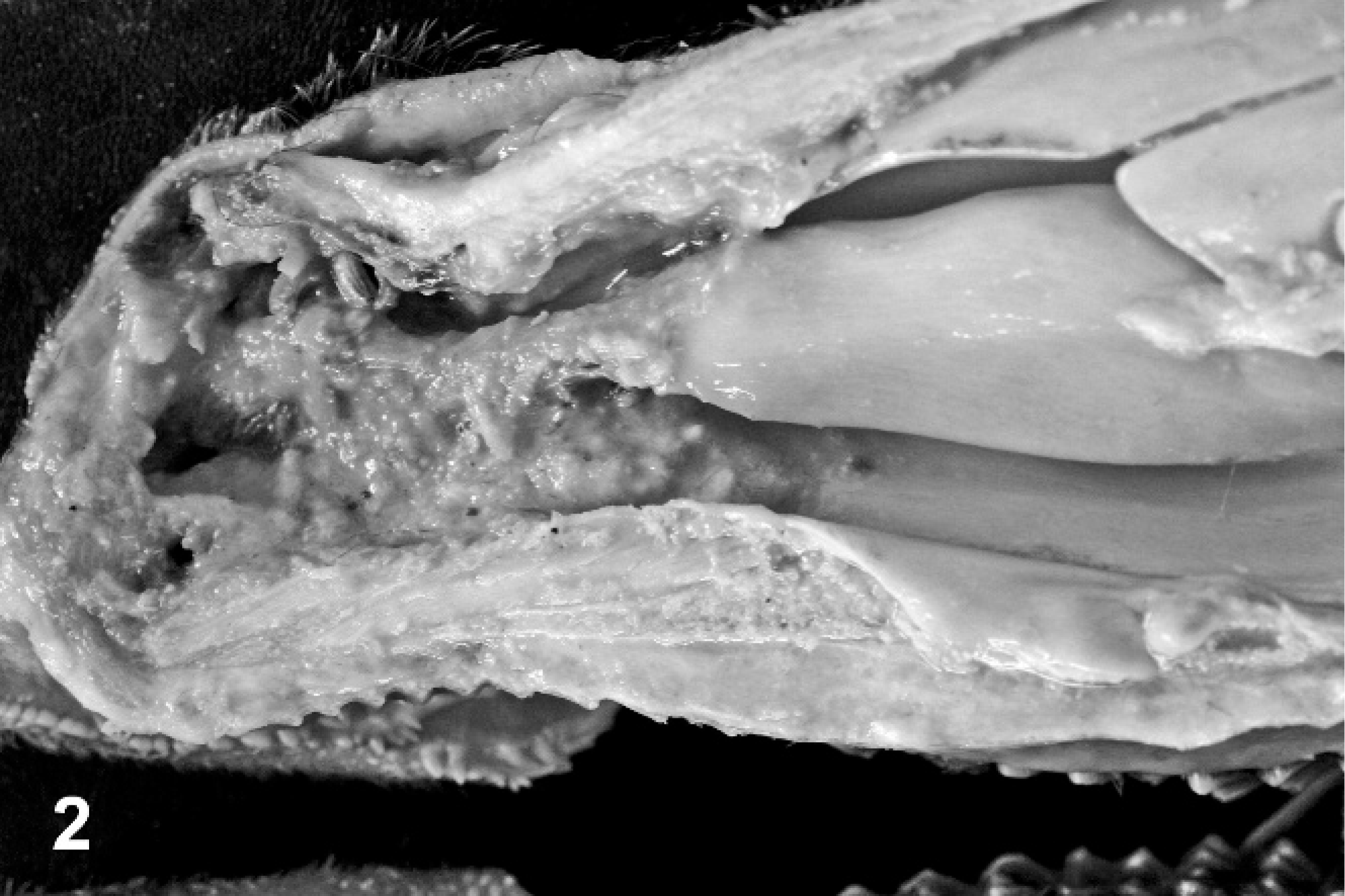
Nasal vestibule; goat. Nasal mucosa from the external nares to the nasal turbinates is ulcerated with fibrinonecrotic surface.
Samples of the cutaneous and nasal lesions and other selected organs collected at necropsy were fixed in 10% buffered formalin, embedded in paraffin, sectioned at 6 μm, and stained by hematoxylin and eosin. Selected sections were also stained by periodic acid–Schiff (PAS) and Gomori methenamine silver. Histologic lesions were necrotizing, pyogranulomatous dermatitis, and rhinitis, with infiltration by lymphoid cells, macrophages, giant cells, and neutrophils (Fig. 3). Fibrous tissue was also observed within and around the lesions. Myriads of ovoid-to-spherical, nonbudding, walled structures (sporangia) that measured 3–15 μm were observed in inflammatory foci and within macrophages (Figs. 3, 4). Some sporangia were internally septated and contained variable number of progeny walled cells (sporangiospores) that formed a morula-like structure that measured 10–15 μm in diameter. Some of these morula-like sporangia had a daisy-like aspect, with a central rounded endospore surrounded by a corona of molded endospores (Figs. 3, 4). The agent was identified as Prototheca. There was no histologic evidence of dissemination of infection in regional lymph nodes, lungs, liver, digestive tract, kidneys, spleen, heart, central nervous system, thyroid gland, adrenal glands, pancreas, or skeletal muscles.
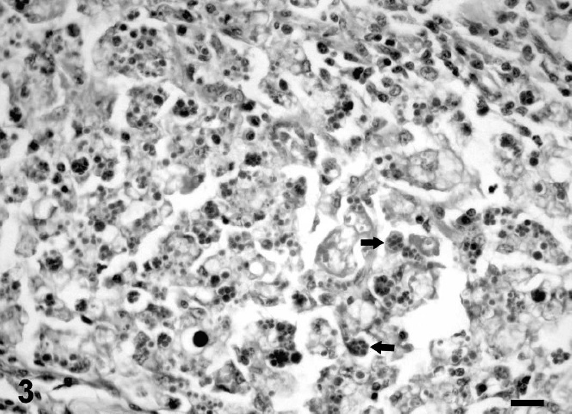
Subcutis of the muzzle; goat. Myriads of oval-to-spherical, nonbudding, walled sporangia, 3–15 μm in diameter, are in the inflammatory exudate. Some sporangia have a daisy-like shape (arrows). HE. Bar = 30 μm.
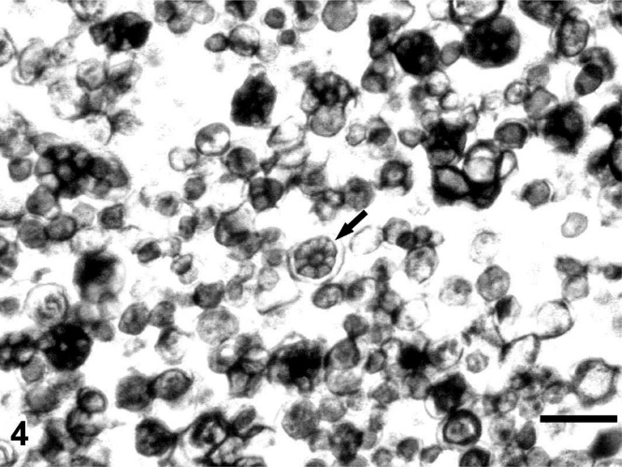
Subcutis of the muzzle; goat. Myriads of oval-to-spherical, nonbudding, walled sporangia are observed. Note the morula-like sporangium with a central rounded endospore surrounded by a corona of molded endospores (arrow). Gomori methenamine silver stain. Bar = 30 μm.
Both P. zopfii and P. wickerhamii reproduce by internal septation that results in the production of infective sporongiospores (endosporulation). P. zopfii is usually larger, 7–30 μm, in contrast to 3–15 μm for P. wickerhamii. 11 The characteristic feature of P. wickerhamii is the presence of sporangia with a central rounded endospore surrounded by a corona of molded endospores, which is described as moruloid, daisy-like, spoke-like, and frambesiform. 9 In this case, the presence of numerous daisy-like morulae that measured 10–15 μm in diameter, is characteristic of infection by P. wickerhamii. Prototheca spp. can be differentiated from Chlorella by the absence of green color in the gross lesions and the absence of PAS-positive starch granules. The chloroplasts present in Chlorella and absent in Prototheca can be observed only by electron microscopy. 6, 9, 11
This appears to be the first report of protothecosis in a goat. It is of interest that the goat disease differs from the disease in cattle (typically mastitis), but is similar to some reported cases in dogs, cats, and immunocompromised humans. In immunodeficient humans, protothecosis usually develops as a localized cutaneous lesion at the site traumatic injury. 9, 11 Evidence of dissemination, as has been described in other species, 3, 9, 10, 12, 13 was not noted, despite the prolonged course of the illness. When the lesions were first observed, the goat was in good condition, with no evidence of immunosuppression or preexisting disease. Because the goat was kept near a large pond for long periods, the infection may have occurred because of exposure of a traumatic lesion to contaminated water. The skin lesions extended into the nasal vestibule, and the consequent obstruction of nasal passages resulted in the severe respiratory signs.
Footnotes
Acknowledgements
We thank Dr. Chris Gardiner and the Armed Forces Institute of Pathology for the examination of paraffin blocks.