
Abstract
Primary renal tumors are rare neoplasms in nonhuman primates. This report describes a mixed epithelial and stromal tumor of the kidney (MESTK) in a 14.5-year-old female ringtail lemur. The well-demarcated, solid, and cystic mass was located in the pelvis of the left kidney and consisted histologically of both epithelial and mesenchymal components. The mesenchymal cells were arranged in fascicles around cysts lined by a well-differentiated epithelium. Neither the mesenchymal nor the epithelial parts showed significant nuclear atypia or mitotic figures. To our knowledge, only 1 similar case, classified as adenoleiomyofibromatous hamartoma, has been reported in a ringtail lemur. In humans this tumor affects predominantly perimenopausal women and can express estrogen and progesterone receptors. However, neither estrogen nor progesterone receptors could be identified by immunohistochemistry in the tumor of the present ringtail lemur. Therefore, a hormonal mechanism could not be demonstrated in this case.
This report describes a mixed epithelial and stromal tumor of the kidney (MESTK) in a 14.5-year-old female ringtail lemur (Lemur catta). In nonhuman primates, primary renal tumors are rare neoplasms.
The ringtail lemur from the Basel zoo was presented for postmortem examination to the Centre for Fish and Wildlife Health in Berne, Switzerland. The ringtail lemur was born in the Basel zoo in 1989. She had been kept in a group and had never been pregnant. Keepers reported sporadic sudden falls from climbing branches and short tonic seizures in her hindlegs during the last 3 years; however, these symptoms did not persist and disappeared before clinical exams were carried out by the veterinarian only minutes later. Symptomatic treatment was performed using oral circulation therapy (Effortil®, etilefrin hydrochloride, Boehringer Ingelheim, Basel, Switzerland). Reduced appetite and weight loss were observed during the lemur's last year, and as a result, a clinical examination under general anesthesia was performed revealing cachexia and dehydration. The main finding at palpation was an intra-abdominal mass that was confirmed radiographically. Hematology was within the normal ranges and serum chemistry showed mildly increased uremia (30.2 mmol/l), the reference ranges for physiological data values for uremia being between 0 and 24.63 mmol/l.9 A renal neoplasia was suspected, and the lemur was euthanized because of poor general condition and prognosis.
Complete necropsy was performed, including examination of the brain. The animal was emaciated and weighed 2 kg. The only significant pathologic lesions were observed in the kidneys. When compared macroscopically with the right kidney, the left kidney was markedly enlarged (4.9 cm × 4.1 cm × 3.5 cm), slightly rounded, pale red, and mottled. On cut section, a well-demarcated, 4.8 cm × 4 cm × 3 cm mass was visible, compressing the rest of the renal parenchyma marginally and causing atrophy. The mass presented solid areas that were firm, homogenous, and pale red with multiple cysts of varying size (up to 1.2 cm in diameter) (Fig. 1).
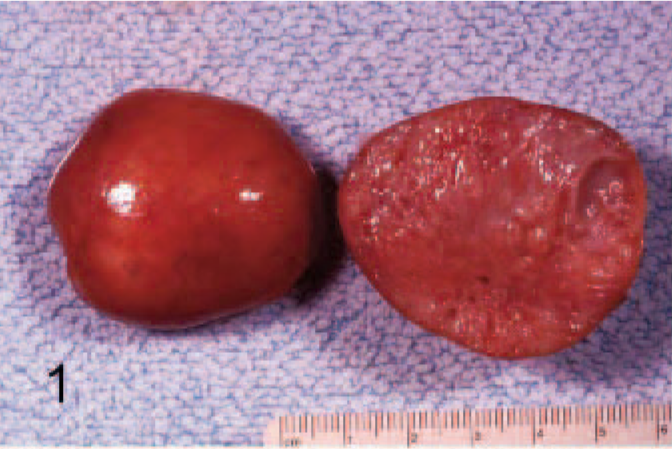
Left kidney; L catta. On cut section, a well-demarcated mass was visible, occupying the entire kidney.
Histologically, the renal mass was a dimorphic tumor composed of solid and cystic areas consisting of a mesenchymal component characterized by a spindle-cell stroma growing in solid septae that surrounded an epithelial component composed of cysts and papilliform structures (Fig. 2). The cysts were lined by a well-differentiated epithelium with multifocal papillary projections and were supported by a rim of collagenous stroma (Fig. 3). The epithelial cells were well differentiated, monolayered to pseudo-stratified, and columnar to cuboidal. Larger cysts were sometimes lined by attenuated epithelial cells. The epithelial cells had an abundant, clear to pale, eosinophilic cytoplasm with vacuoles and a prominent, central, round to oval, basophilic nucleus with stippled chromatin and inconspicuous nucleolus (Fig. 4). The neoplastic mesenchymal cells were well-differentiated spindle cells arranged in interlacing fascicles. These cells were embedded in a fine collagenous eosinophilic matrix; had a plump, elongated nucleus and an abundant, eosinophilic cytoplasm; and were morphologically similar to smooth muscle cells. The neoplastic mesenchymal cells stained brown, and the collagenous fibers of the stroma stained red with the Van Giesson staining. The mesenchymal cells stained magenta and the collagenous fibers of the stroma blue with the Mallory trichrome staining. Neither the mesenchymal nor the epithelial parts showed significant nuclear atypia or mitotic figures. No blastema, typical for nephroblastic tumors, was observed. Sections of the mass were examined immunohistochemically by an avidin-biotin-peroxidase complex (ABC) technique (DakoCytomation, Carpinteria, CA) and the following antisera were used: monoclonal antibodies against vimentin, desmin, smooth-muscle actin, cytokeratin 622, estrogen receptor α, and progesterone receptor. Positive control tissue consisted of the tongue (vimentin, desmin, and cytokeratin) and the uterus (smooth-muscle actin, estrogen receptor, and progesterone receptor) of the ringtail lemur. The slides were counterstained with hematoxylin. Immunohistochemically the spindle cells were positive for vimentin (Fig. 5a), desmin, and smooth-muscle actin (Fig. 5b); the epithelial cells were positive for cytokeratin (Fig. 5c). Immunohistochemically neither estrogen nor progesterone receptors could be identified within the tumor or the kidneys. Additional findings were a severe interstitial nephritis in the right kidney and remainder of the left kidney, characterized by interstitial fibrosis admixed with infiltrates of numerous lymphocytes and few plasma cells. The glomeruli were variable in size with thickened basement membranes and Bowman's capsules and increased mesangial cellularity and were occasionally sclerotic. Some tubules were necrotic; others showed tubular regeneration; and tubular lumina often contained proteinaceous casts, sloughed cells, and rarely crystals and mineralizations (Fig. 2).
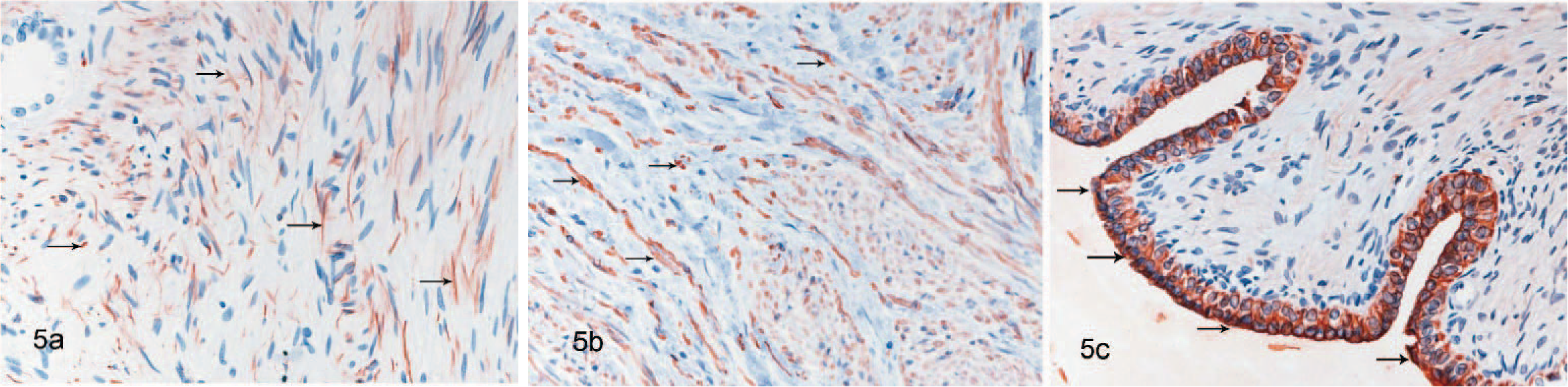
Kidney; L catta. Immunohistochemistry of the MESTK.
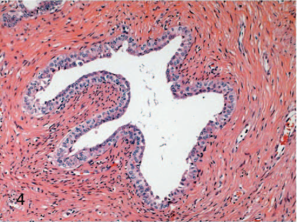
Kidney; L catta. HE staining. MESTK. Cystic structures are lined by well-differentiated epithelium.
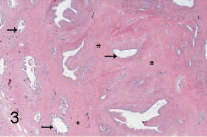
Kidney; L catta. Higher magnification of Fig. 2. The MESTK consists of a mixture of mesenchymal (asterisks) and epithelial components (arrows).
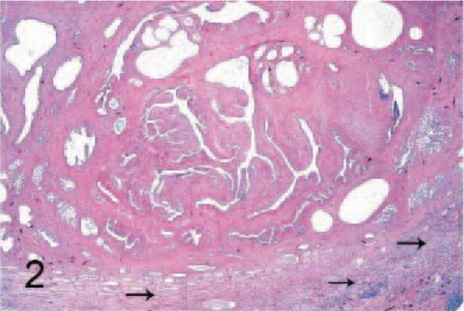
Kidney; L catta. HE staining. Histological features of the MESTK. A well-demarcated mass compressed the renal parenchyma. Arrows indicate the atrophic renal parenchyma with interstitial nephritis.
According to the World Health Organization classification of tumors of the urinary system in human pathology and based on the histopathological appearance,5 the renal neoplasm was diagnosed as MESTK. By definition the mixed epithelial and stromal tumor is a complex renal neoplasm composed of a mixture of stromal and epithelial elements.5 The term MESTK was first introduced by Michal and Syrucek in 1998.11 These tumors often arise centrally in the kidney, grow as expansile masses, and have a typical morphology comprising both cystic and solid areas. Histologically, they are composed of small cysts lined by epithelial cells embedded in a variably cellular stroma of spindle cells that resemble smooth muscle.5,11 Immunohistochemically these spindle cells show strong reactions with antibodies to actin and desmin. In some tumors, the nuclei of the spindle cells react with antibodies to estrogen and progesterone receptors.1,5 The epithelial cells react with antibodies to a variety of cytokeratins and occasionally with antibodies to estrogen receptor.1,5
In human pathology, about 50 cases of MESKT exhibiting similar characteristics have been described in the literature.1 These tumors are described as benign biphasic tumors occurring in adulthood.1,12 There is a 6:1 predominance of women over men,1,5 and tumors occur mostly in perimenopausal age (46 years). Reported symptoms include flank pain, hematuria, or urinary tract infection, but 25% of the cases are incidental findings. Several studies describe the presence of estrogen and progesterone receptors in mixed epithelial and stromal tumors, suggesting that the hormones could play a role in the pathogenesis of this tumor.1,12 The spindled stromal cells frequently express muscle markers and estrogen and progesterone receptors. The epithelium also is occasionally positive for estrogen and progesterone receptors.2 It has been hypothesized that this tumor possibly derives from müllerian/paramesonephric rests.2 The tumor may be an extra ovarian neoplasia arising from paramesonephric remnants, deriving from the müllerian tract with close relation to the kidney and upper urothelial tract.2 The possible trigger for the tumor could be a hormonal change (perimenopausal changes or hormonal therapy) that leads to a proliferation of mesenchymal cells, which in turn stimulate the growth of the epithelial component.2
In a previous review of 30 nonhuman primate renal tumors, only 1 similar case, classified at the time as adenoleiomyofibromatous hamartoma, was described in a female, 11-year-old ringtail lemur.10 Renal carcinoma, transitional cell carcinoma, renal adenoma, adenosarcoma, nephroblastoma, nephroblastomatosis, hamartoma, transitional cell papillomatous hyperplasia, and hemangiosarcoma have been described in nonhuman primates.7,8,10 The true incidence of primary renal tumors in nonhuman primates remains unknown and may vary among species. To our knowledge this is the first report of MESTK in an animal species other than human. Given that the known lifespan of prosimians and lemurs in captivity is about 23 and 30 years, respectively (data coming from compiled material of zoos in the international species inventory system13), the female, 14.5-year-old ringtail lemur could be considered to be middle aged. However, naturally occurring menopause is not described in prosimians. Furthermore, neither estrogen nor progesterone receptors could be identified immunohistochemically in the tumor or in the kidneys, such that no evidence of a hormonal mechanism was found in this case.
The clinical relevance of this tumor is unclear, but according to the human medicine literature, MESTKs are often incidental findings.1 The chronic interstitial nephritis constitutes the only significant pathologic lesion observed besides the tumor. In the literature, chronic interstitial nephritides are described in prosimians without specific causes, and they are considered to be age related.3,4,6 However, despite the marked histopathologic changes of the kidney, the animal was only mildly uremic, and therefore the cause of the clinical symptoms, including the reported seizures, cachexia, and dehydration, could not be definitively established.