
Abstract
A review of the published literature indicates that marine mammal neoplasia includes the types and distributions of tumors seen in domestic species. A routine collection of samples from marine mammal species is hampered, and, hence, the literature is principally composed of reports from early whaling expeditions, captive zoo mammals, and epizootics that affect larger numbers of animals from a specific geographic location. The latter instances are most important, because many of these long-lived, free-ranging marine mammals may act as environmental sentinels for the health of the oceans. Examination of large numbers of mortalities reveals incidental proliferative and neoplastic conditions and, less commonly, identifies specific malignant cancers that can alter population dynamics. The best example of these is the presumptive herpesvirus-associated metastatic genital carcinomas found in California sea lions. Studies of tissues from St. Lawrence estuary beluga whales have demonstrated a high incidence of neoplasia and produced evidence that environmental contamination with high levels of polychlorinated biphenols and dichlorophenyl trichloroethane might be the cause. In addition, viruses are suspected to be the cause of gastric papillomas in belugas and cutaneous papillomas in Florida manatees and harbor porpoises. While experimental laboratory procedures can further elucidate mechanisms of neoplasia, continued pathologic examination of marine mammals will also be necessary to follow trends in wild populations.
In companion animals, the discipline of oncology has seen incredible growth because of an advanced understanding of neoplastic processes. This has resulted in better management of many cancers through the development of new strategies for successful treatment. Having said that, there is still a vast amount to learn about oncologic processes in both domestic species and nondomestic species. In marine mammals, we are early in the process of discovery of the type and the range of neoplastic lesions and potential etiologies. Prognostic information or treatment options have been limited, because most of the reports involve necropsy samples from wild populations. In addition, marine mammals, as top-level predators, may act as environmental sentinels. Probable reasons for the relatively low numbers of reported neoplasms in marine mammals (with the notable exceptions of California sea lions [Zalophus californianus] and St. Lawrence beluga whales [Delphinapterus leucas]) is that many marine mammal mortalities are not observed and that many marine mammals die before reaching old age, when most cancers occur. Even in those mortalities that are detected, there is often a delay in collecting samples. This may result in considerable postmortem autolysis, which hampers diagnosis.
The literature documenting marine mammal neoplasia is expanding gradually (Tables 1–6). Most reports attempt to characterize the types of cancers and the organ systems involved in the different cetacean and pinniped species represented. Overall, the reported incidence of neoplasia in marine mammals appears to be low. As previously mentioned, this may be because many deaths in wild populations go unnoticed and/or that representative tissue samples are not examined from necropsies, because of the extensive autolytic changes that may accompany the common delay in initiation of examination of these species. In addition, most stranded animals do not undergo thorough necropsy examinations. Therefore, the most detailed pathology information is often from single cases involving captive marine mammals, where some historical and treatment information may also be available.
Summary of reported neoplasms in baleen whales (suborder Mysticeti).
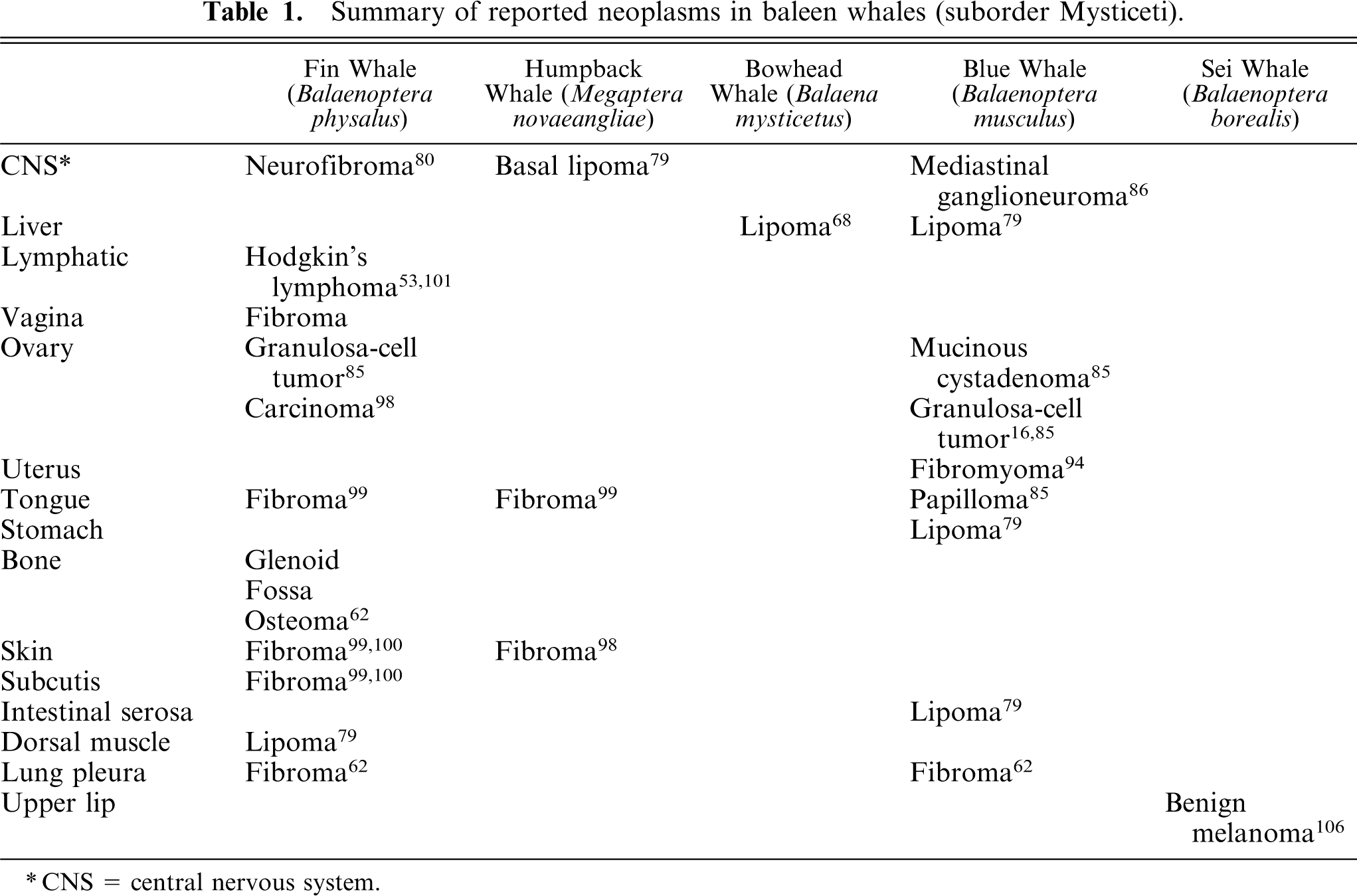
CNS = central nervous system.
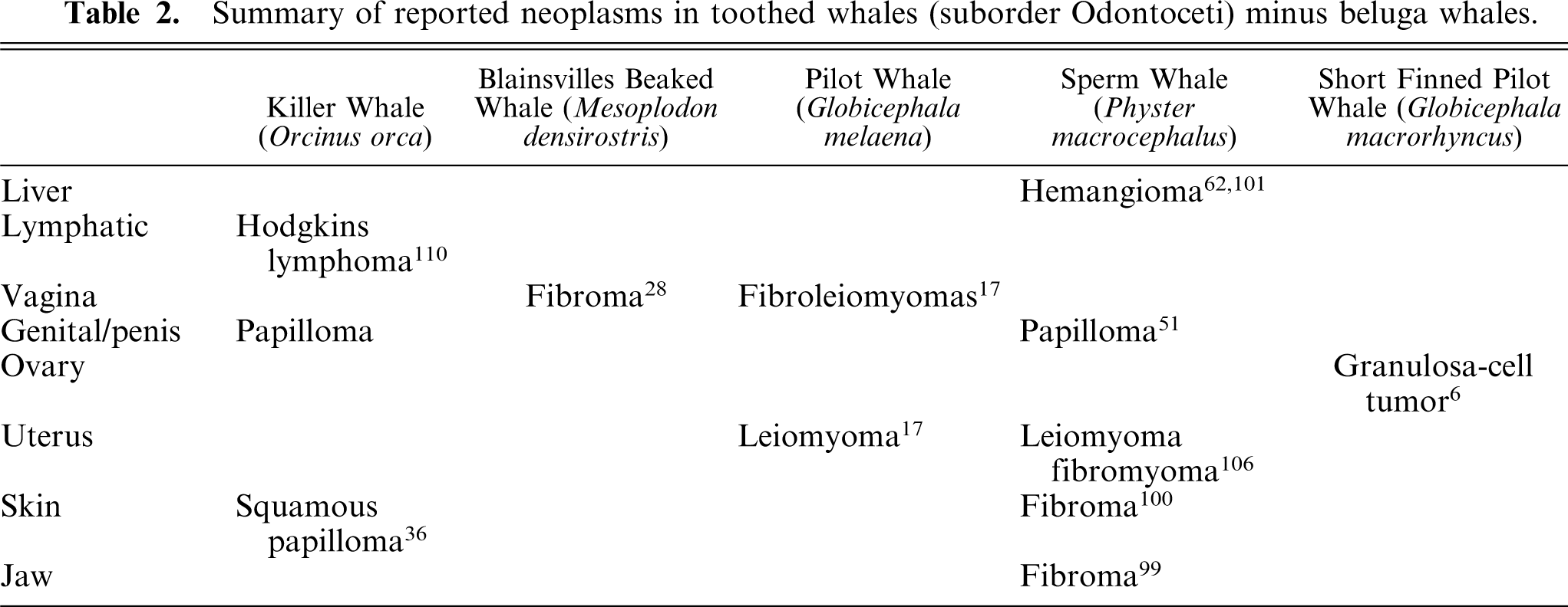
Summary of reported neoplasms in toothed whales (suborder Odontoceti) minus beluga whales.
Summary of reported neoplasms in beluga whales(Delphinapterus leucas).∗
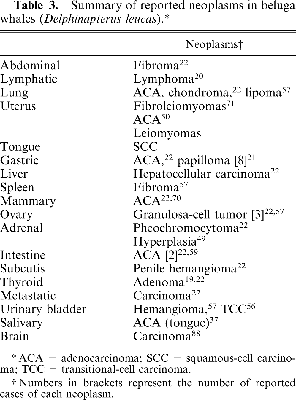
ACA = adenocarcinoma; SCC = squamous-cell carcinoma; TCC = transitional-cell carcinoma.
Numbers in brackets represent the number of reported cases of each neoplasm.
Summary of reported neoplasms in dolphins and porpoises.∗
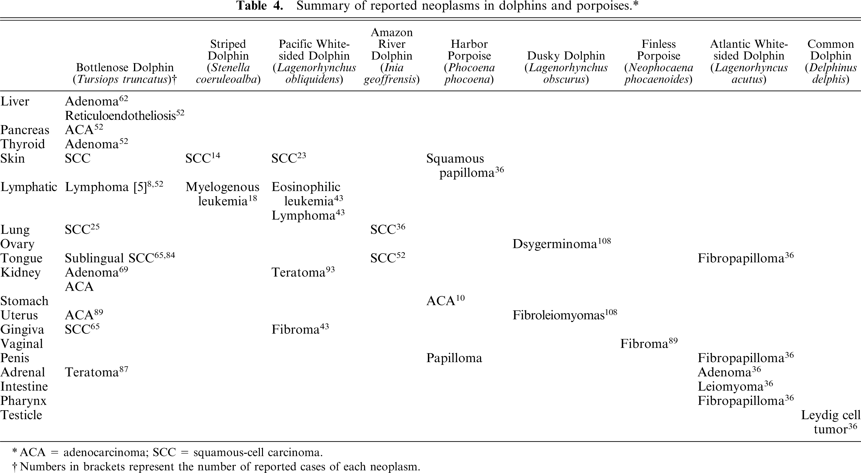
ACA = adenocarcinoma; SCC = squamous-cell carcinoma.
Numbers in brackets represent the number of reported cases of each neoplasm.
Summary of reported neoplasms in sea lions and fur seals (suborder Pinnipedia, family Otariidae).∗
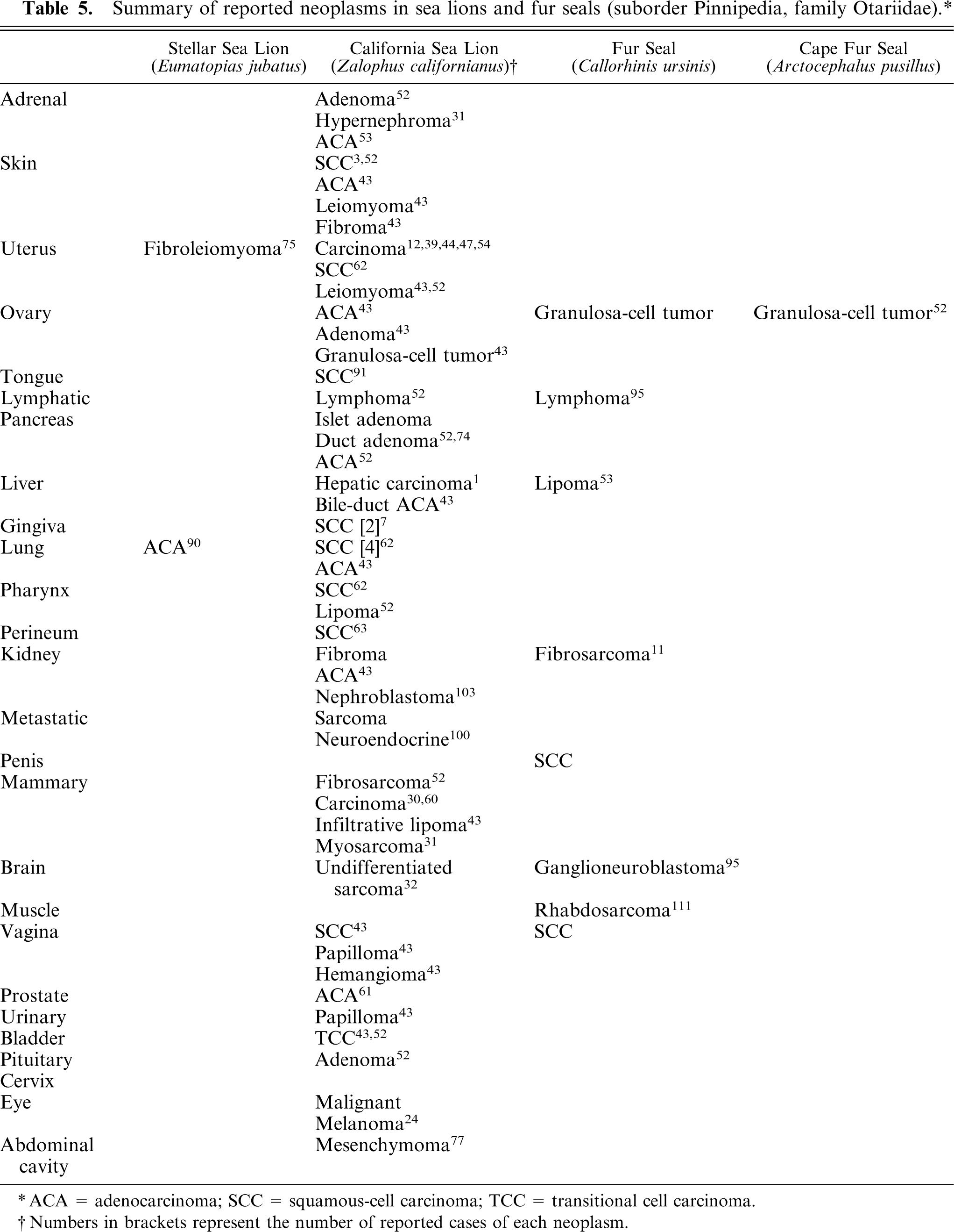
ACA = adenocarcinoma; SCC = squamous-cell carcinoma; TCC = transitional cell carcinoma.
Numbers in brackets represent the number of reported cases of each neoplasm.
Summary of reported neoplasms in seals (Order Pinnipedia, Family Phocidae).∗
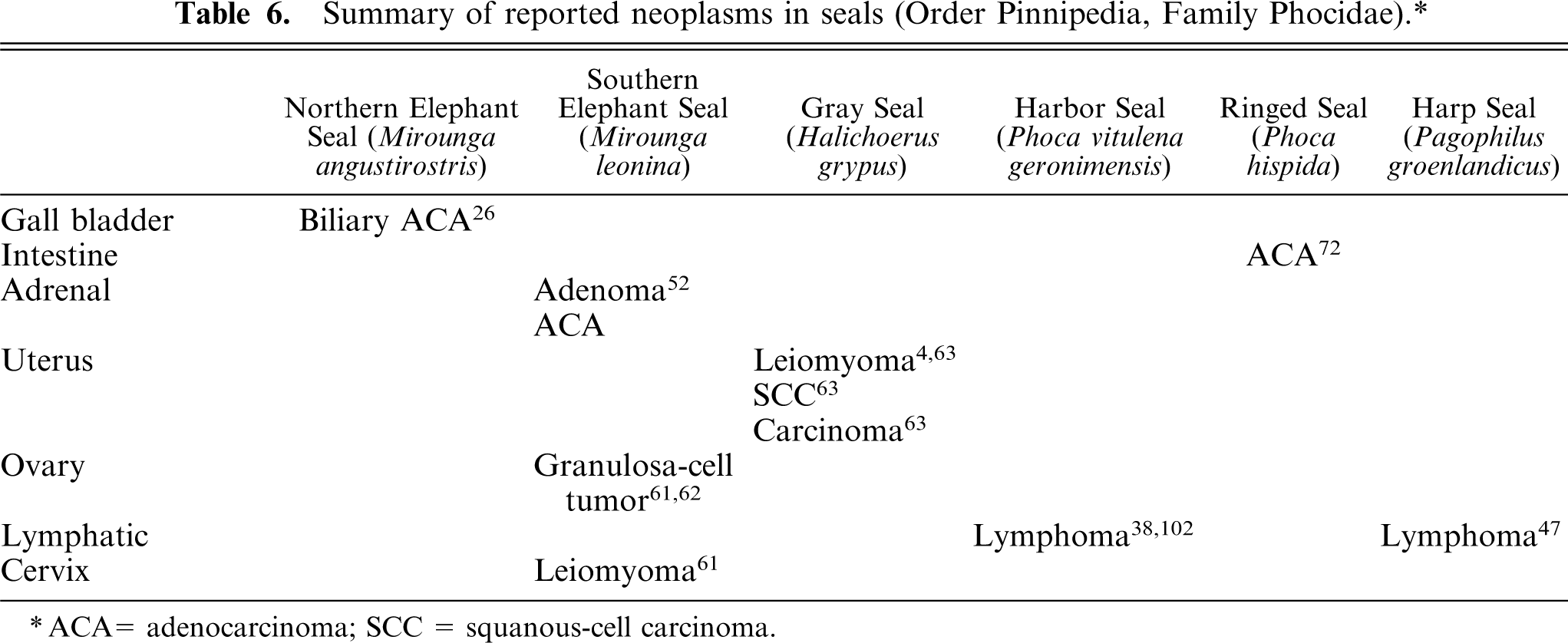
ACA = adenocarcinoma; SCC = squanous-cell carcinoma.
Wild cetaceans undergoing regular examination of mortalities include those from a population of approximately 500 beluga whales (Delphinapterus leucas) in the St. Lawrence estuary. These whales are a source of many of the reported cases of cetacean neoplasia. 22 The incidence of neoplasia in this group has been determined to be high with respect to other mammalian species and was last reported in the literature at 37% (28/75). 22 Proposed causes include environmental exposure to carcinogenic substances or reduced immune function/surveillance because of limited genetic diversity in this cloistered population. Other comparative information on the rate of neoplasia in marine mammals comes from an assessment of 39 marine mammal neoplasms (35 from California sea lions (Zalophus californianus) and 4 from Pacific white-sided dolphins [Lagenorhynchus obliquidens]) from a population of 1,500 animals, such that a 2.5% rate of neoplasia was determined. 43 The cancer-related death rate in this population was documented at just over 1.9% (28 malignancies/1,500 deaths). 43 In addition, 14 tumors were identified from examination of over 1,800 cetaceans. 36 These included a bronchogenic carcinoma in a Amazon River dolphin (Inia geoffrensis), adrenal adenomas in 2 Atlantic white-sided dolphins (Lagenorhynchus acutus), intestinal leiomyomas in 5 Atlantic white-sided dolphins (L. acutus), fibropapillomas in 3 Atlantic white-sided dolphins (L. acutus), and 3 squamous papillomas; one each in a harbor porpoise (Phocoena phocoena), a killer whale (Orcinus orca), and a narwhal (Monodon monoceros). 36 In a report that documented results from a series of 68 necropsy examinations performed on individual marine mammal strandings from along the Oregon coast, there were only 2 cases of neoplasia in the 10 species represented, and these included a lymphosarcoma in a harbor seal (Phoca vitulina) and an endocrine adenocarcinoma in a California sea lion (Z. californianus). 102 Fifty-one stranded California sea lions (Z. californianus) were examined and 2 had neoplastic processes: a nephroblastoma and a squamous-cell carcinoma of undetermined tissue origin. 102 In another study, 15 of 35 examined California sea lions (Z. californianus) had genital tract neoplasia and 60% of those tumors were malignant. 43
The literature documents the occurrence of neoplasia in many species of marine mammals from many geographic areas, including beluga whales (D. leucas) from Hudson's Bay, beluga whales (D. leucas) from the St. Lawrence estuary, 22 manatees (Trichechus manatus latirostris) from Florida, 9 bottlenose dolphins (Tursiops truncatus) from the Atlantic Ocean, 8 and striped dolphins (Stenella coeruleoalba) from the Mediterranean Sea. 14 In a review of cetacean tumors, certain organ systems were more commonly affected by neoplasia and included the gastrointestinal tract (13/41 = 31%), integument (10/41 = 24%), and the reproductive tract (12/41 = 21%). 36
Benign and malignant epithelial and mesenchymal neoplasia in human beings and most companion animals has been reported in virtually all organ systems. The reported distribution of organ involvement is much less extensive in marine mammals, probably because of the overall fewer total necropsy examinations performed. The organs reported to be most commonly affected in whales include the following: ovary, vagina, uterus, skin, tongue, liver, and central nervous system, in decreasing order of frequency (Tables 1, 2). 85, 86, 98– 101, 106 In beluga whales (D. leucas), in particular, the organs reported to be most commonly affected include uterus, stomach, and ovary (Table 3). 22, 58, 70, 104 The organ systems reported to be most commonly affected in dolphins include skin, lymphatic system, tongue, lung, and kidney (Table 4). 9, 36, 65, 69 A wide variety of tumors in many different organ systems have been reported in California sea lions (Z. californianus) (Table 5). Neoplasms are less commonly reported in seals (Tables 5, 6).
Pinnipeds
Sea lions
In 1980, metastatic adenocarcinoma of undetermined origin was reported in a female and in a male California sea lion (Z. californianus). 12 There were widespread metastases involving lung, liver, and internal iliac lymph nodes in the female, and lung, liver, kidney, spleen, and omentum in the male. 12 These authors suggested that, because of the presence of tumor metastases in the internal iliac lymph nodes and mucin secretion by the neoplastic epithelial cells, the urogenital tract might be one possible site of origin for these tumors. Subsequently, a single case report in a California sea lion (Z. californianus) documented widespread metastases of a poorly differentiated squamous-cell carcinoma affecting liver, lung, mesenteric lymph node, kidney, and ovary, but the primary site was not determined. 44 In 1996, a larger study examined 370 California sea lions (Z. californianus) and documented 66 cases (18% incidence) of suspected transitional-cell carcinoma, with widespread metastases similar to the cases previously reported. 39 Typically, these animals had mass lesions surrounding the ureters that often resulted in secondary hydroureter. 39 Metastatic nodules were often noted as caseous masses in adrenal glands, kidneys, uterus, liver, sublumbar lymph nodes, lung, spleen, mediastinum, thoracic and abdominal lymph nodes, omentum, pancreas, pericardium, and myocardium. 39 In another study, histologic assessment of necropsy tissues from 10 California sea lions (Z. californianus) found that the penis and the prepuce were affected in the 3 males, whereas the cervix was affected in all 7 females, the vagina in 5 of the 7, and the uterus in 3 of the 7 females. 54 The histologic lesion, in both male and female sea lions, was characterized by the presence of thickened and dysplastic mucosal epithelium. Distinct cytoplasmic borders, polygonal to round cells with moderate amounts of cytoplasm, and the presence of occasional squamous differentiation/keratinization were also noted. In some of the tumors of the females, there was glandular differentiation. 54 In one of the examined animals, transition from intraepithelial neoplasia to invasive carcinoma was detected. 54 Hence, it was proposed that the metastatic epithelial neoplasms originated from the lower genital tract as intraepithelial neoplasia/carcinoma in situ. 54 Immunohistochemistry confirmed cytokeratin expression by neoplastic cells. 39 In addition, intranuclear inclusion bodies were identified in intraepithelial neoplasia and metastatic carcinoma. 54 Electron microscopy, immunohistochemistry, and polymerase chain reaction identified a herpesvirus. 54 Herpesvirus-like particles had been previously detected in a lesion described as a hyperplastic plaque on the penis of a California sea lion (Z. californianus). 39 Based on sequence analysis of the terminase gene, the sea lion virus was a novel gammaherpesvirus in the Rhadinovirus genus. Human herpesvirus 8, which has been implicated as a possible cause of Kaposi's sarcoma, is the most closely related known virus. 54 The virus has been designated otarine herpesvirus-1. 47 Because gammaherpesviruses are believed to contribute to causation of other malignancies, such as nasopharyngeal carcinoma, the African form of Burkitt's lymphoma and Kaposi's sarcoma, it is possible that otarine herpesvirus-1 might cause or contribute to causing the sea lion carcinoma. Polychlorinated biphenols (PCB) have been suggested as a possible cofactor. 40
Other reported benign epithelial tumors of sea lions (Z. californianus) include an islet-cell adenoma, an adrenal adenoma, an ovarian adenoma, a vaginal papilloma, a urinary papilloma, 42 and a cutaneous papilloma. Malignant epithelial neoplasms include mammary adenocarcinomas; 60 a pulmonary adenocarcinoma; 43 a hepatic carcinoma with neuroendocrine pattern; 1 bile-duct adenocarcinoma; 43 a perirenal neuroendocrine carcinoma; 43 an endocrine adenocarcinoma; 102 an ovarian granulosa cell tumor; 43 a urinary bladder transitional-cell carcinoma; 43 a renal adenocarcinoma; 43 an ulcerated gingival squamous-cell carcinoma, with mandibular lymph-node metastases; 7 and squamous-cell carcinomas of lung, skin, pharynx, perineum, liver, uterus, cervix, and vagina. 43 However, it is now apparent that some of these previously reported carcinomas may have been of genital-tract origin. Reported mesenchymal tumors in California sea lions include renal fibroma, uterine leiomyomas, 43 a cutaneous leiomyoma, 43 a vaginal hemangioma, 43 an infiltrative lipoma of the mammary gland, 43 and an undifferentiated sarcoma of possible histiocytic origin that caused hydrocephalus. 32 Rhabdomyosarcoma originating from the latissimus dorsi muscle, with metastatic foci in the lungs, was reported in a Steller sea lion (Eumatopias jubatus). 111
Immunohistochemistry was performed as a diagnostic test in a case of hepatocellular carcinoma that involved the liver and the spleen of a California sea lion (Z. californianus). 1 The neoplasm was positive for cytokeratin 2 and was negative for epithelial membrane antigen and polyclonal carcinoembryonic antigen. 1 In another report, a California sea lion (Z. californianus) was diagnosed with 2 separate mammary gland carcinomas: a left anterior tumor was a complex mammary carcinoma, whereas a right posterior mass was a simple mammary carcinoma. 60 Metastases were found in lung, pleura, mediastinum, right inguinal lymph node, and kidney. 60
Seals
Many of the documented neoplasms in seals involve the genital tract. Aged Baltic gray seals (Halichoerus grypus) were found to have a 64% (34/53) incidence of uterine leiomyomas. 4 The most common location was the uterine corpus. Ovarian corpus luteum/albicans were not noted in the affected seals, and, thus, it was postulated that the stimulus for smooth-muscle proliferation may have been environmental organochlorines with hormone-like properties. 4 Tissue from these seals contained high concentrations of both dichlorophenyl trichloroethane (DDT) and PCBs. 1 High levels of organochlorines, particularly DDT and, to a lesser degree, PCB may exert estrogenic influences. These compounds are referred to as xenoestrogens.
Leiomyoma in a Baltic gray seal (H. grypus), 61 ovarian granulosa cell tumor in a Cape fur seal (Arctocephalus pusibilus) (T. O'Neill, personal communication), a lymphosarcoma in a northern fur seal (Callorhinis ursinis) (T. O'Neill, personal communication) and a harbor seal (Phoca vitulina geronimensis) 102 have been reported. Similarly, a viral etiology was considered as an explanation for a cluster of leukemic lymphoma in 2 harbor seals (P. vitulina geronimensis) in close proximity. 38
Walrus
A single case each of osteosarcoma of the glenoid process 78 and myelogenous leukemia 53 comprise the neoplastic processes documented in the literature for the Atlantic walrus (Odobenus rosmarus rosmarus). In addition, 18 neoplasms were found during examination of tissues from 107 carcasses of Pacific walrus (Odobenus rosmarus divergens) from Alaskan subsistence hunting over a 10-year period. 27 The neoplasms included 6 uterine leiomyomas, one of which was seen in combination with an ovarian leiomyoma, 1 mesenteric leiomyoma, 2 peripheral nerve sheath tumors in the thoracic cavity, 2 gastric gastrointestinal stromal tumors, 2 ovarian dysgerminomas, 1 intestinal hemangioma, 1 hepatic hemangioma, 1 mammary adenoma, 1 fibrohistiocytic neoplasm in the chest, and 1 scirrhous adenocarcinoma of undetermined origin that involved the lung, the liver, and the mesentery. 27
Cetaceans
Belugas (D. leucas) from the St. Lawrence estuary represent one of the most extensively and consistently studied groups of free-ranging marine mammals to date. In a recent compilation of 17 years of surveillance data on this population, the annual rate of all cancer types was estimated to be 163/100,000 beluga whales. 41, 55, 104 Thirty-three additional cases of cancer have been documented in captive or wild cetaceans other than the St. Lawrence estuary beluga. 59 In another large survey of cetacean mortality, only 14 cases of neoplasia were documented of 1,800 cetacean necropsies. 36 Necropsy results from 50 randomly selected (nonstranded) Arctic beluga whales (D. leucas) showed no occurrence of neoplasia. 22 Only 1 of 55 pilot whales (Globicephala melaena) from Newfoundland had neoplasia, but both a uterine leiomyoma and a vaginal fibroleiomyoma were detected. 17 Two of 2,000 whales from a South African whaling expedition had neoplasia that consisted of a uterine fibromyoma in a sperm whale (Physeter macrocephalus) and a benign lip melanoma in a sei whale (Balaenoptera borealis). 106 One of 10 bottlenose dolphins (T. truncatus) had neoplasia, a myelogenous leukemia. 18 One of 41 harbor porpoises (P. phocoena) from the British coast had neoplasia, an adenocarcinoma of undetermined origin. 5 None of 15 odontocetes stranded on the Oregon shore had neoplasia. 102 Five American east coast dolphins (1 Atlantic spotted dolphin (Stenella frontalis), 1 pantropical spotted dolphin (Stenella attenuata), and 3 Atlantic bottlenose dolphins (Tursiops truncatus)) had lymphosarcoma. The authors speculated that retroviral infection might be the cause.
Beluga whales
The incidence of neoplasia appears high in the examined beluga whale (D. leucas) mortalities from the St. Lawrence estuary. In 1 case series, 21 tumors were identified in 12 of 24 carcasses examined. Fifteen of those cases were designated as benign, and 6 were of a malignant phenotype. 22 Necropsy examination of a total of 75 of these belugas, identified an additional 28 tumors, bringing the total tumor count to 41 cases from this population. 22 In a recent compilation of 17 years of surveillance data on this population, the annual rate of all cancer types was estimated to be 163/100,000 beluga whales. 41, 59, 104 The annual rate of intestinal cancer was estimated as 63/100,000. 59 The crude cancer rate was estimated as 233 per 100,000 for this sequestered population. 59
Beluga whales (D. leucas) are afflicted with a wide variety of neoplasms, many of which are of similar types to those seen in domestic species and in human beings. Epithelial neoplasms are well represented in beluga species (Table 5) and include intestinal adenocarcinomas, gastric papillomas, mammary adenocarcinomas, and uterine adenocarcinomas. In the St. Lawrence estuary beluga population, gastrointestinal cancers appear to be overrepresented. 22, 59 Exposure to chemical carcinogens (e.g., polyaromatic hydrocarbons [PAH]) in the food chain is the primary suspected etiology, although a viral cofactor might play a role, because these whales are also predisposed to the development of viral gastric papillomas. 21 As in other species, gastric papillomas can undergo malignant transformation to form adenocarcinomas. The gross appearance of intestinal adenocarcinoma is a poorly demarcated multinodular annular mass lesion that thickens the wall with homogeneous white firm tissue. 58 This neoplasm often spreads by carcinomatosis in the abdomen and by metastases to local lymph nodes. 58 Histologically, these adenocarcinomas result in thickening of the wall by small randomly distributed poorly-formed tubules and acini lined by simple well-differentiated cuboidal epithelium, often with a moderate mitotic rate. 58
A series of gastrointestinal papillomas was documented in 8 beluga whales (D. leucas) from the St. Lawrence estuary. 21, 57 The papillomas are typically located in the stomach, 21 although some are reported further down the gastrointestinal tract. 59 Gastric papillomas in belugas (D. leucas) number from 1 to 20 and are located principally in the first compartment of the stomach. 21 The gastric papillomas are characterized by exuberant focal exophytic epithelial proliferations with thin submucosal projections and resemble squamous papillomas in other species. 21 Ultrastructurally, virus-like structures characterized as hexagonal particles that measure 40 nm in diameter were observed in the cytoplasm of some epithelial cells, but intranuclear papilloma viruses were not detected. 21 The proposed etiology is an unidentified virus. These papillomas are usually of no clinical significance, because they appear to be self-limiting and transient, although they often occur concurrently with other neoplasms.
There have been 3 reports of 2 forms of mammary adenocarcinomas in St. Lawrence estuary beluga whales (D. leucas). 22, 70 One form was a multicentric mammary adenocarcinoma, whereas the other form was a tubular mammary adenocarcinoma (simple type). 70 Mammary adenocarcinomas may be caused by a retrovirus in mice, but virus was not identified in these beluga whale cases. In addition, strong intranuclear immunoreactivity for estrogen receptors suggests hormonal involvement. Long pregnancy and lactation periods may predispose belugas to mammary tumor development. 70 Increased deoxyribonucleic acid (DNA) adducts have been detected in these tumors, and, hence, xenoestrogens could contribute to tumor progression. 64, 70
Genital-tract neoplasms have been reported with some frequency in St. Lawrence estuary beluga whales (D. leucas). Six genital fibroleiomyomas were identified at necropsy in a group of 9 adult female beluga whales. 71 These masses were pedunculated or submucosal thickenings present within the vagina, pedunculated or intramural thickenings within the cervix, or intramural thickenings within the uterus. The genital fibroleiomyomas were composed of loosely packed interwoven bundles of fusiform cells separated by well-vascularized stroma. 71 By immunohistochemistry, most of the fibroleiomyomas were positive for smooth-muscle actin, fewer for desmin, and even fewer for vimentin. In addition, a single case of uterine adenocarcinoma that presented as a segmental stenotic thickening of the left uterine horn was documented in a St. Lawrence estuary beluga whale (D. leucas). 50 The transmural mass was composed of slightly pleomorphic infiltrative epithelial cells arranged as glandular structures and surrounded by extensive scirrhous stroma. 50 Carcinomatosis involving the mesosalpinx and serosal surfaces of the stomach was noted. This is the only report of uterine malignancy in the beluga whale (D. leucas). 50 A role of endocrine disrupting compounds (xenoestrogens) in the development of this neoplasm was speculated. 50
An unusual epithelial tumor was diagnosed in the brainstem of a beluga whale (D. leucas). 88 Immunohistochemistry supported epithelial differentiation based on cytokeratin positivity and vimentin negativity. Neurologic markers such as glial fibrillary acidic protein and neuron specific enolase were negative. 88 The carcinoma may have arisen from an epidermoid cyst of the cerebellopontine angle or it might have metastasized to the brain from an undetected site. Primary epithelial neoplasia is rare in the central nervous system of any species.
Dolphins and porpoises
Neoplasia has been reported more often in dolphins than in any other marine mammal species. Epithelial tumors are most common. Examples of benign neoplasms in dolphins include renal adenomas, hepatic adenomas, adrenal cortical adenomas, and testicular tumors. 62, 69 Renal adenoma in an Atlantic bottlenose dolphin (T. truncatus) presented as an incidental solitary nodule in the cortical pole of 1 kidney. 69 It was an encapsulated mass composed of well-differentiated cuboidal epithelial cells oriented in a tubular arrangement and resembling normal renal cortex. 69 In addition, single reports of both a renal adenocarcinoma and a metastatic uterine adenocarcinoma are also documented in Atlantic bottlenose dolphins (T. truncatus). 89 A gastric adenocarcinoma with metastases is reported in a harbor porpoise (P. phocoena). 10 An adrenal teratoma was reported in an Atlantic bottlenose dolphin (T. truncatus), and a renal teratoma was reported in a Pacific white sided porpoise (Lagenorhynchus obliquidens). 87, 93
There have been several reports of squamous-cell carcinomas in bottlenose dolphins. A pulmonary squamous-cell carcinoma involving the left lung of an Atlantic bottlenose dolphin (T. truncatus) was characterized by multiple firm white nodules and central regions of coagulation necrosis. 25 Similar neoplastic foci were noted along the right-lung pleura. Histopathology revealed multiple, well-circumscribed, nonencapsulated, expansile, and compressive masses of polygonal cells, with fewer circumferential flattened basaloid cells. 25 Metastases were noted to the kidneys and the lymph nodes. Immunohistochemistry revealed strong positivity for cytokeratin and sporadic vimentin expression.
A single well-circumscribed sternal mass that infiltrated the subcutaneous tissues in a striped dolphin (Stenella coeruleoalba) was diagnosed as squamous-cell carcinoma. 14 In addition, squamous-cell carcinoma was reported in the skin of a Pacific white-sided dolphin (Lagenorhyncus obliquidens), in the lung of an Amazon river dolphin (Inia geoffrensis), and in the oral cavity (2 sublingual and 1 gingival) in 3 separate Atlantic bottlenose dolphins (T. truncatus). 65, 66, 84 Recently, oral squamous-cell carcinoma was postulated as the end stage of infection with a dolphin papillomavirus, although papillomavirus was not demonstrated in the neoplasms. 48, 65, 66
Of the mesenchymal tumors in bottlenose dolphins (T. truncatus), lymphosarcoma is the most commonly reported. 8, 43, 52 Lymphosarcoma in dolphins may cause hepatomegaly; splenomegaly; lymphadenopathy of prescapuslar, mediastinal and mesenteric lymph nodes; and pulmonary infiltrates. Histopathology often revealed effacement of organ parenchyma by sheets of small to large polymorphic lymphoid cells, some of which show plasmacytoid differentiation. The neoplastic cells had distinct cell membranes, scant amorphous cytoplasm, prominent perinuclear halos, and common and occasionally bizarre mitotic figures. 8 Electron microscopic examination revealed plasmacytoid differentiation, and increased serum immunoglobulin G production has been noted. Several of these neoplasms were characterized as “polymorphic immunoblastic lymphoma.” The etiology is not known, but involvement of a retrovirus was speculated. 8
Manatees
Florida manatees (Trichechus manatus latirostris) have been diagnosed with cutaneous papillomas affecting flippers, upper lip, nares, and periorbital regions. 9 Two separate outbreaks were described and suspected to have been caused by 2 slightly different papillomaviruses. 9 The papillomas of 1 outbreak consisted principally of multiple pedunculated papilliferous masses or, less consistently, as diffuse sessile masses along previous scratch lines. 9 Hyperplastic epithelium with thick dermal papillae, koilocytes, and no evident inclusions characterized the initial lesions. 9 In the later outbreak, the lesions developed as focal sessile plaques filled with hyperplastic keratinocytes, rete ridges, and no apparent koilocytes. Electron microscopic examination revealed papillomavirus particles. 9
Sea otter
Neoplasia has been infrequently reported in sea otters. Reports of epithelial neoplasia include 1 cholangiocellular carcinoma and an adrenal pheochromocytoma. 97 Other neoplasms included leiomyomas, 97, 109 lymphoblastic lymphoma, 46 and a malignant seminoma, 83 as well as a series of soft-tissue sarcomas. 13 Incidence of neoplasia for northern sea otters (Enydra lutris) was determined to be 1.8% after assessment of 112 otters. 109 In another study, the estimated incidence of neoplasia was 1.1%. 109 The neoplasia incidence for sea otters from the northern climates seem similar to those in the southern hemisphere, so possible causes may be similar.
A cholangiocellular carcinoma occurred in an adult female sea otter (E. lutris) and originated from the common hepatic duct. 97 Histology revealed ductular and acinar patterns. Metastatic disease was widespread. This malignant neoplasm was accompanied by a benign leiomyoma in the right uterine horn and a pheochromocytoma in the adrenal medulla. 97
Fifteen (1%) of 1401 sea otters (E. lutris) from Alaska were diagnosed with uterine or cervical tumors. 109 Histologic assessments were not performed in these cases. Leiomyomas were also reported in 2 sea otters (E. lutris). 109
Lymphoblastic lymphoma that involved mesenteric lymph nodes and thymus was documented in a 4-year-old male sea otter (E. lutris). 46 The mesenteric lymph node was enlarged and firm, and the thymus showed threefold enlargement. Compact sheets of lymphoblastic lymphocytes, with mild degrees of anisocytosis and a low mitotic rate, effaced nodal and thymic architecture. 46 Because viral etiologies are suspected in other mustelidae lymphomas, such as those in ferrets, a virus was considered as a possible etiology in this case. 46
Malignant seminoma was the cause of enlargement of an intra-abdominal left testicle in a free-living adult male sea otter (E. lutris). 83 The enlarged, retained testicle was connected to a spermatic cord that had undergone three 360° twists. Testicular architecture was effaced by sheets of neoplastic polygonal cells, with moderate eosinophilic cytoplasm, round nuclei, stippled chromatin, occasional binucleation, and a high mitotic rate. 83 The tumor had metastasized to the left sublumbar lymph node. Cryptorchidism may have contributed to the development of this neoplasm, as is documented in some domestic species. 83
Three soft tissue sarcomas, two originating from the last rib and one from the hindlimb, were reported in 3 northern sea otters (E. lutris). 13 One rib mass was consistent with a chondrosarcoma, whereas the other neoplasms resembled peripheral nerve-sheath tumors.
A single epithelial neoplasm, a probable parathyroid carcinoma, has been reported in a southern sea otter (Enhydra lutris nereis). 105 Several tumors, including an osteosarcoma, an oligodendroglioma, a lymphoproliferative disorder resembling lymphosarcoma, a leiomyoma, and a seminoma, have also been reported in southern sea otters (E. lutris nereis). 105 The latter three neoplasms have also been reported in the northern sea otter.
Polar bears
Hepatic neoplasia has been documented in a 15-year-old female and a 19-year-old female polar bear (Ursus maritemus). 73 The hepatic masses were large, obscuring at least 50% of the remaining hepatic parenchyma. 73 Histopathology determined that 1 neoplasm was a hepatocellular carcinoma (trabecular form), and the other was a cholanigocarcinoma. 73 Both types of hepatic neoplasia have been reported previously in polar bears. 15, 42, 82 The specific etiology for this neoplasm in polar bears is not known, but exposure to industrial chemicals, clonorchid flukes, and viral agents (e.g., canine adenovirus) have been postulated. Dietary factors related to continual feeding in zoo environments rather than intermittent feeding related to natural hibernation and subsequent overproduction of cholelytic agents in bile have also been suggested. 73
A pancreatic islet-cell tumor in a 25-year-old polar bear (U. maritemus) presented as multiple, firm, tan 2–8-mm nodules in the pancreatic parenchyma. 2 Neoplastic cells were characterized as a uniform population of polygonal cells, with moderate amounts of cytoplasm and vesicular nuclei. The cells were arranged in small nests, cords, or lobules, with minimal fibrovascular stroma. 2 Although some nodules were well encapsulated, others were not. No extrapancreatic metastases were documented. 2 Agyrophilic granules were detected in the cytoplasm. There was some speculation by these authors as to whether high carbohydrate diets could have predisposed to development of this neoplasm. 2 Hyperinsulinism was not confirmed in this case, and a single antemortem blood glucose sample was within the normal reference range. However, not all islet-cell tumors produce insulin, and some release their stored products intermittently. 2
An osteosarcoma involving the right distal tibia and fibula was reported in a 7-year-old polar bear (U. maritemus). 81 The distal regions of the tibia and the fibula were obscured by a proliferation of neoplastic osteoblasts accompanied by abundant osteonecrosis and hemorrhage. Metastatic lesions were noted in the lungs. 81
Etiology of Marine Mammal Neoplasia
Genetic immune system suppression
Few studies have attempted to characterize the mechanisms of neoplasia induction in marine mammals. The predominance of neoplasia in the heavily scrutinized St. Lawrence estuary belugas (D. leucas) has stimulated investigators to explore potential causes of cancer development in these animals. Inbreeding has lead to a probable loss of genetic diversity, based on results of DNA fingerprinting. This technique revealed a reduced level of genetic variation and a higher level of mean allele frequency in the St. Lawrence estuary belugas (D. leucas) compared with those in the Beaufort Sea. In addition, an increased expression of certain genetic mutations was recorded. There also may be a genetic basis for decreased immune surveillance associated with increased tumor expression. This population is composed of closely related individuals with reduced genetic variability. 29 The decrease in immune surveillance may be manifested as decreased antibody levels, and/or decreased numbers of T cells and, occasionally, as thymic atrophy. 29 Researchers, however, have not found evidence for any hereditary cancer syndrome in the closely related belugas (D. leucas) of the St. Lawrence estuary. 59
Viral
In the case of cutaneous papillomas in the Florida manatees (T. manatus latirostris), 2 separate outbreaks with slightly different tumor morphologies were suspected to be caused by 2 different papillomaviruses. 9 T-cell immunosuppression was thought to play a role in expression of these virally induced tumors. Although not confirmed, gastric papillomas of beluga whales (D. leucas) are suspected to be caused by a virus. Theoretically gastric papillomas in belugas might transform to malignancies of the proximal gastrointestinal tract after exposure to environmental carcinogens in the diet. 55 Other viral etiologies that have been reported to cause proliferative lesions in cetaceans include papillomaviruses, as the suspected etiological agents of venereally transmitted genital tumors in Burnmeisters porpoises (Phocoena spinnipinnis) and dusky dolphins (Lagenorhynchus obscurus) from Peru, and of cutaneous warts in harbour porpoises (P. phocoena) from the North sea. 107, 108 The genital papillomas show hyperplasia of the stratum spinosum and koilocytes. 108 The cutaneous variants of papillomas show elongated dermal papillae, marked epidermal hyperplasia, and abnormal terminal-cell differentiation. 107 In addition, parapoxvirus infections cause proliferative glossal/oral and dermal lesions (localized to flippers, chest, neck, and perineum) in young harbor seals (P. vitulena geronimensis). 76 The histologic lesions were characterized by ballooning degeneration, large eosinophilic cytoplasmic inclusions, perivascular to interstitial lymphohistiocytic infiltration, fibroblastic proliferation, and neovascularization. 76 The association of otarid herpesvirus-1 to the genital carcinomas of California sea lions (Z. californianus) was described above.
Chemical carcinogens
Information related to chemical carcinogenesis and the health of the marine environment is derived mainly from study of the St. Lawrence estuary belugas (D. leucas). Because of documented contamination of the environment in which the St. Lawrence belugas (D. leucas) reside and the apparent high prevalence of tumors in this population, chemical carcinogens have been strongly implicated in these cases. Specifically, the St. Lawrence estuary is known to be polluted by carcinogens accumulated from local industry sources. Beluga whales (D. leucas) consume bottom-dwelling invertebrates in the Saquenay River, where sediments contain high concentrations of polyaromatic hydrocarbons (PAH). 59 Because cetaceans have high levels of noninducible cytochrome P450 (CYP1A) and low levels of inducible cytochrome P450 (CYP2B), chemical contaminants may be more consistently converted to reactive carcinogens within tissues. 57
Researchers have found elevated levels of organochlorines, benzopyrenes, polychlorinated biphenols (PCB), dichlorophenyl trichloroethane, mirex, mercury, and lead in St. Lawrence estuary beluga tissues compared with Arctic populations. 45, 67 Twenty-six St. Lawrence estuary belugas (D. leucas) assessed in 1988 showed high organochlorine content, specifically PCBs and DDT, in various tissues. 55, 57 Benzopyrene adducts were detected in the DNA of the brains of 3 beluga whales (D. leucas). 57 Of 45 St. Lawrence estuary belugas (D. leucas) necropsied more recently, 10 malignant neoplasms were seen in 9 animals that corresponded to an unusually high prevalence of cancer. The total concentrations of PCBs and chlorinated PCB congeners detected in this group were also much higher in the St. Lawrence animals than in the Arctic belugas (D. leucas). 55 In addition, benzoapyrene (BAP) adducts were detected in 10 of 11 St. Lawrence belugas (D. leucas). 55 Benzoapyrene DNA adducts have been detected at concentrations that approached those associated with carcinogenesis in laboratory mammals, hence, BAP might be responsible for some of the cancers observed in this population. 55, 57
Organochlorines have been implicated in the oncogenic process in pinnipeds, because of cases of reproductive failure, immunosuppression, opportunistic infections, and functional and morphologic changes in thyroid glands and adrenal cortices of seals. 4 In addition, levels of PCB and DDT in the blubber of sea lions with metastatic carcinoma were threefold higher than those of sea lions that died from acute trauma. 40 Traces of DNA adducts were documented in sea lion carcinomas and had some similarities to those documented in fish after exposure to PAH. 40 In addition, organochlorine compounds and mercury induced micronuclei in beluga skin fibroblasts. 33, 34 Biomarkers of DNA damage in marine mammals include micronuclei, sister chromatid exchange, and chromosome aberration. 33, 35 DDT, toxaphene, and chlordane compounds were the major pesticide derivatives found in the St. Lawrence beluga whales (D. leucas), and all of these compounds had direct genotoxicity in beluga whale fibroblasts in vitro. 34 Low concentrations of toxaphene may be involved in the etiology of cancer in St. Lawrence beluga whales (D. leucas) by posing a long-term genetic hazard. In vivo risks are considered low at concentrations found in St. Lawrence beluga whales (D. leucas). 34
Polybrominated diphenylethers, a class of chemicals widely used as flame retardants, have accumulated in beluga whale blubber. 96 This group of toxins was suspected of tumor induction in rodent carcinogenicity studies where hepatic and pancreatic adenomas were produced. 64 Mercury methyl hemoglobin is another suspected carcinogen in St. Lawrence belugas (D. leucas) and has been documented to produce multimicronucleation in fibroblasts because of genotoxic effects. 33
Endocrine disruption by PCBs is well documented. A role in the development of thyroid neoplasia may result from a decrease in plasma concentrations of thyroid hormones followed by a compensatory increase in thyroid stimulating hormone (TSH) secretion by the hypophysis;. 19, 92 Permanent stimulation of the thyroid by TSH leads to thyroid follicular hyperplasia and, subsequently, to thyroid follicular neoplastic transformation. Reproductive suppression may also be a result of estrogenic effects of the PCBs.
In addition, genital fibroleiomyomas have been postulated to be caused by exogenous estrogens secondary to prolonged hormonal stimulation associated with long pregnancies and lactation periods in beluga whales (D. leucas). 19, 50 This may be a result of exposure to estrogenic organochlorinated compounds, but, unfortunately, these chemicals were not measured in the 6 affected belugas (D. leucas). Elevated levels have been detected in other St. Lawrence belugas (D. leucas). Mammary carcinomas may result from chronic estrogenic effects associated with the presence of organochlorines or PAHs. In addition, uterine leiomyomas in Baltic gray seals (H. grypus) were suspected to be secondary to chronic exposure to elevated environmental concentrations of DDT and PCBs. 4
Discussion
Because of the difficulty in obtaining adequate samples from wild populations of marine mammals, expansion of the literature, in particular, that associated with neoplasia, has been slow. However, cancer in marine mammals has always been of interest to veterinary pathologists because of the common environmental exposures that can initiate carcinogenesis in animals and people. These animals may be used as sentinels for environmental problems, particularly in regard to toxin exposure. Neoplasms in these species may be related to infectious and carcinogenic chemical exposure. Diligent examination and documentation of neoplastic processes in marine mammal species will be required to establish an understanding of the range of cancerous lesions. This information could furnish the basis for future marine mammal research studies.
Footnotes
Acknowledgements
We gratefully acknowledge the late Dr. Tim O'Neill for making materials of the Armed Forces Institute of Pathology available for inclusion in this study. The authors thank Ms. Teresa Jennings for graphic assistance.