
Abstract
A homologous malignant mixed Müllerian tumor of the uterus occurring in an 8-year-old Persian cat was described with regard to its clinical and pathologic features. A polypoid multinodular mass of the right uterine horn was shown by an ultrasound examination. Grossly, the right uterine horn was enlarged because of a vegetative and infiltrating tumor, grayish-white in color, that penetrated the uterine wall to the level of the perimetrium. Many metastatic nodules were found in abdominal and thoracic cavities. Histologically, the neoplasm had both carcinomatous and sarcomatous components and was diagnosed as an uterine malignant mixed Müllerian tumor. This is the fourth case reported in cats. The histologic features and proliferation rate of this tumor were similar to the corresponding human neoplasms, which occur mainly in postmenopausal women. The possible hormone dependence of the tumor is briefly discussed.
Uterine mixed Müllerian tumors (MMTs) are uncommon in the cat. 2,4,9 MMTs are characterized by carcinomatous and sarcomatous components, and mainly occur in postmenopausal women. 7 Depending on the histologic appearance of the sarcomatous component, MMTs are classified as either homologous or heterologous type; 11 their metastases, as a rule, consist of epithelial cells alone. 12 Both homologous and heterologous MMTs are now regarded as metaplasic carcinomas, 12 arising from a single transformed epithelial cell lineage. 5,6
An 8-year-old female Persian cat was admitted to the Veterinary Teaching Hospital of the University of Messina for evident abdominal enlargement. The case history documented normal estrous cycles, with no breeding or treatment with progestogens. Clinical examination showed a poor nutritional state and a hard palpable abdominal mass. Ultrasound examination revealed moderate ascites and widening of the right uterine horn. A polypoid, multinodular neoplasm of inhomogeneous echogenicity was also recognized, with a lateral nonechogenic area 3 cm in diameter. At laparotomy, an extensive uterine tumor was found, replacing the right horn and involving other extragenital abdominal sites. At the request of the owner, the cat was euthanatized.
At necroscopy, the uterus was asymmetrical because of a considerable widening of the right horn that was 1–4 cm in diameter. A vegetative mass included both the ipsilateral mesometrium and the ovary. In the ovary, a large follicular cyst 3 cm in diameter was also observed (Fig. 1). Grossly, a polypoid, grayish-white tumor was obvious. The tumor completely filled the uterine cavity and deeply infiltrated the uterine wall. The left uterine horn (0.8 cm in diameter) and ovary were apparently uninvolved, and only a protruding ovarian cyst (1.5 cm in diameter) was found (Fig. 2). Many solid tumor nodules of various sizes were present in the right kidney, peritoneum, diaphragm, omentum, middle ligament of the bladder, and lungs. Bilateral hydrothorax was evident.
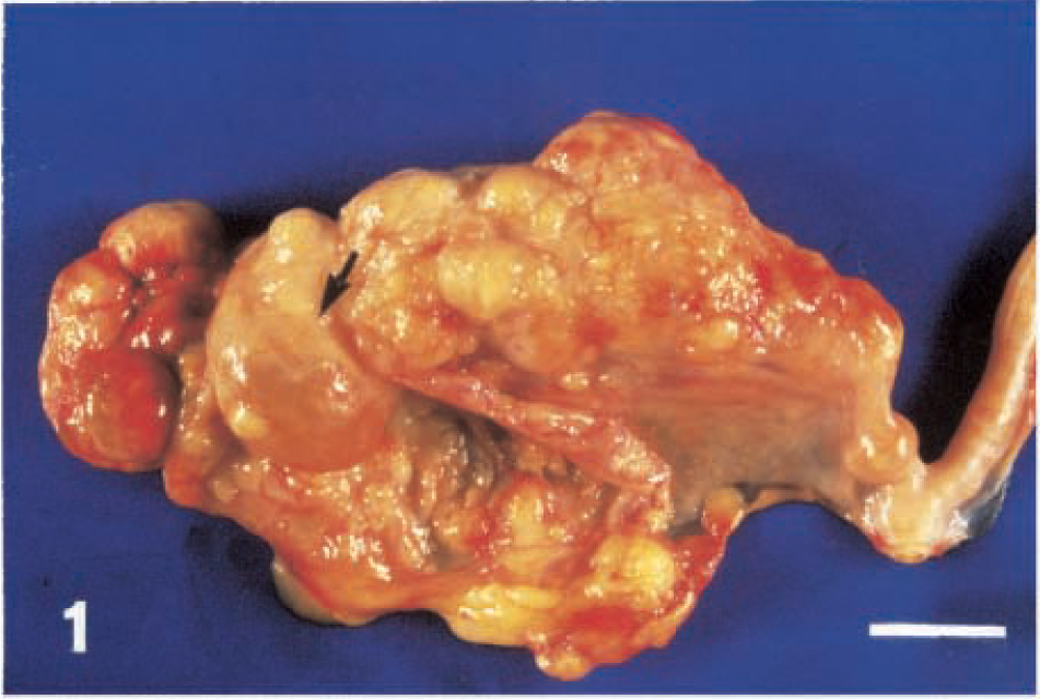
Uterus; cat. A vegetative and infiltrating MMT almost completely involves the right horn. The ovary (arrow) contains a large follicular cyst. Bar = 1.7 cm.
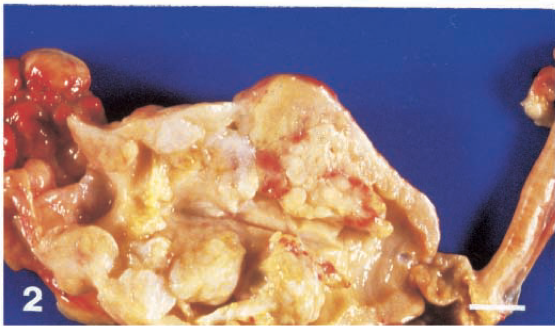
Uterus; cat. A polypoid mass fills the uterine cavity, replacing the uterine wall almost to the level of the serosa. Bar = 1.3 cm.
Several samples of the tumor in the uterine wall and extragenital nodules were excised, fixed in 10% neutral buffered formalin, and embedded in paraffin. Sections (4–5 µm thick) were stained with hematoxylin-eosin-orange and van Gieson's for light microscopy. Immunohistochemical staining was performed on serial sections using primary monoclonal antibodies to vimentin (VIM; clone 3B4), diluted 1:250 (Ylem, Avezzano-Aquila, Italy); cytokeratin (CK; clone AE1-AE3), diluted 1:40 (Ylem); and MIB-1 (trademark of the anti-Ki-67 antibody, clone MIB-1), diluted 1:100 (Amac Inc., Westbrook, ME). For MIB-1 immunohistochemistry, the slides were microwaved twice for 5 minutes. The ABC system (avidin–biotin–peroxidase complex, Vector Laboratories, Burlingame, CA), Vector VIP (Vector Lab) as chromogen substrate, and Mayer's hematoxylin or methyl green as counterstain were employed. Mitotic figures (MFs) per thousand cells and the percentage of MIB-1–positive nuclei were also determined by simple methods, 8 to quantify the mitotic index (MI) and the MIB-1 labeling index (MIB-1-LI).
Light microscopy showed intermingled carcinomatous and sarcomatous components (Figs. 3, 4). Carcinomatous cells were arranged in solid or papillary structures and displayed occasional necrotic areas. The malignant cells were medium or large in size. They had abundant eosinophilic cytoplasm and vesicular nuclei, with prominent nucleoli. Many cells were pleomorphic. This component of the tumor resembled a poorly differentiated endometrioid carcinoma. In contrast, the sarcomatous component consisted of closely packed round and polygonal cells with vesicular nuclei, resembling endometrial stromal sarcoma cells. Necrotic areas and hemorrhages were present in these areas. MFs were frequent in both components, with scores of 14 and 12 per thousand cells, in carcinomatous and sarcomatous areas, respectively. Metastatic endometrial carcinoma cells were found in myometrial lymphatic and blood vessels. Strong diffuse CK-immunostaining was evident primarily in the carcinomatous component (Fig. 5), whereas sarcomatous cells were VIM-positive. The MIB-1-LI scores were estimated to be 13.6 and 29.1, respectively, in the carcinomatous and sarcomatous components (Fig. 6).
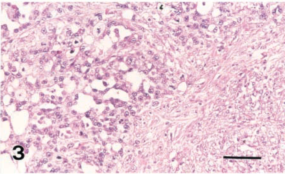
Uterus; cat. Carcinomatous component of MMT. Hematoxylin-eosin-orange. Bar = 65 µm.
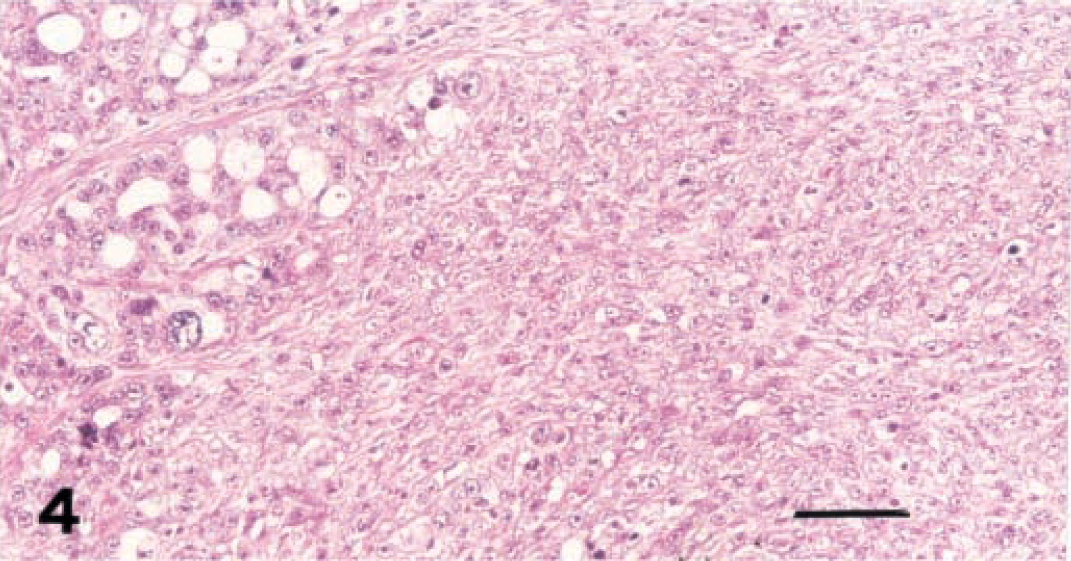
Uterus; cat. Sarcomatous component of MMT. Hematoxylin-eosin-orange. Bar = 65 µm.
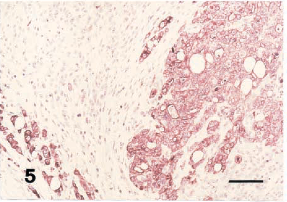
Uterus; cat. Strong cytokeratin immunostaining in the carcinomatous component of the MMT. Avidin–biotin–peroxidase complex method. Mayer's hematoxylin counterstain. Bar = 65 µm.
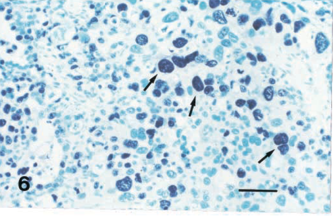
Uterus; cat. Frequent MIB-1–positive nuclei (arrows) can be seen in carcinomatous and sarcomatous components of the MMT. Avidin–biotin–peroxidase complex method. Methyl green counterstain. Bar = 36 µm.
Young female cats commonly undergo ovariohysterectomy, so that the real incidence of uterine tumors cannot be estimated. However, occasional uterine malignancies have been reported in older cats (over 8 years of age); these are mostly endometrioid adenocarcinomas. 13 Uterine carcinosarcomas or malignant MMTs are unusual neoplasms in the cat. To the best of our knowledge, only three such cases have been reported to date; 2,4,9 this is the fourth case of uterine MMT to be reported. The tumor is now regarded as a fast growing metaplasic carcinoma 5,6,12 that deeply invades myometrium and is similar to the corresponding human tumor. It is significant that the MI and MIB-1-LI were found to be very elevated in both the carcinomatous and sarcomatous components in the present case. This suggests a high growth fraction, which is known to predict an unfavorable clinical course. Concurrent follicular cysts, cystic endometrial hyperplasia, and pyometra are often found in feline uterine tumors, 1,10,13 suggesting a possible hormone dependence for such malignancies. Such a dependence has also been implicated in similar tumors of rabbits 3 and of humans. 14 In conclusion, occasional cases of malignant uterine neoplasms previously have been reported in the cat, and most of them were highly aggressive adenocarcinomas. The lesion in the present case was an endometrioid adenocarcinoma with a metaplasic component of endometrial stromal sarcoma. On the basis of its high MI and MIB-1-LI, this case of uterine MMT has to be regarded as a metaplasic endometrial malignancy. The present case, in which there was a high proliferation rate and poor clinical outcome, was very similar to its human counterpart.
Footnotes
Acknowledgements
We thank Rosario Duca for his technical assistance.