
Abstract
In the spring of 2003, a series of sudden deaths in a group of adult lions (Panthera leo) with a previous history of depression, inanition, and lethargy, was investigated. Five animals died within 24 to 36 hours after onset of signs of disease. Serologic screening for viral disease detection was negative, evidence of parasites was not detected, and results of a complete blood count and serum biochemical analysis were within reference intervals in all lions. The most relevant lesions observed were multiple areas of necrosis and hemorrhage in the intestinal outer muscular layer, and cellulitis with an intense bloody edema in the mesenteric and the pericardial fat tissue. On the basis of the fulminant course of the disease, the gross and histologic findings, and the isolation and identification of Clostridium sordellii, a diagnosis of infectious myositis and cellulitis associated with acute clostridiosis was made. To the authors' knowledge, this is the first report of sudden death associated with C. sordellii in felines.
Keywords
The common natural habitats for Clostridium spp. are soil, water, and the gastrointestinal tract of animals and humans. 14, 15 Various species such as C. tetani, C. perfringens, C. botulinum, C. septicum or C. bifermentans are commonly isolated as infective agents from human and animals. 2 Most enteric clostridial diseases caused by toxigenic types of C. perfringens usually develop in herbivores. Occasionally, other members of the genus can be associated with disease of the alimentary tract, causing blackleg-like myositis. 2 Only a few descriptions of clostridial disease in felines have been reported in veterinary literature. Clostridium perfringens was identified from an enterotoxicosis outbreak in a collection of cheetahs in Florida; 3 dual infection with C. piliforme and feline panleukopenia virus (FPLV) in three kittens has been described; 8 C. botulinum has been isolated from a group of cats fed pelican carrion; 6 and recently, C. difficile has been associated with acute diarrhea in adult cats. 18
Clostridium sordellii is a gram-positive, anaerobic bacillus reported as an unusual cause of clostridial myositis in humans and animals. 1, 4, 10, 13, 16, 19 It has been associated with malignant edema, myositis, hemorrhagic enteritis, and sudden death in cattle and sheep. 1, 10, 16, 17, 19 To our knowledge, previous descriptions of clostridiosis associated with C. sordellii have not been reported in felines.
We describe the clinical and pathologic features of acute clostridial enteritis associated with myositis in a group of captive lions. The disease resulted in sudden death of all affected animals.
Between March and April of 2003, five of nine adult lions (two males, three females) located in The Tabernas Desert Zoological Reserve (Almería, Spain) developed signs of depression, lethargy, loss of appetite, and prostration. Despite medical treatment with intravenously administered fluid therapy and antibiotic therapy (Ringer's Lactate solution, Braun, Melsungen, Germany; marbofloxacine, Vétoquinol Laboratories, Lure, France) death occurred within 24 to 36 hours after onset of clinical signs of disease. The other four lions received a preventive oral antibiotic (marbofloxacine) and did not manifest evidence of systemic disease.
Blood samples were collected from all lions (n = 9). The complete blood count and serum clinical biochemical parameters for the affected group were within reference intervals. Serologic screening for feline leukemia virus, feline immunodeficiency virus, feline coronavirus, feline panleukopenia virus, feline calcivirus, feline herpesvirus, and Toxoplasma gondii was performed (n = 9), but results were negative. Examination of freshly obtained fecal smears was negative for presence of parasites (n = 9).
Complete necropsy was performed immediately after the five lions died. The serosal surfaces were pale gray and glistening, with obvious edema located in the subserosal spaces (n = 5). Multifocal to moderately extensive dark-red-to-black hemorrhagic areas associated with the duodenal and jejunal external muscular layer and serosa were clearly demarcated (n = 4; Fig. 1a). The intestinal mucosa was dark-red in color, with a bloody mucous material in the lumen (n = 4). The pericardial and mesenteric fat tissue was hemorrhagic and edematous. There was marked congestion in liver, lungs, kidneys (n = 5), and adrenal glands (n = 2).
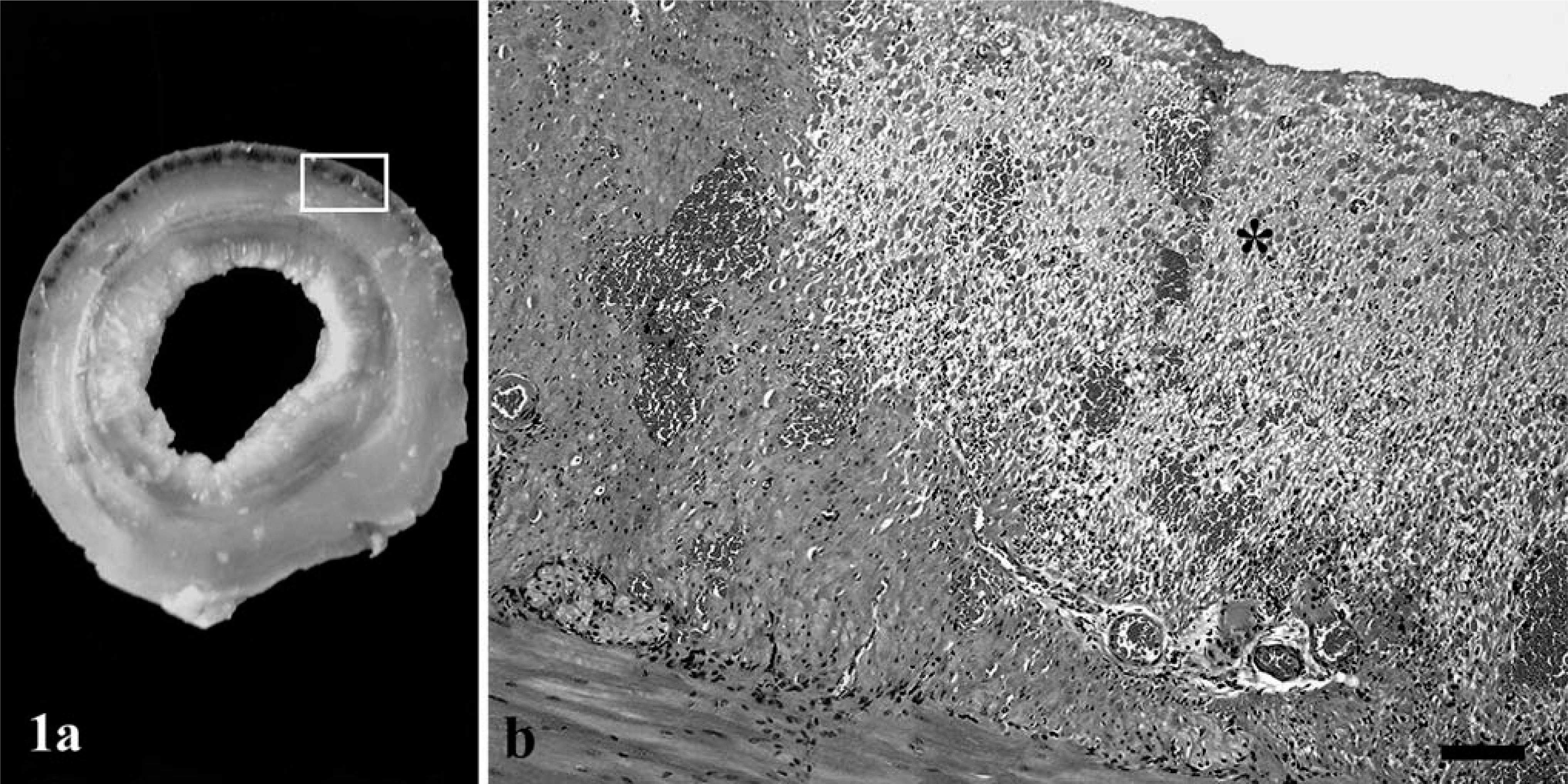
Small intestine; lion (Panthera leo).
Several tissues including small and large intestines, stomach, liver, spleen, kidney, heart, and lung were collected for bacteriologic culture. All samples were removed from the carcass, inoculated onto blood agar base (added to 5% defibrinated blood), and MacConkey's, Baird Parker, and Saboureaud agars by use of standard methods, and were incubated under aerobic conditions at 37°C for 24 hours. Anaerobic culture was performed as well. The specimens were grown on fastidious anaerobic agar and trypticase sulfite neomycin (TSN) agar (Becton, Dickinson and Company, Franklin Lakes, NJ) and were incubated at 37°C for 48 hours and 6 days, respectively. Tissue specimens were also fixed in neutral-buffered 10% formalin, trimmed, processed in routine manner, embedded in paraffin wax, sectioned at 5-µm thickness, and stained with hematoxylin and eosin (HE) and Gram's stain (Brown and Brenn modified method). In addition, impression smears were made from the gastric and the intestinal mucosa and were stained by use of Gram's method.
Histologic evaluation of sections from the small intestine revealed extensive superficial and a few deep areas of necrosis in the mucosa of the duodenum and jejunum (n = 4; Fig. 2). The intestinal villi were edematous, with mild-to-intense infiltration of neutrophils, macrophages, and plasma cells (n = 4). A few lymphocytes also were present in deeper areas of the mucosa. The intestinal epithelium was missing in the apical two-thirds of the villi (n = 4; Fig. 2). Occasionally, dilatation of the intestinal crypts and marked congestion of the mucosal and submucosal blood vessels were observed as well. The necrotic mucosal lesions in the small intestine were associated with the presence of large gram-positive rods, which also were numerous in the lumen (n = 5). The stomach had large colonies of the same organisms in the gastric glands and free in the lumen (n = 5). Multifocal to locally extensive areas of necrosis in the external muscular layer of small intestine associated with multiple areas of hemorrhage that occasionally coalesced were consistent features (n = 4; Fig. 1b). A clear edema poor in neutrophils was noted among the necrotic muscle fibers and was intensely stained by eosin (n = 4; Fig. 1b). Similar poor-cell edema was observed in the fat tissue of mesentery (n = 5). An intense inflammatory exudate characterized by fibrin-rich edema and numerous neutrophils was observed on the pericardial fat tissue (n = 5; Fig.3). The inflammation also extended into the adjacent epicardium penetrating along the connective tissue septa between the muscular bundles (n = 3). Other minor findings were a discrete microvacuolar degeneration of hepatocytes and hemosiderin storage in Kupffer cells (n = 1). Impression smears from the intestine and stomach showed the massive presence of gram-positive bacilli.
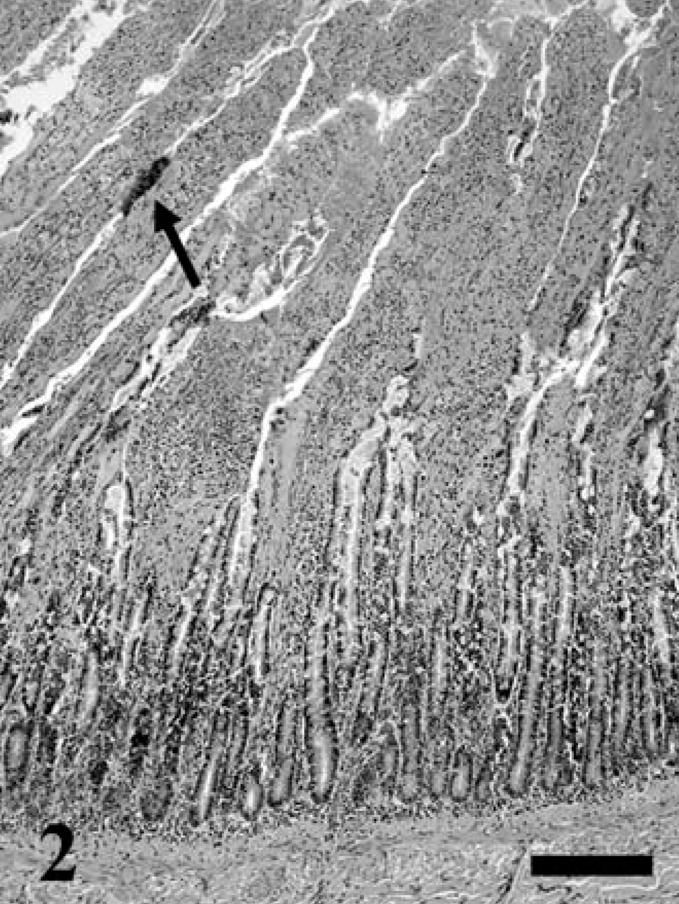
Small intestine; lion (Panthera leo). Photomicrograph of a section of the jejunal mucosa showing edema and infiltration of inflammatory cells in the lamina propria of the intestinal villi with necrosis and loss of the intestinal epithelium. Notice the bacterial colonies associated with the intestinal villi (arrow). HE. Bar = 80 µm.
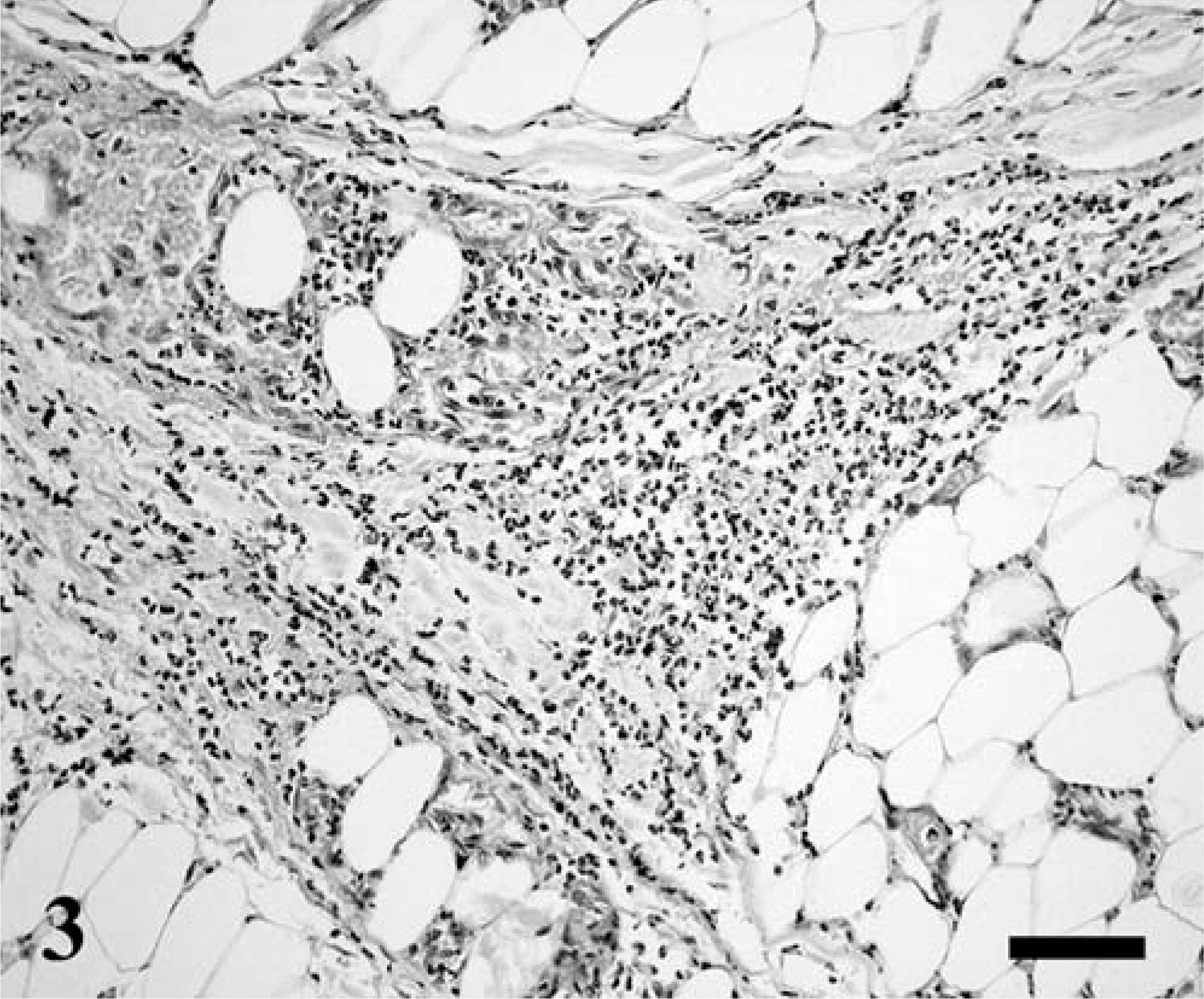
Pericardial fat tissue; lion (Panthera leo). Photomicrograph of a section showing the edema and the neutrophil-rich exudate in the connective septa of the pericardial fat and among the adipocytes. HE. Bar = 60 µm.
Aerobic culture failed to yield any known pathogens in all cases (n = 5). However, several black bacterial colonies were observed after 72 hours in TBS cultures inoculated with samples of stomach, small and large intestines, liver, and kidney. These bacterial colonies were invariably present in culture from samples of the stomach and small intestine in every animal of the affected group. The colonies were subcultured for purity and were identified by use of an enzymatic identification system for anaerobes (Rapid ID 32 A, Bio Mérieux, Marnes-la-Coquette France) in the Microbiology Service of the Dr. Negrín Hospital, Las Palmas GC, Spain. All colonies were identified as C. sordellii, with reliability close to 99% (positive test responses to indole production, urease presence, and activity for proline arylamidase). Food samples (raw meat) and water administered to the animals also were analyzed. All food samples were negative for bacteria, but a large concentration of C. sordellii was found in the drinking water.
On the basis of the gross and histologic findings and isolation of C. sordellii from all five lions, a diagnosis of sudden death associated with acute clostridial enteritis, myositis, and cellulitis was made.
C. sordellii was first isolated from a case of human postoperative gas gangrene by Sordellii in 1922. 10 Since then, it has been reported to cause sporadic cases of intestinal disease or infectious myositis, or both. 1, 4, 10, 12, 13 Ruminants, horses, and swine are highly susceptible, whereas carnivores are rarely affected. 7 Two forms of clostridial myositis can be distinguished: 1) a nongangrenous form or malignant edema characterized by serous exudate and cellulitis rather than by myositis; and 2) a gangrenous form or gas gangrene with extensive disintegration of muscles and gas production. 7, 11 The muscles, especially if devitalized in some manner, are highly susceptible to bacteria of the genus Clostridium, and these organisms, when they proliferate, are highly toxigenic and cause cellulitis, extensive necrosis of muscle with blood-stained edema, and formation of gas. 7, 11 Gangrene or gas production in the tissues was not found in the lions at necropsy. In addition, necropsy was performed immediately after each animal died. Thus, the lesions observed were induced by the isolated pathogen rather than being attributable to postmortem changes. On the other hand, the nongangrenous form is more common than the gangrenous form. 7 The findings in the affected group of lions were more consistent with a nongangrenous or edematous form, especially the lesions found in the fat tissue of the pericardium and mesentery.
C. sordellii produces two toxins, the hemorrhagic toxin and the lethal toxin, which contribute to the progressive edema, myonecrosis of skeletal muscle, and refractory shock frequently associated with these infections. 10, 16, 19 C. sordellii organisms were not seen in the nondigestive tract lesions. A possible explanation for this fact might be a nonbacteremic (toxigenic) presentation of the disease in the animals. Several cases, either bacteremic C. sordellii infection or nonbacteremic infection, have been reported. 16 Usually, clinical signs of toxemia are lethargy, prostration, circulatory collapse, and sudden death. 4, 7, 10, 16 These signs were noted in all lions affected (n = 5) within 24 to 36 hours before death. In fact, it is not uncommon for animals to die within 24 hours of the onset of local signs as a result of systemic intoxication. 7 Mortality is correlated with shock at presentation and affects about 50% in the bacteremic form and 71% in the nonbacteremic form. 16 Moreover, the pathogens were isolated from liver and kidney as well. This may suggest systemic invasion with consequent bacteremia. Invasion, sometimes massive, of the circulation usually occurs shortly before death; thus, the offending organisms can then be isolated from most tissues. 7
The isolation of C. sordellii from the stomach, intestine, and other tissues in the absence of other pathogens, in concert with the gross and microscopic pathologic changes, might suggest it to be a primary pathogen. Large amounts of gram-positive, clostridial-shaped bacteria seen in the stomach of the affected animals, bacterial colonies grown in TSN cultures inoculated with samples of this organ, and bacteria isolated from the drinking water of the affected group suggested a possible external origin. C. sordellii is ubiquitous and distributed widely in the environment similar to other clostridia. 9, 10, 12 Their natural habitats are soil, water, and the gastrointestinal tract of animals and humans. 14, 15 Although several species of clostridia can be part of the normal flora in the distal portion of the gastrointestinal tract of various animals, the failure to isolate C. sordellii from many cases of clostridial disease and sudden death suggests that it is not a consistent inhabitant of the gastrointestinal tract. 10
On the other hand, exogenous diseases caused by clostridia from an external source are well known historically and are clinically important. 9, 12 They have even been isolated from diets including raw meat fed to animals. 5, 18 We were not able to find C. sordellii in the food samples analyzed, but an important number of them were isolated from the drinking water of the affected group. Careful examination of these cases suggests that the lions ingested contaminated water containing the bacteria. C. sordellii is not a member of the normal gastric bacterial flora; 10 thus, the presence of this organism in the stomach may be indicative of oral ingestion. Because the lions had showed aggressive behavior among them, they were previously separated into two groups. C. sordellii was isolated only from the drinking water of the group affected. Other drinking water places or food samples analyzed did not indicate presence of pathogens, which means that the water could be the most probable vehicle for C. sordellii, as has been reported previously. 14
This report represents, as far as we have been able to find, the first case of myositis and sudden death associated with C. sordellii in felines. C. sordellii are agents to be suspected as pathogens in animals with signs of infectious myositis.