
Abstract
A healthy, 3-year-old, intact male alpaca (Llama pacos) died suddenly and unexpectedly and had an unusual pattern of hypertrophic cardiomyopathy that was restricted to the interventricular septal and right ventricular myocardium. Grossly pale areas in the affected myocardium corresponded histologically to thickened and disorganized myofibers with excessive branching and marked nuclear pleomorphism. Mitoses were absent. Inflammation and fibrosis were minimal. A few thickened muscular arteries were scattered in the affected myocardium.
South American camelids are becoming increasingly popular in the United States for their use in a wide range of products and services. 7 In 2007, more than 13,000 alpacas (Llama pacos) were raised on approximately 2,500 farms throughout the United States (2007 Census of Agriculture—State Data, National Agricultural Statistics Service, U.S. Department of Agriculture, available at http://www.agcensus.usda.gov/Publications/2007/Full_Report/index.asp). Reports of sudden death associated with cardiovascular disease in alpacas are rare. In the current report, an alpaca that died suddenly with evidence of cardiomyopathy is described.
A 3-year-old, 72-kg, intact male alpaca was submitted for necropsy with a history of unobserved death in late spring 2007. The alpaca had been seen alive and well 12 hr prior to being found dead in a dry lot. Four other alpacas in the same lot and 49 other alpacas on the farm with fence-line contact with this alpaca were clinically unremarkable. This alpaca had been sedated and shorn approximately 2 weeks prior to death and had no history of health problems. Diet consisted of dried grass hay, mostly orchard grass, a small amount of commercially prepared grain supplement without coccidiostats, and free-choice mineral block. Water and feed sources had not changed in more than 2 months prior to death. The body condition score of this alpaca and of other alpacas on the farm was excellent (4/5). All adults on the farm were treated every 3 months with a parasiticide a and a commercial Clostridium vaccine.
A full necropsy was performed. The heart weighed 495 g, with a heart-to-body weight ratio of 0.69% (0.82% is normal for alpacas slaughtered in the Andean highlands). 3 In the unopened heart, the apical two thirds of the right ventricular wall was mottled pale (Fig. 1). The right ventricular free wall was moderately thickened. The average wall thickness of the right ventricular free wall was 1.3 cm, compared with the left ventricular free wall, which had an average thickness of 2.5 cm (left ventricular: right ventricular ratio of <2:1). On cross section, the interventricular septum was similarly thickened. Both right ventricular free wall and the anterior and posterior portions of the interventricular septum contained transmural, poorly demarcated pale areas that did not bulge on cut surface and had the same consistency as more normally colored myocardium of the left ventricular free wall (Figs. 1, 2). Other areas of the heart, including endocardium, epicardium, epicardial vessels, both atria, the pulmonary outflow tract, pulmonary artery, left ventricular myocardium, aortic outflow tract, aorta, and both atrioventricular valves, were unremarkable. Pulmonary arteries were dissected to 2-mm branches and were free of fibrin thrombi. The lungs were diffusely dark red, wet, and heavy, and pink froth partially filled the trachea. Clear fluid oozed from the cut surfaces of the lung. No gross lesions were identified in other tissues or organs.
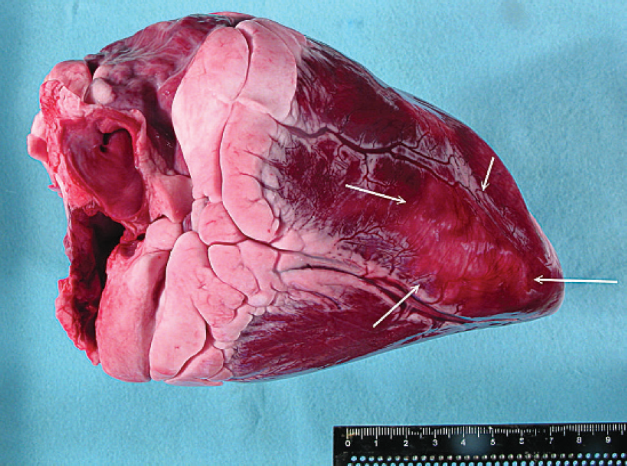
Heart, alpaca (Llama pacos). Pale area outlined by arrows in the right ventricular myocardium.
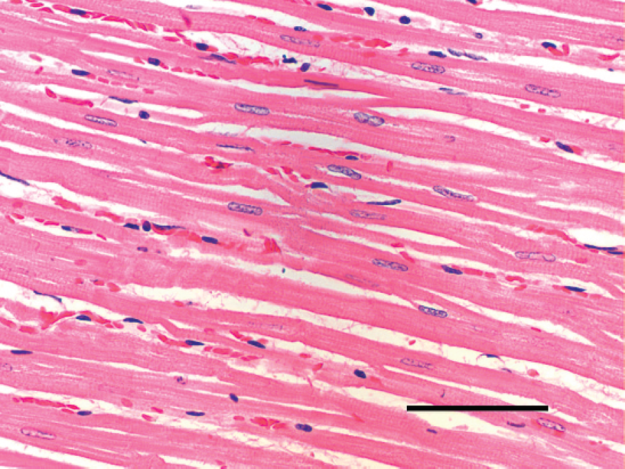
Histologically, sections of the pale areas of the right ventricular free wall and interventricular septum appeared similar. The primary change was marked disorganization of enlarged myocytes. Myocytes were in disarray, intermixed at oblique angles, excessively branched, and minimally separated by clear spaces (edema; Fig. 3). Longitudinal myofibers in the affected areas varied markedly in diameter but were often enlarged with myofiber diameters >15 μm, compared with the unaffected left ventricular myocardium, which had typical myofiber diameters <12 μm. Similarly, the myofiber diameters from 8 other alpaca hearts from historical controls (alpacas that died of nonheart conditions such as trauma and sepsis) were generally <12 μm (Fig. 4). Affected areas had moderate anisokaryosis. Myocyte nuclei were usually more rounded, vacuolated, or hyperchromatic and generally >7 μm in diameter compared with nuclear diameters of 5 μm or less in the unaffected left ventricular myocardium (Fig. 5). Myocyte nuclei from the 8 control hearts were generally <5 μm in diameter. In affected and unaffected myocytes, mitoses were absent. Fibrosis between myocytes, based on Masson trichrome staining of the myocardium, was minimal, and inflammation was absent. A few medium-sized intramural coronary arteries had mild medial hypertrophy with luminal narrowing. No significant changes were identified in the right and left atrial myocardium. Pale eosinophilic proteinaceous fluid filled most pulmonary alveoli, but there was no cellular response to the fluid, indicating pulmonary edema was an acute terminal event.
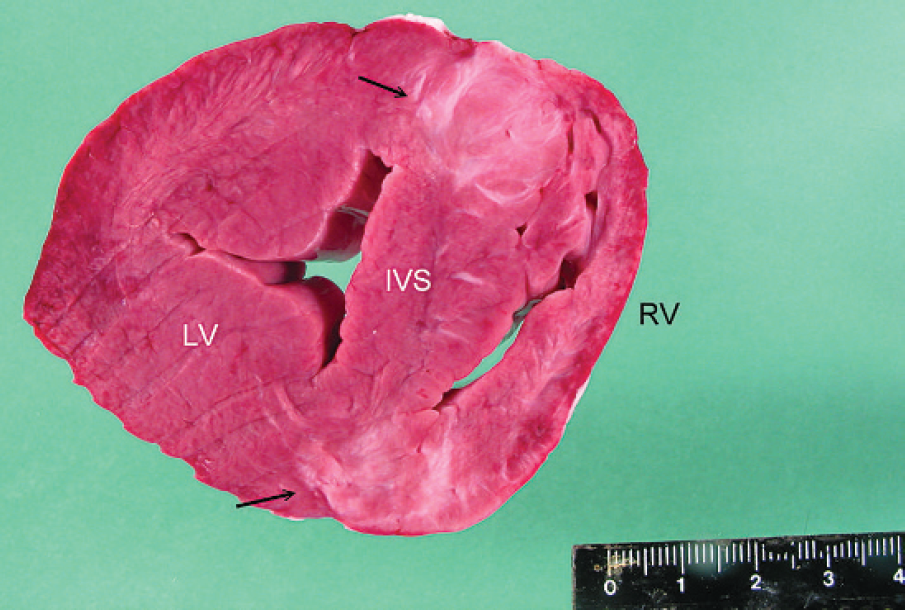
Heart, alpaca (Llama pacos). Cross section at the level of the right papillary muscle. Overall, the right ventricular myocardium (RV) and interventricular septum (IVS) had a variegated pattern, with some areas of the anterior and posterior septal wall (arrows) more pale than others. The left ventricular myocardium (LV) was unremarkable.
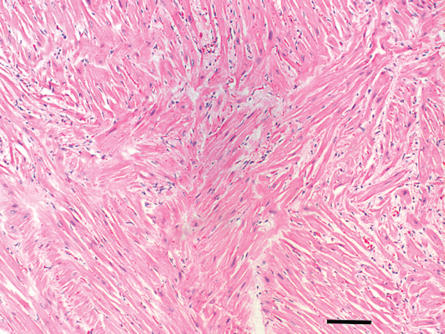
Heart, alpaca (Llama pacos). The affected right ventricular myocardium has marked myocyte disarray characterized by thickened myocytes intermixed at oblique and, sometimes, right angles. Hematoxylin and eosin. Bar = 100 μm.
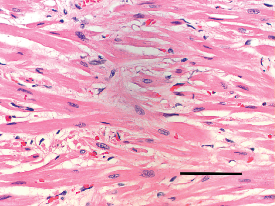
Heart, alpaca (Llama pacos). Affected right ventricular myocardium. Variably sized, often enlarged, myocytes branch excessively and are separated by scant clear spaces (edema). Pleomorphic nuclei are often rounded and enlarged compared with nuclei in the unaffected left ventricular myocardium. Hematoxylin and eosin. Bar = 100 μm.
Heart, alpaca (Llama pacos). Left ventricular myocardium with fibers in orderly longitudinal arrangement. Hematoxylin and eosin. Bar = 100 μm.
Cardiovascular diseases are uncommonly reported in South American camelids. 7 The most commonly reported conditions are congenital heart diseases (septal defects, valve dysplasia, and vascular anomalies) and acquired cardiac diseases (nutritional myopathy, cardiotoxicity, and bacterial endocarditis and myocarditis). 7 Primary disease of the myocardium is rare. Two cases of dilated cardiomyopathy were reported in young crias. 7 To the authors' knowledge, this is the first report of asymmetrical hypertrophic cardiomyopathy (HCM) in an alpaca.
Hypertrophic cardiomyopathy is a relatively common cardiac disease in many species, including humans and cats. 2,4,5 In ruminants, HCM has been reported in 2 adult Holstein cattle. 6 In humans, cats, cattle, and most domestic species with HCM, hearts are usually large and rounded with biventricular involvement, and the most severe hypertrophy is on the left side of the heart. Fibrofatty changes that are predominantly, but not exclusively, on the right side of the heart occur in arrhythmogenic right ventricular cardiomyopathy, a familial cardiomyopathy in Boxer dogs. 1 The lesions in the alpaca in the present report were unique compared with other species. In this alpaca, myocardial alterations were restricted to the interventricular septum and the right side of the heart. Microscopically, myocyte hypertrophy and disarray are hallmark features of HCM in all species. Myocardial fibrosis, endocardial thickening, and mineralization are also very common changes in most species but were not important features in this alpaca.
Similar to the clinical course observed in this otherwise healthy young adult alpaca, HCM is often a clinically silent disease in humans and domestic animals, resulting in sudden, unexpected cardiac death, especially in young adult athletes. 4 In humans, HCM is an autosomal dominant genetic disease with incomplete penetrance. 4 An autosomal dominant mode of inheritance has been demonstrated in cats. 5 On the well-maintained farm in the current case, herd health was high, and health records were sufficient to confirm that no other siblings or half siblings from the sire and dam of this alpaca were reported to have died unexpectedly or suffer from symptoms of congestive heart failure. Whether HCM is an inherited condition in camelids is as yet unknown.
Footnotes
a.
Dectomax®, Pfizer Animal Health, Groton, CT.