
Abstract
A spontaneous case of renal tumor was observed in a 7-year-old ovariectomized female pet ferret (Mustela putorius furo). Clinical signs included exhaustion, emaciation, anorexia, and stooping position. At necropsy, a solid and cystic mass replaced the left kidney and adrenal gland. The tumor was composed of pleomorphic epithelial cells with a large number of giant cells. Metastases were recognized in the lung, liver, greater omentum, right renal pelvis, and systemic lymph nodes. immunohistochemical stains revealed that the tumor cells were positive for CD10, cytokeratin (CAM 5.2), and Ki-67 (MIB-1). on the basis of morphologic and immunohistochemical features, the tumor was diagnosed as a pleomorphic renal adenocarcinoma. This type of neoplasm is very rare in all species and has never been reported in a ferret.
The ferret (Mustela putorius furo) has become increasingly popular as a pet in Japan. Endocrine tumors are the most common neoplasm in ferrets, 1, 2, 4, 6 and renal tumors are very rare; only a few cases have been reported. 3, 6 The purpose of this report is to describe what is believed to be the first case of spontaneous, pleomorphic renal adenocarcinoma in a ferret.
An ovariectomized female pet ferret was caged singly and provided with a general diet and tap water. The animal died naturally at 7 years old. Clinical signs included exhaustion, emaciation, anorexia, and stooping position. The animal was autopsied directly after death. Several organs and tissues were removed and fixed in 10% phosphate-buffered formalin at room temperature, embedded in paraffin, sectioned, and examined histopathologically.
Sections were routinely stained with hematoxylin and eosin (HE), and histochemically with periodic acid–Schiff (PAS), colloidal iron, and alcian blue. The sections were examined by standard avidin–biotin complex immunoperoxidase assays. After endogenous peroxidase activity had been blocked, deparaffinized sections were pretreated in 10 mM citrate buffer (pH 6.0) by microwaves, and incubated for 30 minutes at room temperature with primary antibodies diluted as follows: CD10 (1 ° 50; Novocastra, Newcastle upon Tyne, UK, anti-human, mouse monoclonal antibody, clone; 56C6), cytokeratin (CAM 5.2, diluted, 1 ° 1; Becton, Dickinson and Company, Franklin Lakes, NJ), Ki-67 (MIB-1, 1 ° 200; Immunotech, Westbrook, ME), myoglobin (1 ° 2,000; Dako, Carpinteria, CA), vimentin (diluted, 1 ° 1; Dako), desmin (diluted, 1 ° 1; Dako), chromogranin A (1 ° 1,000; Dako), SP-A (surfactant apoprotein A, 1 ° 100; Dako, anti-human, mouse monoclonal antibody, clone; PE10), 34βE12 (cytokeratin, 1 ° 50; Dako, anti-human, mouse monoclonal antibody, clone; 34βE12), AE1/3 (cytokeratin, 1 ° 50; Dako, anti-human, mouse monoclonal antibody, clone; AE1 and AE3), CEA (carcinoembryonic antigen, 1 ° 200; Novocastra, anti-human, mouse monoclonal antibody, clone; 12-140-10), CA125 (ovarian cancer antigen, 1 ° 100; Novocastra, anti-human, mouse monoclonal antibody, clone; Ov185 ° 1), calretinin (diluted, 1 ° 1; Zymed Laboratories, San Francisco, CA), insulin (diluted, 1 ° 3; Dako), glucagon (diluted, 1 ° 2; Dako), somatostatin (diluted, 1 ° 2; Dako), and serotonin (1 ° 50; Dako). Peroxidase-labeled polymer conjugated to goat anti-rabbit or mouse immunoglobulin (EnVision + System-HRP, Dako) was then applied for 30 minutes at room temperature. To visualize immunoreactivity, 10% 3,3′-diaminobenzidine tetrachloride in phosphate-buffered saline containing 0.02% hydrogen peroxide was used. The sections were then washed, counter-stained, dehydrated, cleared in xylene, and mounted. Positive immunohistochemical controls included appropriate cells of normal ferret organ with all primary antibodies evaluated.
Grossly, a mass, composed of a large solid mass (3 × 2.3 × 2 cm) and a cyst (3 cm in diameter), replaced the left kidney and adrenal gland. The border between the solid mass and the cyst was indistinct. The solid mass was spherical, grayish white, and firm. The cyst was spherical and covered by a thick capsule, and contained a yellow translucent fluid. No mass or nodules were seen in the right kidney or adrenal gland. A large number of multifocal nodules (1–5 mm in diameter) were found in the visceral pleura and parenchyma of both lungs, and hardly any normal lung tissue remained. A mass (1.5 × 1.2 × 0.7 cm) in the greater omentum and several multifocal nodules (2–3 mm in diameter) in the liver were found. All of these nodules were similar in gross appearance. Swelling of systemic lymph nodes (hilar, hepatic, portal, and lumbar) because of identifiable metastases was observed.
Histopathologically, the solid mass in the left kidney and adrenal gland was nonencapsulated, and was composed of several lobules showing a tubular pattern with lumen including cell debris and secretions and a sheetlike pattern (Fig. 1). The tumor cells were pleomorphic. The nuclei were large, spherical to polygonal, and hyperchromatic, and contained a single nucleolus or a number of nucleoli. The cytoplasm was abundant with indistinct margins. No vacuoles or lipid droplets were seen in the cytoplasm. A large number of giant cells were seen diffusely over an area of 30–40% of the solid mass, and most of these showed karyomegaly (Fig. 2). Mitoses and heterotypic mitoses were also seen diffusely. PAS-positive basement membrane was seen in the tubular pattern (Fig. 3). No smooth muscle was seen within the tumor. The large cyst was lined by columnar epithelium and supported by a thick fibrous capsule. The cyst was thought to originate from chronic hydronephrosis because of the gross content, the connection with the solid mass, and the columnar epithelium. Microscopic metastases, which had similar morphologic features as the primary tumor, were seen in the lungs, greater omentum, liver, right renal pelvis, and lymph nodes (hilar, hepatic, portal, and lumbar).
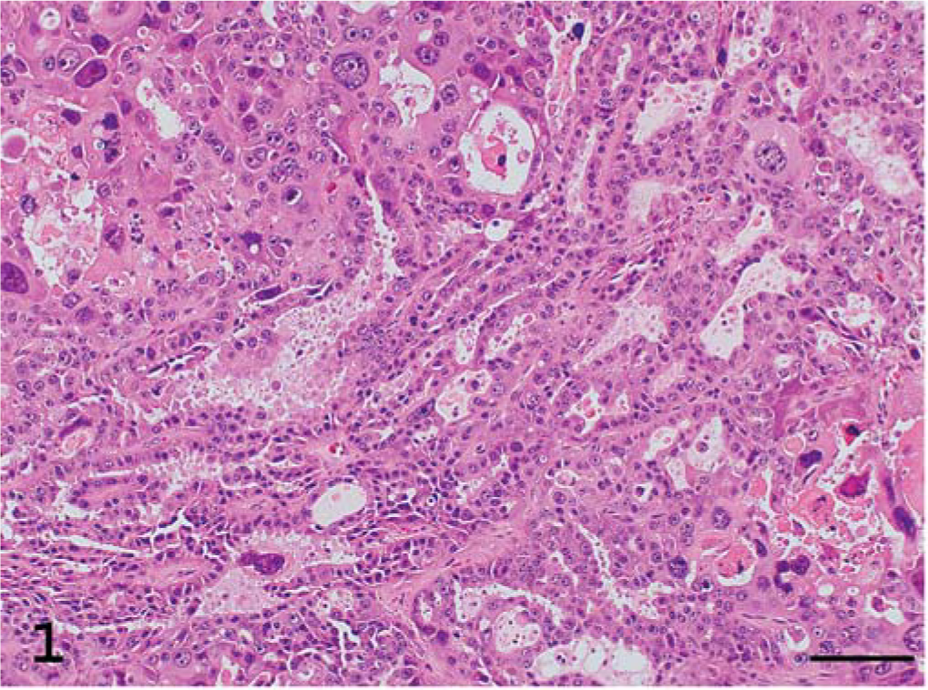
Kidney; ferret. Renal adenocarcinoma. The tumor is tubular and pleomorphic. HE. Bar = 100 µm.
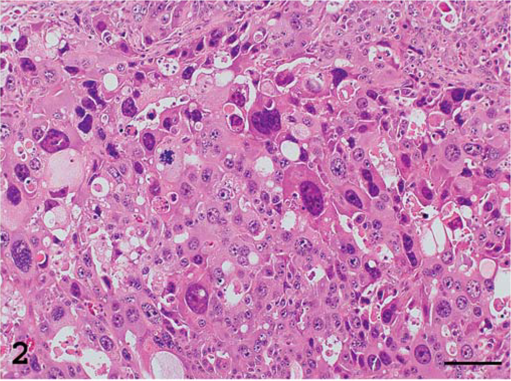
Kidney; ferret. Renal adenocarcinoma. Area with sheetlike pattern includes giant cells with karyomegaly, marked pleomorphism, and strong atypism. HE. Bar = 100 µm.
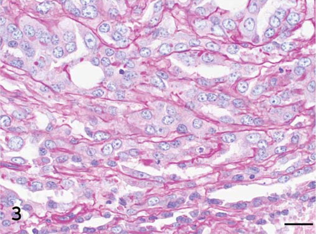
Kidney; ferret. Renal adenocarcinoma. The tumor is tubular with PAS-positive basement membrane. PAS. Bar = 20 µm.
Immunohistochemical and histochemical examinations (Table 1) revealed strong positive staining of CD10 and cytokeratin in the cytoplasm of epithelial tumor cells and giant cells, and positive staining of Ki-67 in approximately 60–70% of the nuclei of the epithelial cells and giant cells. Positive immunostaining was observed in the corresponding cells in a normal ferret organ with all primary antibodies evaluated, but myoglobin, vimentin, desmin, chromogranin A, SP-A, 34βE12, AE 1/3, CEA, CA125, and calretinin immunoreactivities in the tumor cells were negative. PAS was slightly positive, and colloidal iron and alcian blue staining were negative in the tumor cells.
Immunohistochemical and histochemical stainings of pleomorphic renal adenocarcinoma.∗
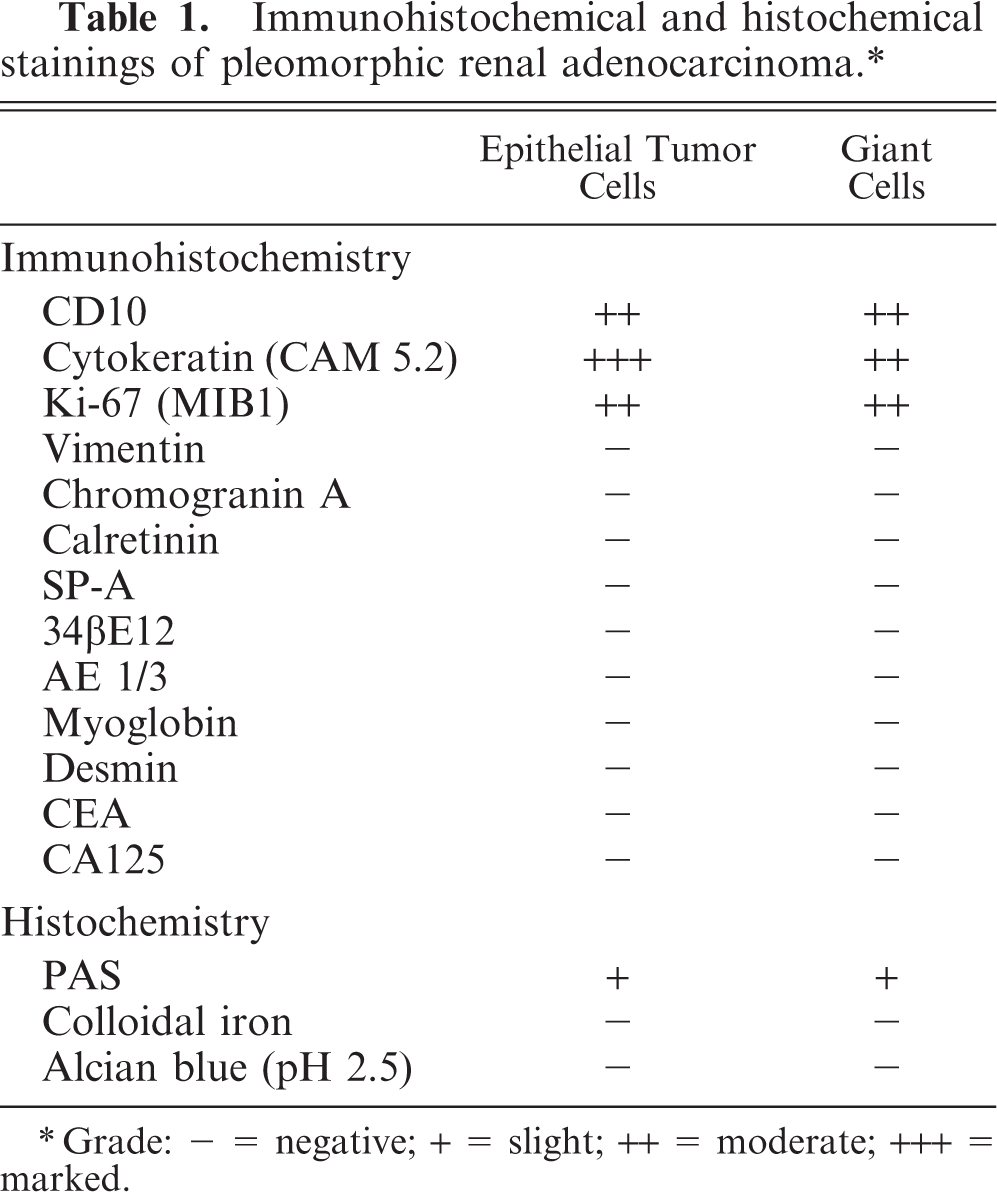
Grade: − = negative; + = slight; ++ = moderate; +++ = marked.
This tumor was confirmed to be a renal neoplasm based on strongly positive expression of CD10 and cytokeratin (Fig. 4), which is recognized in renal neoplasms, 5, 7, 10 and on the tubular pattern with PAS-positive basement membranes, which is a consistent histopathologic pattern. This was not thought to be an adrenal cortical tumor because no vacuoles or lipid droplets were seen in the cytoplasm of the tumor cells, no smooth muscle was seen within the tumor, and vimentin immunoreactivity was negative. 10 Adrenal medullary tumor, primary lung neoplasm, and mesothelioma were excluded based on immunohistochemical and histochemical features. Moreover, immunohistochemistry supports origination of the giant cells from epithelial cells that were transformed after becoming cancerous. Accordingly, using criteria for domestic animals, 9 the tumor was diagnosed as a renal adenocarcinoma. However, the histologic features of the epithelial cells in this tumor were considered similar to those seen in human renal granular cell carcinomas, and the giant tumor cells with karyomegaly were similar to the pleomorphic or bizarre features that are sometimes seen in the human renal cell carcinoma. 8 Subsequently, the tumor may be diagnosed as a pleomorphic renal adenocarcinoma in agreement with the strong pleomorphic features seen in this variant of renal carcinoma.
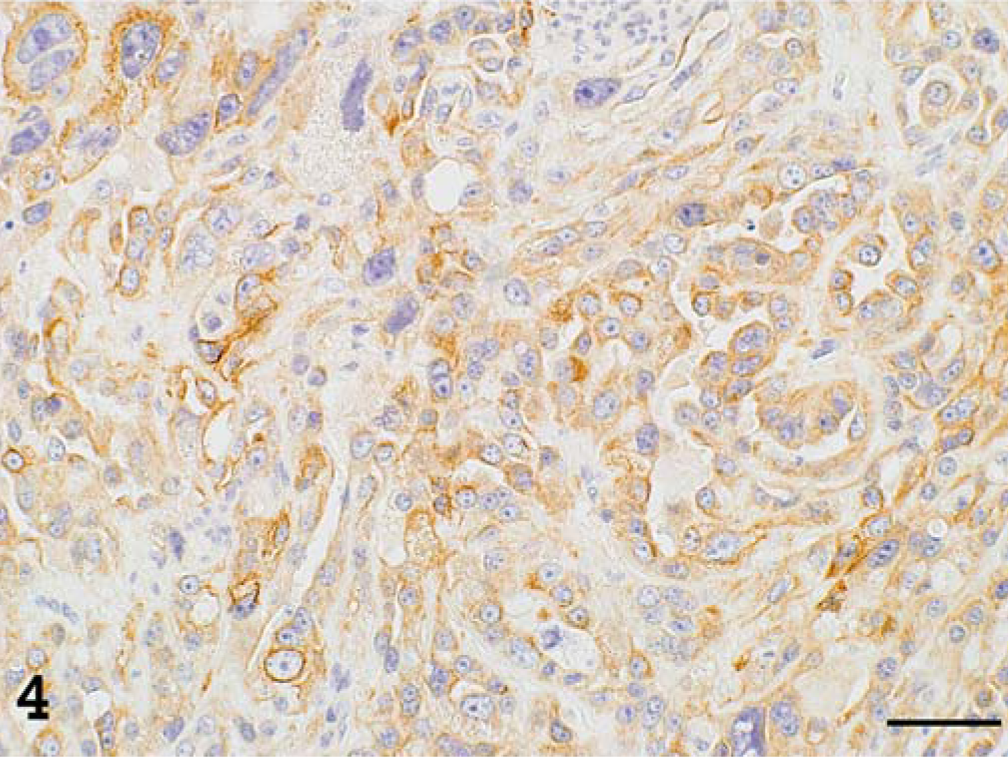
Kidney; ferret. Cytokeratin (CAM 5.2) immunoreactivity is strongly positive in the tubular cells and giant cells. Cytokeratin (CAM 5.2), avidin–biotin complex peroxidase method, hematoxylin counterstain. Bar = 50 µm.
In addition, an adenoma in the pancreatic islets and cortical hyperplasia in the right adrenal gland were found. The adenoma of the pancreatic islets was completely encapsulated by fibrous tissue and well-differentiated. Insulin immunoreactivity was strongly positive, glucagon immunoreactivity was negative, and both somatostatin and serotonin immunoreactivities were slightly positive in the adenoma cells.
In conclusion, pleomorphic renal adenocarcinoma is very rare in all species and never has been reported in a ferret. Moreover, in renal tumors in ferrets, a high Ki-67 index may be associated with metastasis and malignancy, and CD10, cytokeratin, and other immunoreactivities may be useful in diagnosis.