
Abstract
Neoplasia is one of the main causes of euthanasia in geriatric captive nondomestic felids. However, few studies have examined oral tumors in these animals. We describe here the clinicopathologic features of gingival squamous cell carcinoma (SCC) in 2 lions (Panthera leo) from separate zoologic collections. In both cases, the lions had a history of sialorrhea, bloody oral discharge, and anorexia. Autopsy findings in both lions were similar and were characterized by poorly circumscribed, friable, and bloody gingival masses with grossly apparent invasion of the mandibular bone; a pathologic fracture was observed in 1 case. Histologically, the masses consisted of poorly circumscribed, unencapsulated, densely cellular proliferations of neoplastic epithelial cells arranged in irregular islands, cords, and anastomosing trabeculae with formation of keratin pearls, which, coupled with positive immunohistochemistry for pancytokeratin, were diagnostic for SCC. Although no metastases were found in either animal, both lions were ultimately euthanized because of poor prognosis.
Recent advances in zoologic animal medicine have had a positive impact on the management of captive species, particularly in relation to the diagnosis of disease. This has contributed to an increase in the life expectancy of these animals, which in turn may in part have contributed to the higher prevalence of neoplasia observed over the past decades. 4 Neoplasia can represent up to 50% of diagnoses in nondomestic felids, and neoplasia of the digestive, reproductive, hemolymphatic, integumentary, and endocrine systems are among the most common reasons for euthanasia of Panthera spp.6,12,14 In lions, lymphoma, mammary adenocarcinoma and carcinoma, reproductive leiomyoma, and mesothelioma are among the most frequently reported tumors.10,14,17,20
Oral neoplasia is an important cause of illness in captive nondomestic felids and can represent up to 51% of lesions in the oral cavity. 21 These neoplasms have variable morphologic characteristics and clinical behavior, which may pose a diagnostic challenge for veterinarians. Squamous cell carcinoma (SCC) is one of the most common malignant oral tumors in both domestic and nondomestic felids.18,21 Predisposing factors for the development of oral SCC have not been well studied in nondomestic felids. In humans, there is a strong relationship between periodontitis and the development of oral SCC.15,22 Dental disease, which is one of the most commonly reported causes of morbidity in captive nondomestic felids, often goes undetected for extended periods of time.16,20 Dental disease, which is a broad term that typically refers to various lesions of the teeth (e.g., abrasion, tooth fracture, dental calculus), is often associated with other types of oral pathology, such as inflammation of the surrounding soft tissue(s) (e.g., periodontitis, gingivitis). Given the prevalence of chronic inflammatory conditions within the oral cavity of domestic felids, a similar relationship between chronic inflammation and oral neoplasia in other cats has been postulated. 28 We highlight here clinicopathologic features of gingival SCC in 2 lions and discuss potential predisposing factors for oral SCC in nondomestic felids, which, to our knowledge, has not been addressed.
Two geriatric lions from separate zoologic collections (referred to as L1, a 14-y-old Asian lion [Panthera leo], and L2, a 20-y-old African lion [P. leo]) were evaluated by veterinary staff for episodes of bleeding from the oral cavity. L1 had a history of gingivitis and dental disease with several extractions; differential diagnoses before examination included eosinophilic granuloma, trauma, progression of dental disease, and tooth root abscess. Oral examination of L1 while under anesthesia revealed marked loss of bone in the area of the mandibular symphysis; the incisors were held loosely in place by gingival tissue, not bone. The necrotic bone was covered by soft friable tissue, and the bulk of the lower right canine tooth was exposed. Radiographic findings were suggestive of a soft tissue neoplasm with invasion of bone. The abnormal tissue was biopsied, and histologic findings were consistent with SCC. This lion was subsequently euthanized given the poor prognosis.
For L2, when bleeding from the mouth was initially noted, the primary differential was trauma. When the bleeding did not resolve after 10 d, an oral examination was performed by veterinary staff. L2 had a 2.5 × 1.1 × 0.3-cm gingival neoplasm at the level of the left mandibular canine tooth (304). Over the course of the following 3 mo, radiographs revealed focally extensive invasion of the neoplasm into the mandible and, given the progressive wasting of the lion and the poor prognosis, this lion was also euthanized. Both lions were referred for autopsy immediately after death.
On autopsy, L1 was determined to be in excellent nutritional condition. A deep invasive 5.0 × 5.0 × 3.0-cm mass with raised and irregular margins was observed in the gingiva over the right rostral mandible and mandibular symphysis. All of the mandibular incisor teeth and right mandibular canine tooth were absent. A 2.0 × 1.5 × 1.0-cm wound was centered over a second firm, irregularly shaped gingival mass (5.0 × 3.5 × 2.0 cm), located in the right mandibular gingiva, caudal to the incisors.
L2, in contrast, was in poor nutritional condition and, on examination of the oral cavity, a 15 × 10 × 7.0-cm firm, white, ulcerated mass was noted on the oral mucosa adjacent to the mandibular left canine and incisor teeth (Fig. 1A). A biofilm covered the teeth. In the canine and incisor teeth, this alteration was mild-to-moderate and was mostly observed at the neck of the teeth. The premolar and molar teeth were completely covered by a brown-to-black biofilm. The left mandibular canine and incisor teeth were loose, and a complete transverse pathologic fracture at the level of the mandibular mental bulge was noted (Fig. 1B). No other significant gross abnormalities were observed in either carcass.
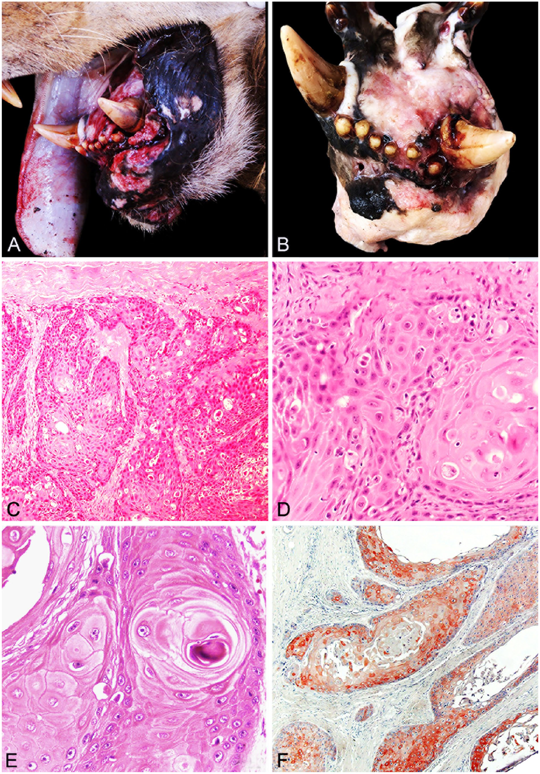
Gingival squamous cell carcinoma in 2 lions.
Samples of the masses, adjacent gingiva, regional lymph nodes, thyroid glands, parathyroid glands, lungs, stomach, small and large intestine, liver, pancreas, spleen, adrenal glands, kidneys, and brain were collected, fixed in 10% neutral-buffered formalin, and routinely processed for the production of H&E-stained sections. Immunohistochemistry for pancytokeratin (1:100, mouse anti-pancytokeratin, clone LU5, CM043C, BioCare; HIER steamer [S1699], 30 min; PBS-Tween, NHS) was also performed on selected specimens. Slides were counterstained with Mayer hematoxylin (Millipore Sigma). Negative controls were prepared by substituting the primary antibody with non-immune mouse serum.
The microscopic features of the gingival masses from both lions were similar and consisted of poorly circumscribed, non-encapsulated, densely cellular proliferations of neoplastic epithelial cells arranged in irregular islands, cords, and anastomosing trabeculae embedded in a dense fibrovascular stroma (Fig. 1C). The neoplastic cells were polygonal and had abundant pale eosinophilic cytoplasm (estimated nuclear:cytoplasmic ratio 1:3) with round-to-oval vesicular nuclei often containing 1 or 2 prominent nucleoli. Anisocytosis and anisokaryosis were moderate-to-marked (Fig. 1D). Mitotic count was 6 and 8 mitotic figures per 10 high-power fields in L1 and L2 (2.37 mm2), respectively. Keratin pearls and scattered dyskeratotic keratinocytes were prominent (Fig. 1E). Neoplastic epithelial cells infiltrated the surrounding bone. No identifiable odontogenic epithelium was observed. The cytoplasm of neoplastic cells labeled strongly for pancytokeratin using immunohistochemistry (Fig. 1F). Papillomavirus testing was not performed on either lion, either antemortem or postmortem.
SCC is the most common malignant oral neoplasm reported in domestic and nondomestic felids, accounting for > 60% and 80% of oral malignancies in these species, respectively.18,21 We conducted a literature search via Google Scholar, using the following search terms: oral, squamous cell carcinoma, tumor, neoplasia, lion, Panthera, and Panthera leo. In contrast to other large felids, oral SCC appears to be far less common in lions with only 4 cases of oral SCC reported in this species (Table 1).3,14,21 Papillomas are among the most common oral lesions reported in a number of captive nondomestic felids, including lions (Table 1). 21 Age, calculus deposition, papillomavirus infection, and concurrent dental and periodontal disease are considered to be the main predisposing factors for oral SCC development in Panthera spp.1,21,27 Most animals live longer in captivity than they do in the wild, and hence have a wider window of opportunity to develop neoplasia than their wild free-ranging counterparts. 4 Dental disease has been proposed to be a predisposing condition for SCC based on the fact that the region immediately adjacent to the maxillary and mandibular incisor teeth is an important site for the development of both periodontal disease and oral neoplasms.5,8,19,20 The formation of dental plaque, calculi, and periodontal disease appears to be more prevalent in captive tigers and lions fed a diet of raw beef, compared to free-living species. 13 The mechanical properties of food consumed by captive and wild nondomestic felids varies considerably; with the diet of wild nondomestic felids involving a larger amount of bone and connective tissue which may provide abrasion of the teeth that prevents the accumulation of dental plaque. 13 Therefore, the increased incidence of dental disease can likely be attributed to differences in diet of captive nondomestic felids compared with their free-living counterparts.5,13 Although we were not provided with information regarding the diet of L1, L2 often ate ground beef, beef bones, and whole dead rabbits. L1 had a history of chronic dental disease prior to diagnosis of oral SCC.
Cases and location within the oral cavity of oral tumors reported in African lions.
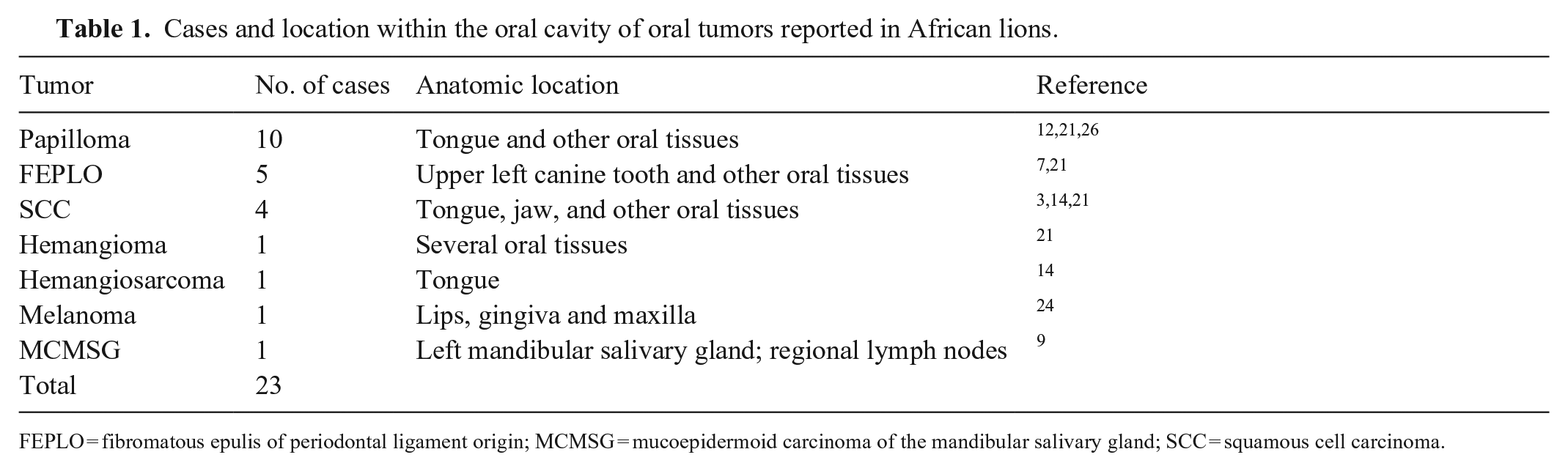
FEPLO = fibromatous epulis of periodontal ligament origin; MCMSG = mucoepidermoid carcinoma of the mandibular salivary gland; SCC = squamous cell carcinoma.
The tongue and mandible are important anatomic sites for the occurrence of SCC in felids.14,23,25 As is true for our cases, gingival SCCs often manifest grossly as one or more hemorrhagic and ulcerated masses, which rapidly increase in size and progressively infiltrate the surrounding soft tissues and bone(s).
In domestic and nondomestic felids, oral SCCs are generally considered to be locally invasive and poorly responsive to chemotherapy and radiation therapy. Surgery is the mainstay of treatment in domestic felids; however, extensive surgical revision (e.g., maxillectomy or mandibulectomy) is often not well-tolerated because cats typically do not adapt adequately post-operatively. 18 Although SCCs usually have a relatively low metastatic rate, oral SCC often leads to euthanasia due to clinical complications inherent to the location of the mass, such as pathologic fractures. In our cases, there was no gross or microscopic evidence of metastases to regional lymph nodes or other more distant organs, such as the lungs. In sporadic reports of radical surgical excision in other large carnivore species, treatment was often unsuccessful due to recurrence of the neoplasm. 2
Information regarding survival time in cases of oral SCC in lions is scant. However, as observed in the 2 lions reported here, oral SCCs can be extremely invasive and promote rapid clinical decline. The mean survival time of domestic cats with oropharyngeal SCC is reported as 151 d, which was longer than the surviving time for maxillary, mandibular, and sublingual SCC, for which the average survival times were 51, 34, and 33 d, respectively. 11 Hence, the anatomic site of occurrence might influence the survival rate for nondomestic felids with SCC. Metastases are rare in cases of oral SCC in nondomestic felids, although they may occur. 3 As in our cases, oral SCC is not typically detected in captive nondomestic felids until there is extensive local tissue invasion and destruction. Systemic evaluation of the oral cavity (including examination awake and under anesthesia, and intraoral dental radiographs) may facilitate early detection of dental disease and SCC. 16
Footnotes
Acknowledgements
We acknowledge and thank the animal care staff for maintaining these animals.
Declaration of conflicting interests
The authors declared no potential conflicts of interest with respect to research, authorship, and/or publication of this article.
Funding
This research was funded by Coordination for the Improvement of Higher Education Personnel (Brazil) grant 001.