
Abstract
A 10-year-old, female, mongrel showed hemorrhage from vulva. By magnetic resonance image (MRI) and endoscopic examination, a multipapillary mass with a grape-like appearance was found around the urethral opening. Histologically, the mass consisted of variable-sized round-, spindle-to-polygonal-shaped tumor cells including many multinuclear cells. Mitotic figures were also frequently observed. In some areas, that tumor cells were loosely arranged, with intercellular myxoid components. immunohistochemically, these tumor cells were strongly positive for vimentin and focally positive for desmin but negative for myoglobin. Thus, the case was diagnosed as a relatively poorly differentiated botryoid rhabdomyosarcoma by the macroscopic, histopathologic, and immunohistochemical identification. This is the first report of botryoid rhabdomyosarcoma developing in the vagina of a dog.
Rhabdomyosarcoma is a malignant tumor of striated muscles, and it rarely occurs in domestic animals. On the basis of histologic features, rhabdomyosarcomas are classified into four categories: embryonal, botryoid, alveolar, and pleomorphic. 3 Of these subtypes, botryoid rhabdomyosarcoma has been regarded as a variant of embryonal rhabdomyosarcoma and is characterized by polypoid, grape-like growth pattern. It has been reported to occur most commonly in the urinary bladder of large-breed dogs aged less than 2 years and has also been reported to occur in the urinary bladder of a small-breed dog (Maltese) and a horse. 7, 9 There is only one report of its occurrence in an organ other than the urinary bladder, i.e., in the uterus of a horse. 8
In this article, we describe the histopathology of botryoid rhabdomyosarcoma of the vagina in dogs. To our knowledge, this is the first report of a botryoid rhabdomyosarcoma observed in an organ other than the urinary bladder in dogs.
We report the case of a 10-year-old, spayed female mongrel who had continuous hemorrhaging from the vulva. The dog was referred to a veterinary hospital; endometritis was diagnosed, and the dog was ovariectomized. However, the hemorrhage did not stop, and a mass was found at the vestibulum vaginae. This mass was removed and diagnosed as a malignant tumor of unknown origin. A few months later, recurrence was observed at the same site, and hemorrhage was observed again. The dog was brought to the Animal Medical Center, College of Bioresource Sciences, Nihon University. Endoscopy revealed a grape-like, multipapillary mass (approximately 1–3 cm in diameter) around the urethral opening (Fig. 1). This mass was fragile and hemorrhaged easily, and the urethral opening was difficult to find. Magnetic resonance imagining (MRI) revealed that the urethra had translocated, and the mass had developed invasively but had a relatively clear border between it and the neighboring tissues. This mass was surgically removed, and recurrence and metastasis were not observed after this operation.
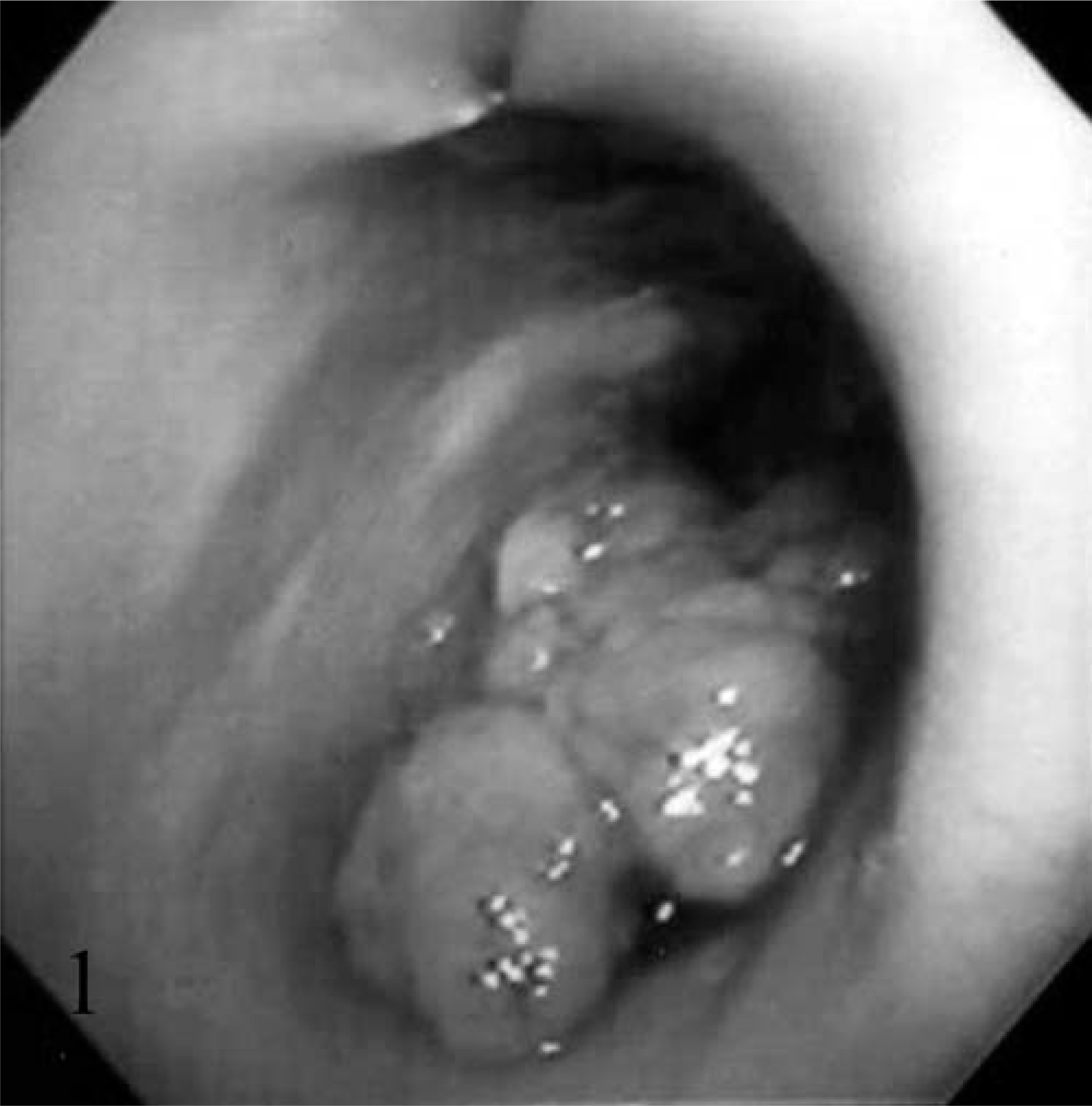
Botryoid rhabdomyosarcoma, vagina; dog. By endoscopy, a multipapillary mass was found around the urethral opening.
The tissue samples were fixed in 10% neutral buffered formalin, and 4-µm-thick sections were stained with hematoxylin and eosin (HE), alcian blue and periodic acid-Schiff (AB-PAS), Van Gieson, and phosphotungstic acid hematoxylin (PTAH). Histologically, the tumor was covered by vaginal mucosa and consisted of variable-sized, round-, spindle-, and polygonal-shaped anaplastic cells, which had clear nuclei with prominent nucleoli and abundant eosinophilic cytoplasm (Fig. 2). Mitotic figures were frequently observed. Many multinucleated cells were also observed; however, strap cells were not observed. Moreover, no cross-striations were detected within the neoplastic cells with Van Gieson and PTAH staining. In some areas where the tumor cells were loosely arranged, an AB-positive myxoid component was observed in the intercellular area (Fig. 3). In addition, hemorrhage, necrosis, and infiltration of the lymphocytes and plasma cells were also focally observed beneath the mucosal epithelium.
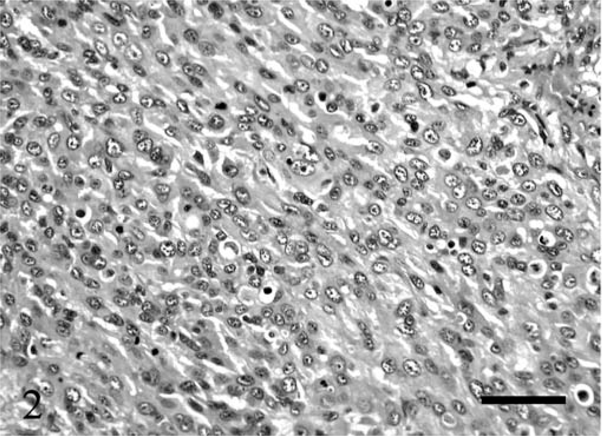
Botryoid rhabdomyosarcoma, vagina; dog. Proliferating, variable-sized, round-, spindle-, and polygonal-shaped anaplastic cells. HE. Bar = 40 µm.
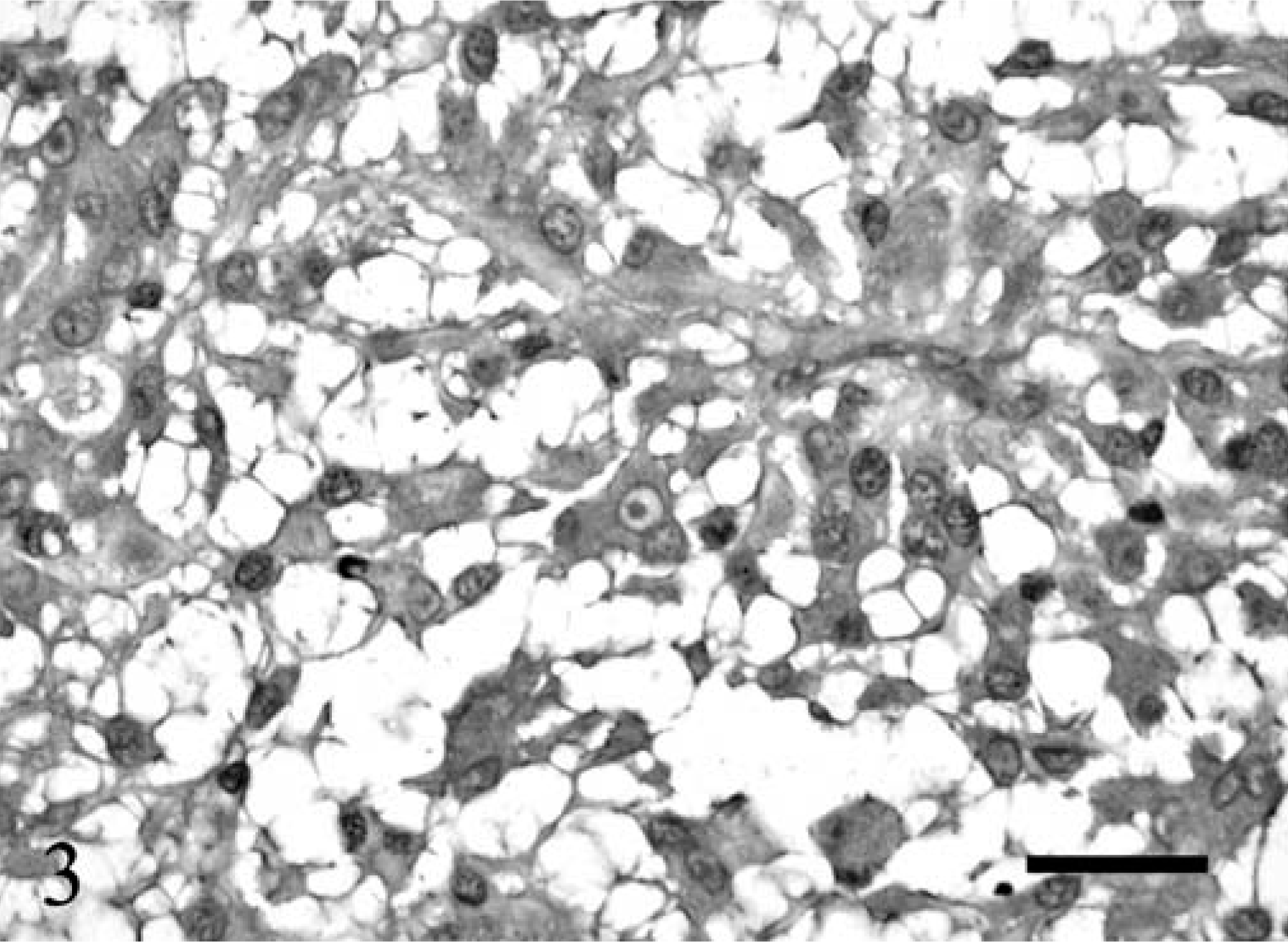
Botryoid rhabdomyosarcoma, vagina; dog. In the loosely arranged area, an intercellular myxoid component can be observed. AB-PAS. Bar = 20 µm
Immunohistochemistry was performed by streptavidin–biotin (SAB) method using an LSAB kit (Dako, Carpinteria, CA). The primary antibodies used were anti-vimentin, anti-human cytokeratin, and anti-human smooth muscle actin, mouse monoclonal antibodies and anti-human lysozyme, anti-desmin, anti-human myoglobin, and anti-S100 protein, rabbit polyclonal antibodies. All primary antibodies were provided by Dako. The tumor cells were observed to be strongly positive for vimentin and focally positive for desmin (Fig. 4).
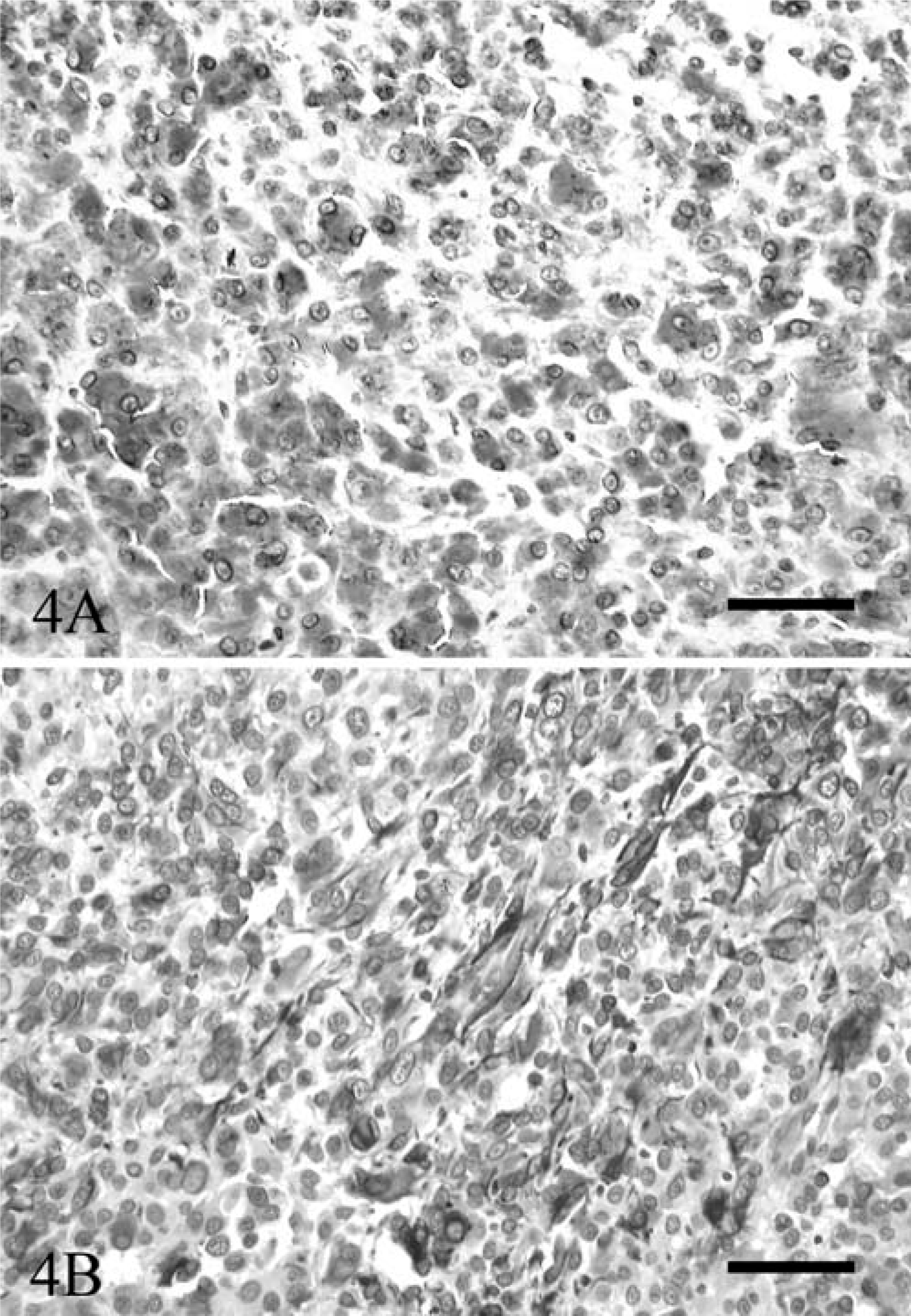
Botryoid rhabdomyosarcoma, vagina; dog.
It is well-known that the immunohistochemical stainability changes depending on the degree of cell differentiation. In the development process of striated muscle, vimentin, desmin, and myoglobin are expressed in that order. 2 Vimentin is expressed in the early phase but is lost later as muscle fiber develops, whereas desmin expression starts in the early phase and persists. 10 Moreover, because myoglobin is expressed after desmin, poorly differentiated rhabdomyosarcomas may have desmin expression but lack myoglobin expression. 1 In addition, we also tried to detect cross-striations by using PTAH staining; however, even on careful examination, no cross-striations were detected within the neoplastic cells. Based on the above-mentioned findings and the knowledge that the lack of cross-striations is frequently observed in poorly differentiated rhabdomyosarcomas, it was suggested that the tumor was a poorly differentiated rhabdomyosarcoma. Moreover, in the literature, a loose arrangement with a myxoid matrix and the presence of a densely cellular cambium layer have been described as the histologic features of botryoid rhabdomyosarcoma. 3 Because these findings were observed in this mass, it can be included as a botryoid subtype.
Botryoid rhabdomyosarcomas normally develop in neonatal and young children, but the development in adults has been reported rarely in the medical field. 5 Furthermore, botryoid rhabdomyosarcoma of the vagina has been reported in young children. 4, 6 Therefore, it can be speculated that botryoid rhabdomyosarcoma of the vagina may develop in adult dogs. Based on the finding that pluripotent stem cells developing from primitive urogenital ridge remnants were thought to be the origin of botryoid rhabdomyosarcoma of urinary bladder, 8 it can be considered that this tumor also originated from that cell population.
In conclusion, based on macropathologic, histopathologic, and immunohistochemical identification, this mass can be diagnosed as botryoid rhabdomyosarcoma. To our knowledge, this is the first case report of botryoid rhabdomyosarcoma of the vagina in dogs.