
Abstract
Two Syrian hamsters developed marked swelling of the ventral neck. Histologic examination of both masses revealed that the submaxillary salivary glands were effaced by large numbers of neoplastic plasma cells. In one hamster, neoplastic cells had infiltrated the adjacent lymph node. The neoplastic cells expressed CD79a antigen and were negative for CD3, lambda, and kappa light chains. Ultrastructural features of neoplastic cells in the salivary gland of one hamster included abundant cytoplasmic rough endoplasmic reticulum profiles, and peripherally displaced nuclei that contained marginated heterochromatin, consistent with plasma cells. Salivary gland plasmacytomas are extremely rare in humans and have not previously been reported in nonhuman species. The occurrence of such neoplasms in two hamsters suggests that this species may be predisposed to developing tumors of this type.
An extramedullary plasmacytoma is a neoplastic proliferation of plasma cells that occurs outside the bone marrow. 10 Extramedullary plasmacytomas have been reported in multiple locations in various animal species, but occur most commonly in the skin and oral mucosa of older dogs. 8 Extramedullary plasmacytomas are uncommon in humans and have a predilection for the head and neck. 10
This is the first report of two extramedullary plasmacytomas in the submaxillary salivary glands in two Syrian hamsters (Mesocricetus auratus). The microscopic, immunohistochemical, and ultrastructural features of these neoplasms are described. Salivary gland plasmacytomas are extremely rare in people and, to the authors' knowledge, have not previously been reported in nonhuman species.
Hamster No. 1 was a male 1-year-old laboratory-raised BIO FIB strain Syrian hamster. The animal had not commenced experimental manipulation. The hamster was serologically negative for antibodies against Sendai virus, pneumonia virus of mice, reovirus type 3, lymphocytic choriomeningitis virus, Encephalitozoon cuniculi, and simian virus 5. A mass was observed in the intermandibular region. This mass enlarged slowly over 6 weeks until significant loss of body condition prompted euthanasia of the hamster. Necropsy examination revealed a 2 × 3 × 2 cm encapsulated oval mass within the area of the left submaxillary salivary gland. The adjacent mandibular lymph node was approximately twice the size of the right mandibular node. No other significant lesions were observed on gross examination of the hamster. Representative samples of the mass, both mandibular lymph nodes, contralateral salivary gland, liver, and spleen were fixed in formalin and submitted to the Athens Diagnostic Laboratory (ADL) for histologic examination.
Hamster No. 2 was a male 2-year-old hamster that was kept as a pet. He presented to a private veterinary practice after developing a large mass within the subcutaneous tissues of the ventral neck. The owners did not report clinical evidence of systemic disease. The hamster was anesthetized and the mass was surgically removed, fixed in formalin, and submitted to the ADL for histologic examination. Unfortunately, the owner could not be contacted and no follow-up information is available for this animal.
Examination of the mass from hamster No. 1 revealed that the submaxillary salivary gland had been heavily infiltrated by sheets of round cells (Fig. 1). The neoplastic cells were loosely packed and surrounded by large amounts of amorphous eosinophilic material. Numerous residual glandular acini and ducts were visible throughout the sections examined. No evidence of infiltration of the salivary gland capsule was visible histologically. The neoplastic cells were large and had well-defined cell borders. They had large quantities of lightly basophilic cytoplasm and most had a prominent perinuclear clearing that was interpreted as Golgi apparatus. Cell nuclei were round, were eccentrically placed, and contained chromatin that was peripherally condensed, producing a “clock-face” appearance. Small numbers of binucleate cells were visible as well as occasional multinucleate giant cells. Eleven mitoses were visible per 10 400× fields within the neoplastic cell population. Examination of the enlarged mandibular lymph node revealed expansion by a similar population of neoplastic cells (Fig. 2). The neoplastic plasma cells were present as a cohesive mass, and the remaining lymph node retained normal architecture with germinal centers and medullary chords visible. Sections of contralateral submaxillary salivary gland, right mandibular lymph node, liver, and spleen were within normal histologic limits.
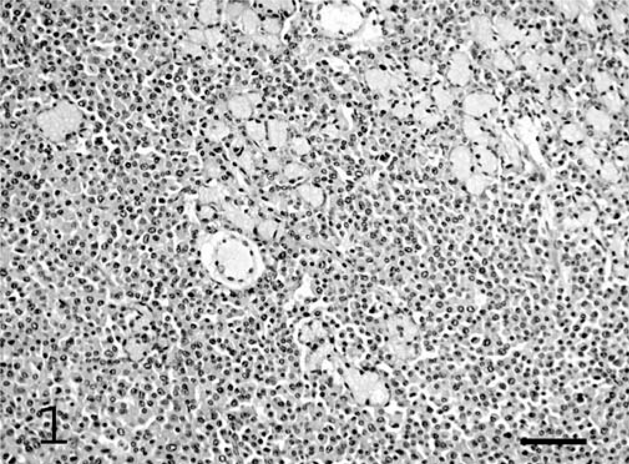
Salivary gland plasmacytoma; hamster No. 1. The salivary gland contains large numbers of round cells. Residual salivary gland ducts and acini are visible at the top right of the figure. HE. Bar = 45 µm.
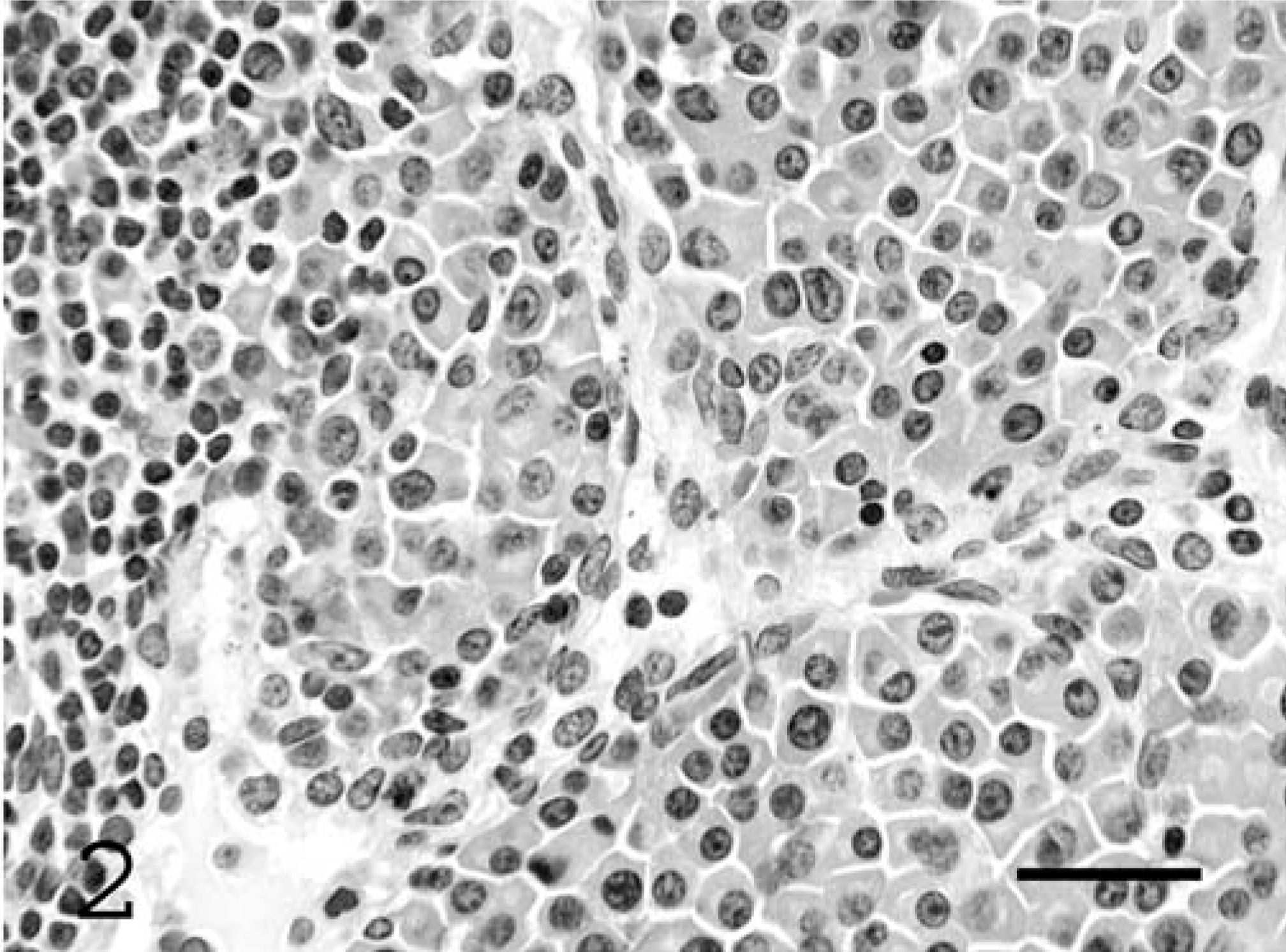
Adjacent mandibular lymph node; hamster No. 1. The lymph node contains a cohesive mass of plasma cells. The interface between the plasma cells and the remaining lymph node cortex is visible at the left of the figure. HE. Bar = 21 µm.
Histologic sections of the mass from hamster No. 2 also revealed a salivary gland that had been diffusely infiltrated by sheets of round cells (Fig. 3). When compared with the neoplasm from hamster No. 1, the neoplastic cells were more tightly packed and were surrounded by small quantities of amorphous eosinophilic material. The neoplasm had more greatly effaced the salivary gland and, although salivary gland ducts were visible, few glandular acini remained. Approximately 20% of the neoplastic cells were necrotic. Cells within the neoplasm removed from hamster No. 2 demonstrated more atypia than those from hamster No. 1. However, approximately 70% of the neoplastic cells were histologically consistent with plasma cells, with basophilic cytoplasm, prominent Golgi apparatus, and a round nucleus with peripherally condensed chromatin. The remaining neoplastic cells were more variable with indented nuclei, binucleate cells, and moderate numbers of multinucleate cells. Three mitoses were visible per 400× field within the neoplastic cell population, including large numbers of atypical mitoses.
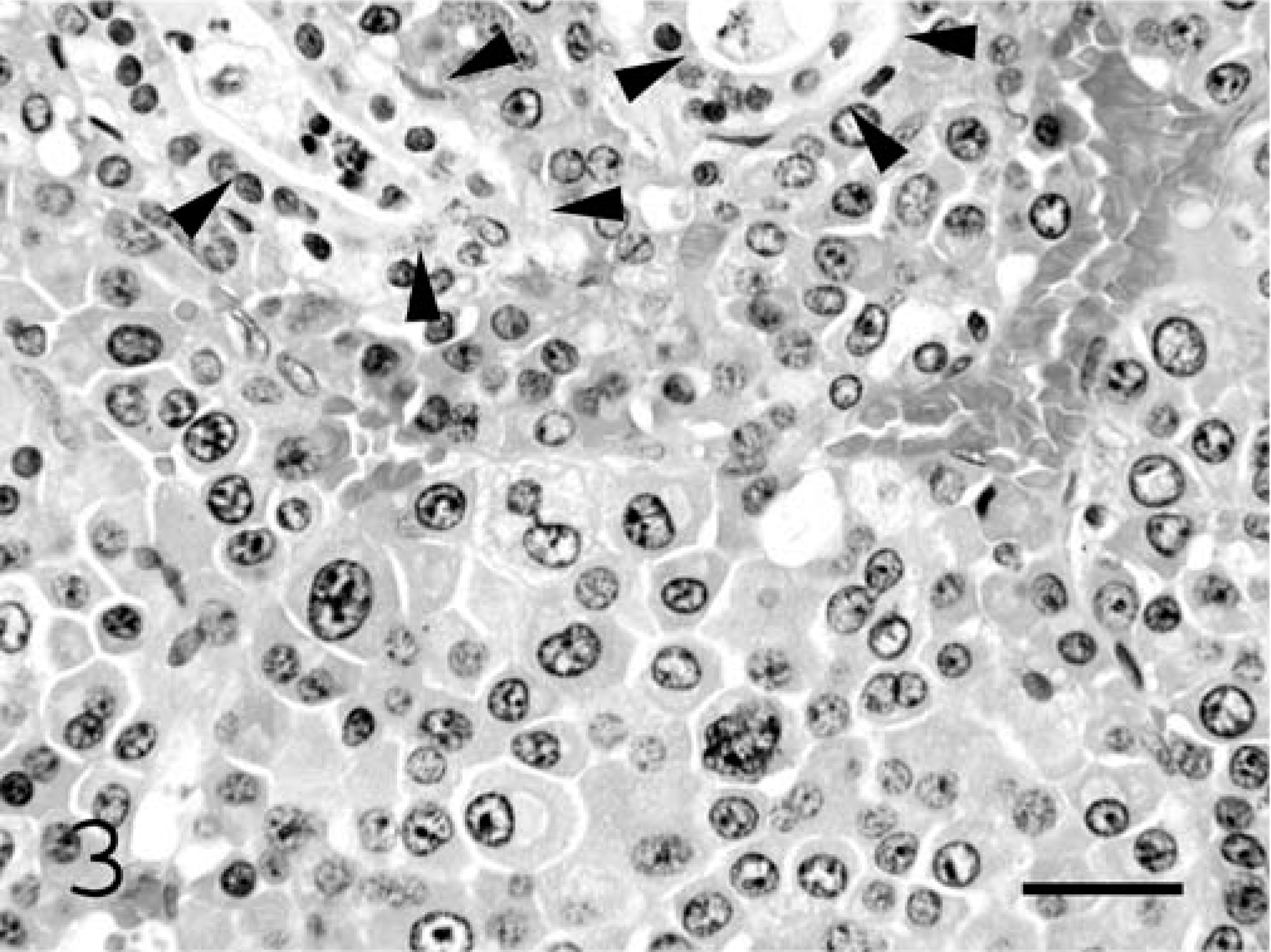
Salivary gland plasmacytoma; hamster No. 2. The salivary gland has been effaced by large numbers of round cells. Most of the cells have a prominent Golgi apparatus and marginated chromatin. Small numbers of residual salivary gland ducts are visible (arrowheads). HE. Bar = 19 µm.←
A Congo red stain was applied to sections of both masses. However, the amorphous eosinophilic background did not react positively within either neoplasm, suggesting that this material was not amyloid.
Immunohistochemistry revealed that approximately 90% of the neoplastic cells within the mass removed from hamster No. 1 and 70% of the cells within the mass from hamster No. 2 reacted positively with antibodies against CD79a (Dako Cytomation, Carpenteria, CA) (Fig. 4). Cells within neither neoplasm reacted with antibodies against BLA.36, CD3, kappa immunoglobulin chains, or lambda immunoglobulin chains (all Dako Cytomation, Carpenteria, CA). The contralateral unaffected lymph node from hamster No. 1 was used as a control. Interestingly, plasma cells within the node reacted to antibodies against CD79a, but not BLA.36. Plasma cells within the node also reacted positively to antibodies against the immunoglobulin light chains. Monoclonal murine antibodies against CD79a, BLA.36 and kappa immunoglobulin chains and polyclonal rabbit CD3 and lambda immunoglobulin chain antibodies were used.
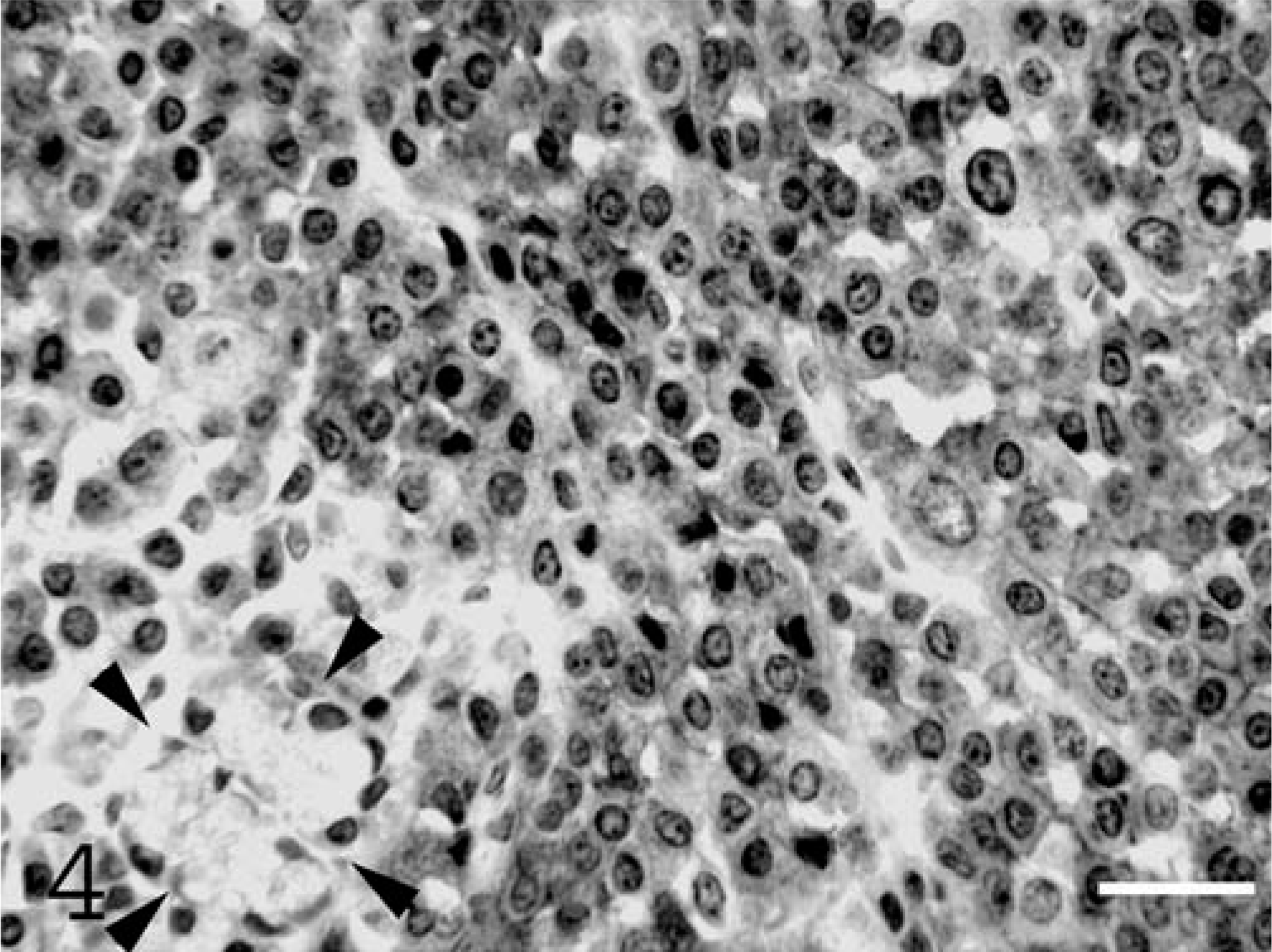
Salivary gland plasmacytoma; hamster No. 1. Neoplastic cells uniformly express CD79a. Residual salivary gland acini do not react with these antibodies (arrowheads). Streptavidin–biotin–peroxidase complex system and Gill's hematoxylin. Bar = 19 µm.
Samples of tumor from hamster No. 1 were postfixed in osmium tetroxide. Ultrathin sections were then cut using an ultra microtome with glass knives and stained with uranyl acetate and lead citrate. Samples from hamster No. 2 were not examined ultrastructurally because only paraffin-embedded tissue was available from this case. Ultrastructurally, the neoplastic cells were round to oval and had a smooth cytoplasmic membrane (Fig. 5). The cytoplasm was extensive and almost entirely filled by variably-sized rough endoplasmic reticulum profiles. Golgi apparatus and small numbers of mitochondria were visible within many cells. Nuclei were round, were eccentrically placed within the cell, and contained condensed, marginated heterochromatin. Small numbers of multinucleate cells were visible within the neoplastic cell population. These cells contained two to four nuclei that were eccentrically placed and contained condensed, marginated heterochromatin. Multinucleate cells also contained large numbers of variably-sized rough endoplasmic reticulum profiles, Golgi apparatus, and mitochondria (Fig. 6). The neoplastic plasma cells were surrounded by granular electron-dense material.
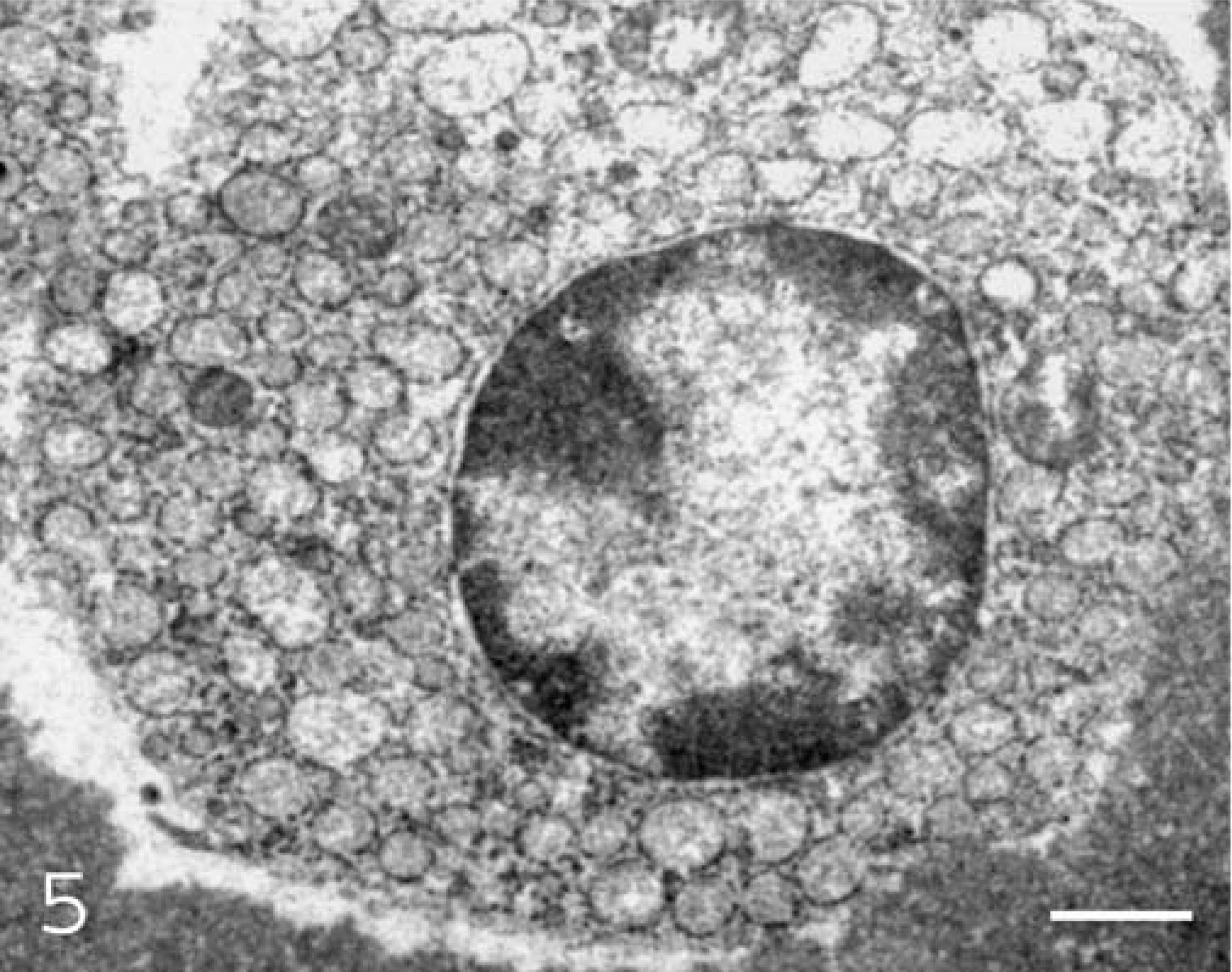
Salivary gland plasmacytoma; Hamster No. 1. Cells contain large numbers of variably-sized rough endoplasmic reticulum profiles and a round central nucleus that contains marginated heterochromatin. Uranyl acetate and lead citrate. Bar = 0.8 µm.
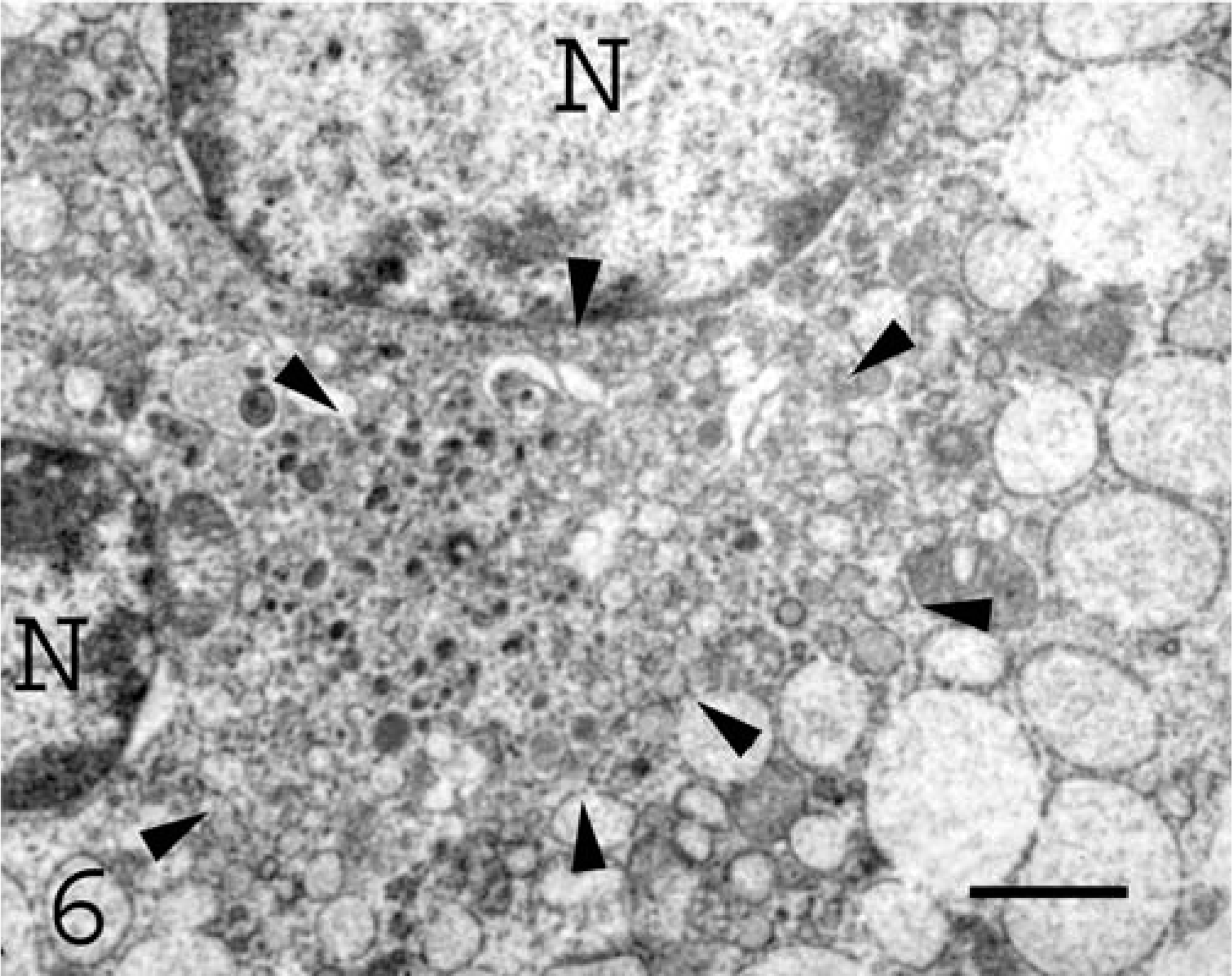
Salivary gland plasmacytoma; Hamster No. 1. This cell contains two nuclei (N) and a prominent Golgi apparatus (arrowheads) Uranyl acetate and lead citrate. Bar = 0.4 µm.
Cells within both tumors were histologically, immunohistochemically, and ultrastructurally consistent with plasma cells. Plasma cell neoplasms are classified as medullary if the neoplasm is present within the bone marrow and extramedullary if the tumor develops in a location outside the bone marrow. 10 Multiple myeloma designates a plasmacytoma that is present in multiple locations within the bone marrow. 10 In the presently described case, the accumulation of neoplastic plasma cells outside the bone marrow is consistent with a classification of extramedullary plasmacytoma.
Two extramedullary plasmacytomas have previously been reported in hamsters. 3 Interestingly, one was located within the soft tissues of the neck and was adhered to the submaxillary salivary gland. 3 The other developed within the mesenteric lymph nodes. 3 Within the domestic species, extramedullary plasmacytomas are most common in dogs, typically affecting the skin or oral cavity. 8 However, canine extramedullary plasmacytomas have also been reported in the gastrointestinal tract, 15 trachea, 1 and brain. 16 Within other domestic species, extramedullary plasmacytomas have also been reported in cats, 8 a ferret, 13 a horse, 12 and a sheep. 14 Although spontaneous extramedullary plasmacytomas are rare within the laboratory animal species, peritoneal plasmacytomas can be induced by pristane in BALB/c mice. 11 Interleukin-6 has been found to be important in the development of these murine plasmacytic neoplasms. 11 Two rhesus macaques were reported to develop spontaneous extramedullary plasmacytomas. 20 In humans, extramedullary plasmacytomas constitute 3–5% of all the plasmacytic neoplasms. 6 Although extramedullary plasmacytomas have been reported in a variety of locations, greater than 80% of human neoplasms involve mucosa-associated sites within the upper respiratory tract. 6
Syrian hamsters develop high rates of spontaneous neoplasia. 9 A survey of hamsters that were used as controls for carcinogenicity studies revealed that 69% of the animals had neoplastic lesions. 9 Adrenal cortical neoplasms were most common, with 66% of male and 38% of female hamsters developing tumors in this location. 9 Lymphoreticular tumors and endometrial and pancreatic islet cell neoplasms were also common. 9 Juvenile hamsters that are infected with polyomavirus are predisposed to multicentric lymphosarcoma. 17 This virus is also associated with trichoepithelioma development in older hamsters. 17
Spontaneous salivary gland neoplasia is rare in hamsters. 19 Experimental exposure of hamster salivary glands to carcinogens most commonly results in fibrosarcoma development. 19 Salivary gland neoplasia is infrequent in the domestic species. 7 In the laboratory animal species, salivary gland neoplasia is common only in mice. 4 Salivary gland neoplasia is also rare in humans. 18 Extramedullary plasmacytomas of human salivary glands are extremely rare, with only approximately 20 cases reported. 5 Of these, around 25% subsequently developed metastases. 5
Immunohistochemically, cells within both neoplasms reacted to antibodies against CD79a. This antigen is expressed almost exclusively in B-lymphocytes and B-lymphocytic neoplasms. 2 This antigen is reported to be expressed by plasma cells more consistently than other B-lymphocyte markers. 2 Cells within neither neoplasm expressed BLA.36, another B-lymphocyte marker. Interestingly, plasma cells within the unaffected lymph node reacted with antibodies against CD79a, but not against BLA.36. This suggests that as B-lymphocytes mature, they retain CD79a but not BLA.36 expression. As expected, neoplastic cells did not express CD3. Neither lambda nor kappa light chains, commonly identified in canine plasmacytomas, were observed within the hamster plasmacytomas. Antibodies specific for hamster immunoglobulins were unavailable. Considering the ultrastructural evidence of protein production within the cells, it is hypothesized that immunoglobulin production was present, but no identifiable light chains were produced by the neoplastic cells.
In conclusion, salivary gland plasmacytomas are extremely rare within all species. Therefore, it was surprising that two tumors of this type, both in hamsters, were observed within a 2-year period at the ADL. Furthermore, of the two plasmacytomas that had previously been described in hamsters, one was reported to be adhered to the submaxillary salivary gland. 3 It appears that hamster salivary glands may be predisposed to developing plasmacytic tumors. The possibility that a viral infection could have promoted neoplasia was considered; however, no evidence of viral involvement was observed. Local lymph node involvement was visible in hamster No. 1, but there was no evidence of widespread dissemination. However, the behavior of tumors of this type cannot be predicted from the observations in these cases.