
Abstract
Primary canine gastrointestinal lymphoma has been believed to be of B-cell origin based on the morphology and behavior of the neoplastic cells and the evidence from the human medical field. However, the neoplasms have not to date been characterized as to the origin of the cell population. Forty-four cases diagnosed as canine gastrointestinal lymphoma were retrieved from the records of the Veterinary Teaching Hospitals at the University of Minnesota and the University of Wisconsin-Madison. Four of the cases have been previously identified as epitheliotropic T-cell gastrointestinal lymphoma. Twenty-three of the dogs were female, with 11 intact and 12 neutered, and 21 of the dogs were male, with 12 intact and nine neutered. Sixteen breeds as well as individuals of mixed breeding were represented. The Boxer and the sharpei were the most commonly represented breeds with six individuals each. The age range of the dogs was 1.5–14.66 years, with two dogs identified as adult and two of unknown age. Archived tissue blocks of gastrointestinal samples were sectioned in duplicate and prepared for immunohistochemical staining with CD3 (T-cell marker) and CD20 (B-cell marker). In 75% of the cases examined under light microscopy, 50–95% of the neoplastic cells stained positively with CD3 and exhibited marked epitheliotropic behavior. In three of the cases, from 10% up to 50% of the neoplastic cells stained positively with CD20, with widely scattered CD3(+) cells. In the remainder of the cases, few to none of the neoplastic cells stained with either of the markers. This retrospective study shows that canine primary gastrointestinal lymphoma is more commonly of T-cell origin, rather than B-cell origin.
Keywords
Malignant lymphoma is one of the most common tumors in dogs, with estimates of occurrence ranging from 13 to 24 per 100,000 annually. 15 The two anatomic forms of the disease that predominate are the multicentric and the alimentary. 14 Gastrointestinal lymphoma accounts for approximately 5–7% of all canine lymphomas. Primary gastrointestinal lymphoma in dogs occurs over a wide range of ages and breeds and affects males more often than females. 3 Canine primary gastrointestinal lymphoma typically does not affect the superficial lymph nodes or the spleen, unlike the multicentric form in which these organs are almost always involved. 14 The majority of canine gastrointestinal lymphomas appear to be primary, with involvement in descending order of frequency of the small intestine, stomach, and colon. Soft to firm cream-colored masses are present in the gastrointestinal submucosa and may extend into the lumen and transmurally to the serosa. Typically, several segments of the intestine are involved, and often there is tumor in regional nodes and the liver. 11
Human primary gastrointestinal lymphomas are those typically confined to the alimentary tract, with no evidence of other systemic involvement (i.e., liver, spleen, bone marrow, or peripheral lymph nodes). 4 In humans, the majority of non-Hodgkin's lymphomas are of B-cell origin, and there is a widely varying prognostic index for these tumors. 1 Identifying lymphoma immunophenotype can be useful for treatment and prognosis because this can affect both the therapy selected and its outcome. 12 Although these neoplasms are commonly thought to be of B-cell origin, 5 characterization of the neoplastic cell type (B lymphocyte versus T lymphocyte) in primary canine gastrointestinal lymphoma has rarely been examined, with one report identifying the cell of origin as T cell. 17
Materials and Methods
Forty-four cases diagnosed as canine gastrointestinal lymphoma were retrieved from the databases of the Veterinary Teaching Hospitals at the University of Wisconsin–Madison (35) between 1984 and 1997 and from the University of Minnesota (9) between 1994 and 1997; four of the Minnesota cases were biopsies rather than necropsies.
Archived blocks of gastrointestinal sections, identified as stomach, duodenum, jejunum, ileum, or small intestine, were sectioned at 4 µm and prepared for immunohistochemical staining according to the avidin–biotin–peroxidase complex method. 10 Sections were deparaffinized, rehydrated through graded alcohols, and washed in buffer (0.05 M Tris-buffered saline, pH 7.6). Microwave pretreatment in 0.01 M citrate buffer, pH 6.0, was carried out twice for 5 minutes each, and then slides were cooled to room temperature for 20 minutes. Endogenous peroxidase was quenched by a 15-minute incubation in 3% hydrogen peroxide. To block nonspecific binding, sections were then incubated with normal goat serum (1 : 20, Vector Laboratories, Burlingame, CA) for 10 minutes. Sections were then incubated for 30 minutes at room temperature with the primary antibodies CD20 (1 : 50, BioGenex, San Ramon, CA.) and CD3 (1 : 50, DakoCytomation, Carpinteria, CA). After this primary incubation, sections were incubated in biotinylated antiserum LSAB-2 link antibody (DakoCytomation) for 15 minutes at room temperature. A second incubation for 15 minutes at room temperature with LSAB2-HRP streptavidin reagent was carried out. Sections were then stained at room temperature for 10 minutes with the chromogen 3-amino-9-ethylcarbazole (DakoCytomation) and counterstained for 3 minutes with Mayer's hematoxylin. Positive controls were sections of normal canine tonsil, and negative controls consisted of substitution of the primary antibodies with mouse ascites fluid (Sigma Chemicals, St. Louis, MO) on the case tissue. Slides were examined with light microscopy using a Nikon microscope at 200×, and four to five fields were examined for each slide.
Results
The most commonly represented breeds were the Boxer and sharpei with six individuals each (6/44 each). Other breeds with more than one individual were Golden Retriever with four (4/44), Springer Spaniel with four (4/44), Doberman Pinscher with three (3/44), German Shepherd with three (3/44), Labrador Retriever with three (3/44), Cocker Spaniel with two (2/44), Schnauzer with two (2/44), and Scottish Terrier with two (2/44). Three dogs were mixed breeds (3/44), and the following breeds were each represented by one individual: Bouvier, Elkhound, Fox Terrier, Maltese, Samoyed, and shorthair (1/44 each). Twenty-three dogs (52%) were female (11 intact and 12 neutered), and 21 dogs (48%) were male (12 intact and nine neutered). The age range was from 1.5 to 14.7 years, with a mean of 7.7 years, with two dogs listed as adults and two as age unknown (Table 1).
Breed and sex distribution of cases.
∗ One animal of unknown age.
† Two animals of unknown age listed as adult.
In 33 (75%) of the cases, the neoplastic lymphocytes exhibited epitheliotropic behavior. Neoplastic cells were commonly found infiltrating gastric and intestinal superficial mucosal and glandular epithelium (Fig. 1). In all these animals, most to nearly all the neoplastic lymphocytic cells stained positively for the CD3 antigen (Fig. 2). In these cases, the neoplastic cells were also arranged in nearly solid sheets replacing and compressing surrounding parenchyma. Nuclear morphology was variable, occurring as round to polygonal to reniform, with moderate to marked anisokaryosis. Chromatin varied from diffusely dense to stippled granular and basophilic. Nucleoli were uncommon. Cytoplasm was amphophilic to basophilic and ranged from scant to moderate. Cell borders were usually distinct. Mitotic figures were common at three to four per 400× field and frequently had bizarre morphology.
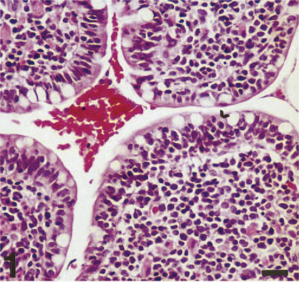
Small intestine; canine; dog No. 22. Epitheliotropic behavior of neoplastic lymphocytes (arrowhead). HE. Bar = 20 µm.
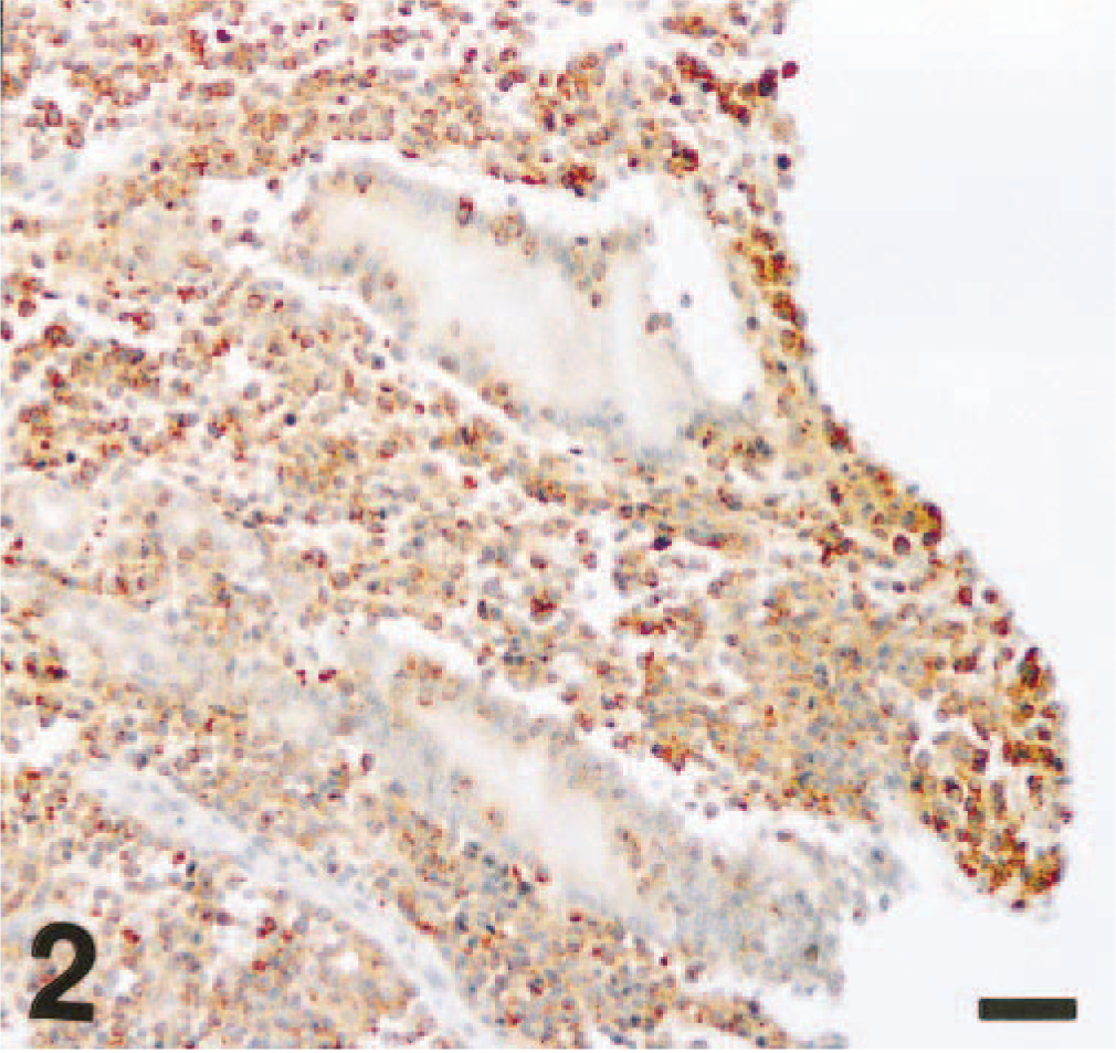
Small intestinal glands; canine; dog No. 22. CD3-positive staining of neoplastic lymphocytes. Avidin–biotin–peroxidase complex method. Hematoxylin counterstain. Bar = 15 µm.
In three (6.8%) of the cases (dog Nos. 36–38), approximately 10–30% of lymphocytic cells stained positively for the CD20 marker, whereas only widely scattered cells stained positively with CD3. In these three cases, the neoplastic cells occurred in solid sheets. Cells had round to ovoid nuclei with moderate anisokaryosis. Chromatin was deeply basophilic although in some cells chromatin was paler staining and was clumped. Cytoplasm was scant in neoplastic cells. In dog No. 37, there was a “starry sky” appearance to the neoplasm because of the presence of many tingible body macrophages. Neoplastic cells in dog No. 36 exhibited some follicular architecture within the solid sheet of cells. Mitotic figures were uncommon in these three cases at less than one per 400× field.
Two cases (4.5%) had widely scattered individual cells positive for either CD3 or CD20 (dog Nos. 34 and 35). The neoplastic cells in dog No. 35 were anaplastic, with marked anisokaryosis. Nuclei were round to ovoid to polygonal with clumped chromatin, and a nucleolus was visible in some cells. Cytoplasm was scant, and mitotic figures were common with some bizarre forms at two to three per 400× field. In dog No. 34, the cells were slightly more uniform with moderate anisokaryosis. Nuclei were slightly more basophilic than in dog No. 35, but the shape still varied from round to ovoid to polygonal. Chromatin was clumped with an occasional nucleolus. Cytoplasm was also scant. Mitotic figures were less common at less than one per 400× field.
In the remaining six cases (13.6%), no immunostaining was evident (dog Nos. 39–44). In these cases, nuclear morphology varied widely from round to ovoid to polygonal with marked anisokaryosis and the presence of scattered karyomegalic cells. Chromatin was moderately basophilic and clumped. Many nuclei had one or two prominent nucleoli. Cytoplasm remained scant and mitotic activity was moderate with approximately one per 400× field. (Table 2).
Immunophenotype of gastrointestinal lymphoma cases.
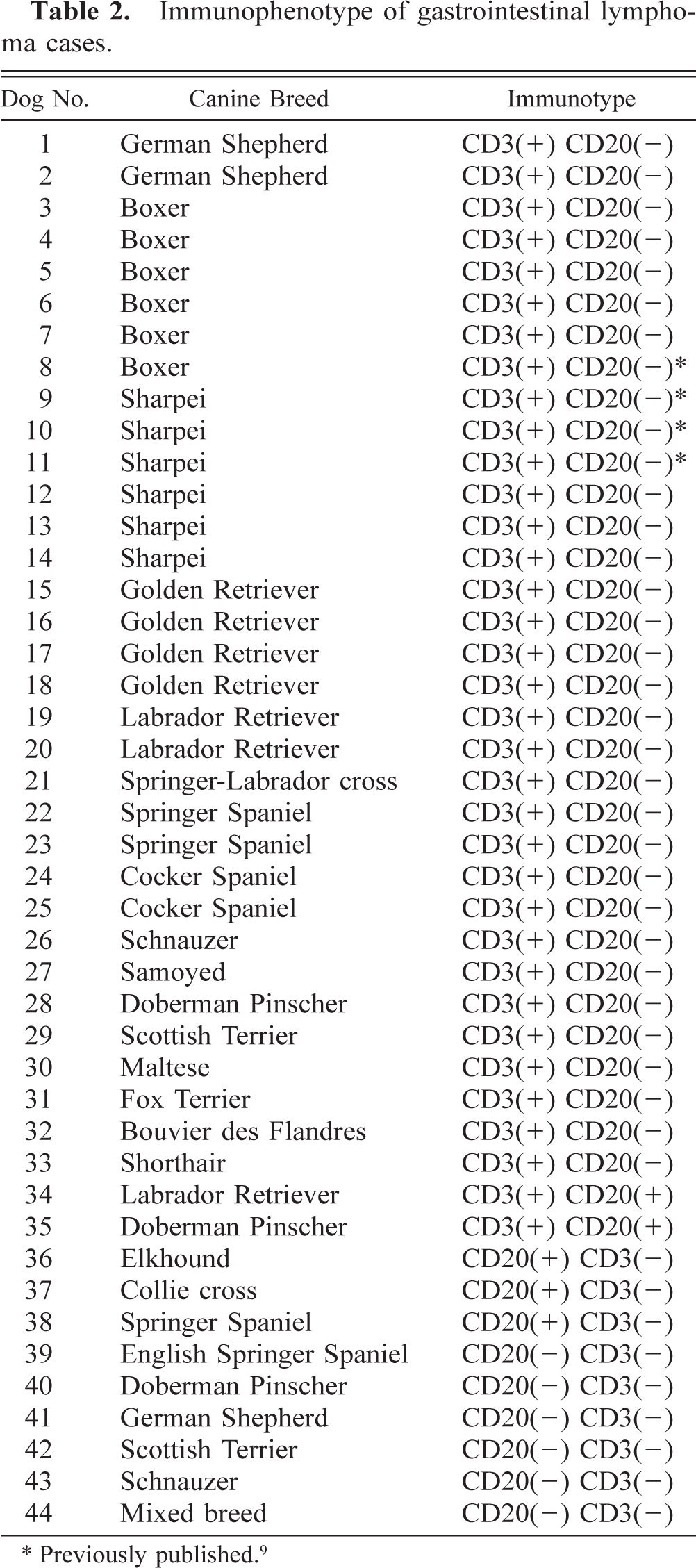
∗ Previously published. 9
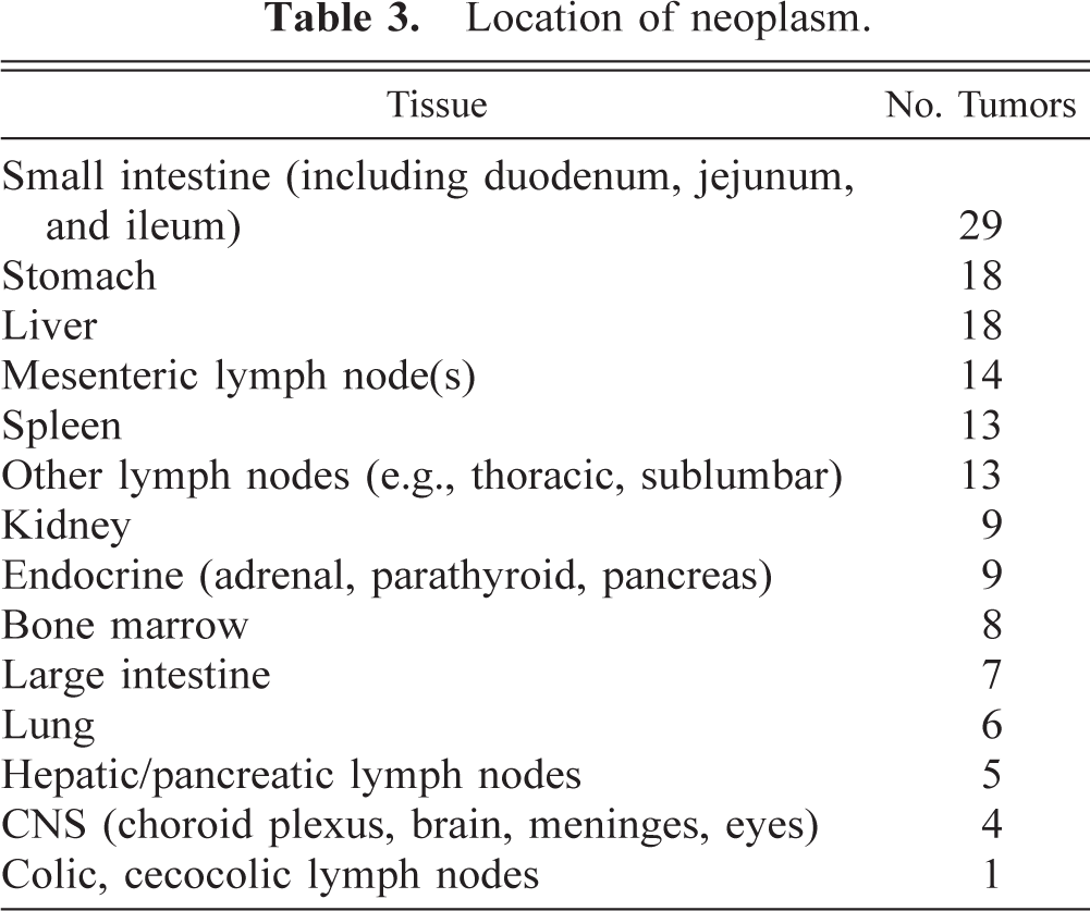
The most common location for lymphoma was the small intestine with 29 tumors. The next most common locations were the stomach and liver with 18 occurrences each. Other locations in descending order were the mesenteric lymph nodes, other lymph nodes (thoracic and ileac), spleen, kidney, endocrine, bone marrow, large intestine, lung, hepatic/pancreatic lymph nodes, central nervous system (choroid, brain, meninges, and eyes), and colonic lymph nodes (Table 3). The total number of occurrences adds up to more than 44 because most cases had tumors in more than one location.
Location of neoplasm.
The CD3(+) lymphoma cases were almost evenly divided between 18 males (41%) and 17 females (39%). The three CD20(+) cases comprised one female (2.3%) and two males (4.5%). The cases with no immunostaining of cells were evenly divided between three females and three males (6.8% each). For the cases with mixed staining, both were females (4.5%).
The CD20(+) tumors occurred in a Collie cross, an Elkhound, and a Springer Spaniel. The mixed staining cases were in a Doberman Pinscher and a Labrador Retriever, whereas the null staining cases were represented by a Scottish Terrier, an English Springer Spaniel, a Doberman Pinscher, a German Shepherd, a Schnauzer, and a mixed breed dog. The remaining breeds all had CD3(+) neoplasms.
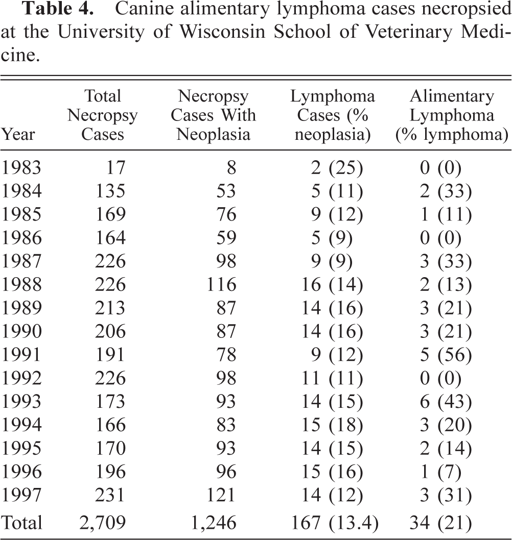
Six percent (167/2,709) of the canine necropsies done at the University of Wisconsin Veterinary Medical Teaching Hospital were lymphoma cases, which also represents 13.4% (167/1,246) of all canine neoplasia necropsies performed (Table 4). Because the records at the University of Minnesota School of Veterinary Medicine have only recently been computerized, cumulative histories to evaluate prevalence are unavailable.
Canine alimentary lymphoma cases necropsied at the University of Wisconsin School of Veterinary Medicine.
Discussion
The breed and age distribution of the lymphoma cases in this study are similar to those reported in other studies, 3,15,18 except for the relatively high number of sharpei in our study. This can be attributed both to the increased number of sharpei owned by people over the last several years as well as to the population in the geographic areas represented by this study. Although males are generally reported to be more often affected with lymphoma than females, in our study, the ratio was equal. This could be attributed to the relatively small study size or reflect the population presented for treatment at the two teaching institutions.
Seventy-five percent of the alimentary lymphomas in this study stained positively for the CD3 marker, indicating a T-cell origin for these tumors in dogs, whereas only 10% stained positively for the B-cell marker, CD20. This is opposite to the pattern seen in humans wherein a B cell is the origin of nearly all non-Hodgkin's lymphomas. Several studies on humans have found the incidence of B-cell lymphoma to be from >50% up to 100% when compared with T-cell lymphomas; 8,16,20,21 primary gastrointestinal lymphomas of T-cell origin have been described as “extremely rare.” 9 Primary gastrointestinal T-cell lymphomas are so rare in humans that several studies either do not address T versus B cell as a prognostic indicator, even though tumors were identified as to immunotype, 13,16 or all the tumors in the study were of B-cell origin. 22 A recent study 6 reported that almost 33% of canine lymphoma cases evaluated immunophenotypically were T-cell lymphomas. However, this study did not identify the location of lymph node samples examined nor did it identify the anatomic type of lymphoma that was diagnosed so that gastrointestinal lymphoma specifically was not addressed.
This was a preliminary study using archived materials. The samples identified as large intestine were not further subdivided into specific locations as the small intestinal samples were in either the biopsy or final pathology reports. Although the microarchitecture of the large intestinal samples had been effaced by the lymphoma to a degree that did not allow further identification, it would provide useful information in a future study to assess identified large intestinal segments for frequency of involvement. We contemplated the use of either CD79a or HM57 as the B-cell marker but decided to use CD20 because this is what the immunohistochemical laboratory that performed the staining typically uses for a B-cell marker. CD20 is acquired as a surface marker later than CD79a 14 so its use may have missed more immature B cells. Had the staining pattern in this study been more equivocal, the use of either CD79a or HM57 could have served as a more discerning marker for cells that had not stained with CD20. In normal dogs, a subset of CD8(+) T cells are most numerous in both the splenic red pulp and intestinal epithelium. 14 It may provide useful information to further characterize the neoplastic T cells in these tumors by staining sections with CD8. Nonstaining neoplastic lymphocytes could be either immature B cells or true null cells, and further immunostaining studies would assist in defining these populations.
The dog has been proposed as an animal model for human lymphoma. 7,19 This proposal is based on several assumptions: 1) the tumors are spontaneous and are apparently not virally induced; 2) dogs share a common environment with humans; 3) dogs are relatively large and tractable with a sufficiently long lifespan such that treatment protocols used in humans are feasible for dogs; 4) owner compliance with treatment is usually quite good and owners are interested in enrolling their dogs in experimental protocols; 5) many canine lymphomas respond well to treatment; and 6) the cell type of origin follows the same pattern as that found in humans.
The last assumption may be the most problematic as far as using the dog as a model for human gastrointestinal lymphoma, given the results of this study. Primary gastrointestinal lymphomas in humans are described as being overwhelmingly of B-cell origin (> 95%), and the rare T-cell tumors that occur are nearly always in association with preexisting enteropathy and malabsorption problems such as celiac disease. 4,13 In humans, malignant T-cell disorders are recognized as peripheral disorders and are often associated with viral infections such as human T-cell leukemia virus type 1 or with immune system dysfunction such as that seen in celiac disease. Mycosis fungoides is another manifestation of T-cell lymphoid neoplasia and is dominated by cutaneous involvement. The microscopic hallmark of this disease is the epithelial association of the neoplastic lymphocytes that cluster within the superficial dermis and invade the epidermis singly or in groups. 1 In all the CD3(+) cases in this study, the neoplastic lymphocytes exhibited a marked epitheliotropism. This would appear to be similar to the behavior of the neoplastic T lymphocytes in human and canine mycosis fungoides but is occurring in a gastrointestinal location in the canine.
Chemotherapy represents the mainstay of treatment for canine lymphoma. In the early stages of this treatment, both B-cell and T-cell lymphomas respond to various chemotherapeutic agents. However, those of T-cell origin have a more aggressive clinical behavior, and although there may be an early positive response to chemotherapy, these tumors nearly always show a rapid proliferation of neoplastic cells, which overwhelms any early response to chemotherapy. 2 Thus, the cell type of origin is an important factor affecting the prognosis in primary canine gastrointestinal lymphoma.
Footnotes
Acknowledgements
We thank Ms. Jan Shivers of the Minnesota Veterinary Diagnostic Laboratory at the University of Minnesota for the immunohistochemical staining. Funds for this study were provided by the Minnesota Veterinary Diagnostic Laboratory.