
Abstract
Tritrichomonas foetus is a venereal pathogen of naturally bred cattle. In domestic cats, T. foetus colonizes the colon, resulting in chronic, large-bowel diarrhea. The infection is prevalent among young, densely housed cats, and there is no effective treatment. To the authors' knowledge, the characteristic microscopic lesions of T. foetus infection in naturally infected cats have not been described. The aim of the study reported here was to characterize the histologic changes in the colon of seven cats with T. foetus infection and chronic diarrhea. All cats were 1 year old or younger (mean, 6.7 ± 1.7 months), and a diagnosis of T. foetus infection was made on the basis of direct fecal smear examination (five cats), fecal culture in InPouch™ TF medium (four cats), single-tube nested polymerase chain reaction (PCR) analysis of DNA extracted from feces (two cats), or observation of trichomonads in sections of colon followed by PCR confirmation on DNA extracted from paraffin-embedded tissue (two cats). The presence of colonic trichomonads was the most diagnostic histologic feature. Organisms were identified in all cats, but in only 24 of 43 (56%) sections of colon. Trichomonads were generally present in close proximity to the mucosal surface and less frequently in the lumen of colonic crypts. The presence of colonic trichomonads was consistently associated with mild-to-moderate lymphoplasmacytic and neutrophilic colitis, crypt epithelial cell hypertrophy, hyperplasia and increased mitotic activity, loss of goblet cells, crypt microabscesses, and attenuation of the superficial colonic mucosa. In two of the cats, histologic lesions were more severe and were associated with invasion of trichomonads into the lamina propria and/or deeper layers of the colon.
Keywords
Tritrichomonas foetus is an important venereal pathogen of naturally bred cattle, in which the organism is transmitted from the prepuce of the bull to the vagina and uterus of the cow. The principal clinical manifestation of infection in cattle is infertility, with occasional abortion during the first half of gestation. 1 The organism has also been described as an inhabitant of the porcine gastrointestinal and nasal mucosa, where its pathogenicity is uncertain. 7, 26 Tritrichomonas foetus and Tritrichomonas suis are considered to be strains of the same species on the basis of morphology, ultrastructural analysis, random amplified polymorphic DNA analysis, enzyme homogeneity, and rRNA gene sequence identity. 3 – 5, 14, 20, 21, 27 Studies of cross-transmission between cattle and swine with T. foetus or T. suis have indicated little host specificity. 7, 17
Beginning in 1996, several reports have documented the presence of large numbers of trichomonads in fecal specimens from young, densely housed cats with chronic large-bowel diarrhea. 10, 22, 23 On the basis of sequence identity of 18S rRNA, the feline organisms have been identified as T. foetus. 18 Following experimentally induced infection, T. foetus colonizes the feline ileum, cecum, and colon, resulting in diarrhea characteristic of the natural infection. 12 Naturally acquired infection is prevalent among purebred cats 13 and is prolonged, 8 and there is no effective antimicrobial treatment.
To the authors' knowledge, the microscopic lesions associated with T. foetus infection have not been described in naturally infected cats. The aim of the study reported here was to characterize the histologic changes in the colon of seven cats with natural T. foetus infection and chronic diarrhea.
Materials and Methods
Case material
Forty-three sections of colon (range, 2–17 sections) were evaluated from seven cats with chronic diarrhea and T. foetus infection. Colonic tissue specimens were obtained at necropsy (five cats), surgery (one cat), or colonoscopy (one cat). At the time specimens were collected, all cats were 1 year old or younger and ranged in age from 10 weeks to 12 months (mean, 6.7 ± 1.7 months). Breeds represented were Persian (n = 2), domestic shorthair (n = 2), and Bengal, Singapura, and Sphinx (n = 1 each). All cats had history of large-bowel diarrhea for three or more months, had received a variety of antimicrobials, and had consumed a variety of diets prior to biopsy or necropsy.
Antemortem diagnosis of T. foetus infection was based on the identification of motile trichomonads on direct fecal smear examination (five cats), cultivation of trichomonads from feces, using In PouchTM TF media (Biomed Diagnostics, White City, OR) 11 (four cats), or demonstration of T. foetus rDNA in feces using a specific single-tube nested PCR 9 (two cats). In two cats, the diagnosis was established at necropsy. Trichomonads were observed on microscopic examination of the colon, and the identity of the agent was confirmed by use of polymerase chain reaction (PCR) analysis of DNA extracted from paraffin-embedded tissue. Additional laboratory diagnostics for affected animals included fecal flotation (four cats), enzyme-linked immunoassay for Giardia antigen (three cats), and fecal bacterial culture (one cat). Evidence for concurrent enteric infection was not identified in any of these cats.
For comparison with those from T. foetus-infected cats, sections of colon from five age-matched control animals selected from the general population of felids submitted to the Iowa State University Veterinary Diagnostic Laboratory were evaluated. The following criteria were used to select negative-control animals: 1) age (under 1 year old), 2) no history of signs of gastrointestinal tract dysfunction, 3) trichomonads not observed on microscopic examination of colon, and 4) no detection of an enteric pathogen. Animals fulfilling these criteria included two cats with ethylene glycol poisoning, one cat with acute renal tubular necrosis presumed due to lily toxicosis, and two cats with pneumonia.
Light microscopy
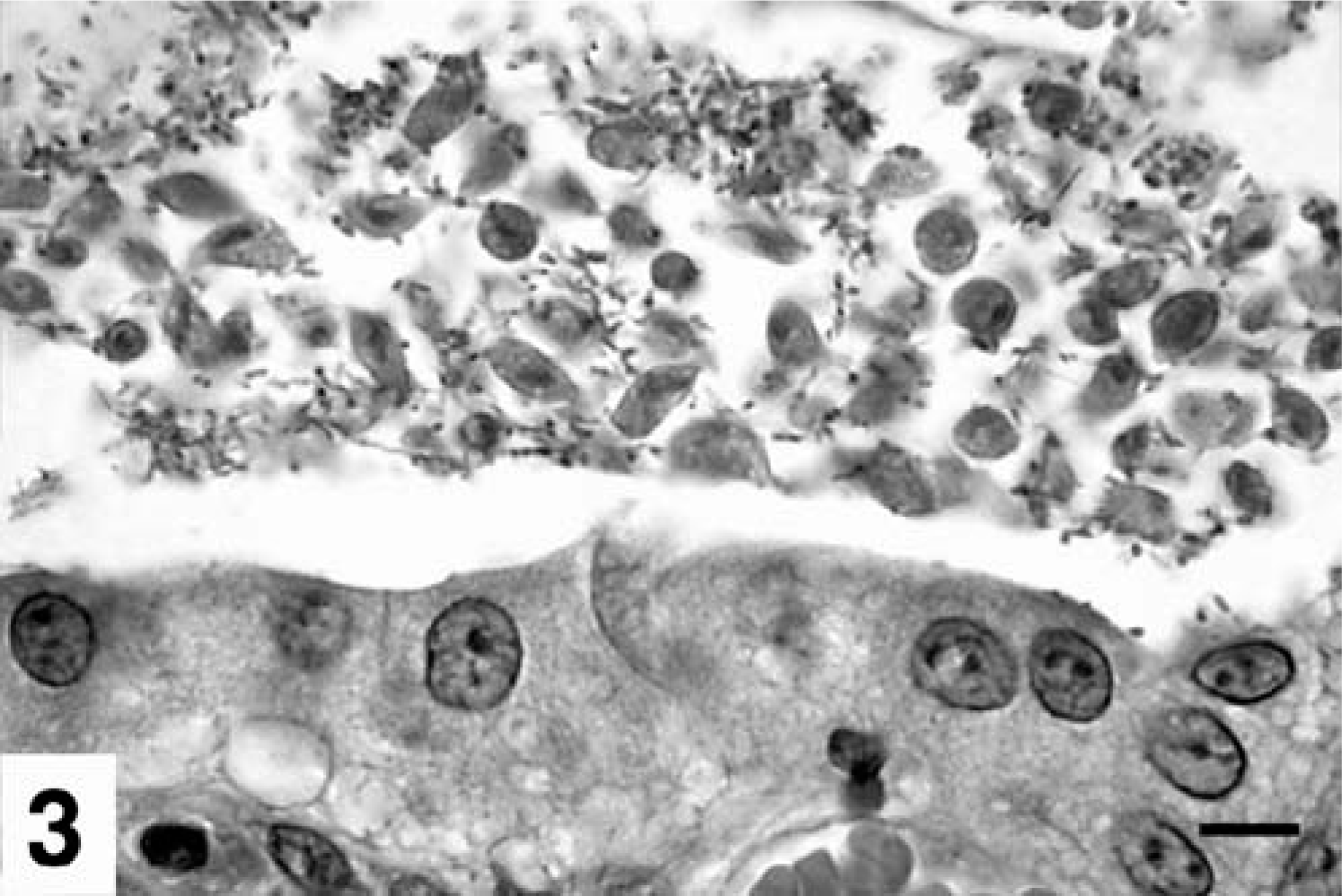
Tissues were fixed in neutral-buffered 10% formalin, trimmed, embedded in paraffin, sectioned at 5-µm thickness, and stained with hematoxylin and eosin (HE). Sections of colon from each cat were assessed for the presence and location of trichomonads. Numbers in 10 random, high-power fields (HPF; 40× objective) from each animal were averaged, and lesions were graded as mild (1), moderate (2), or severe (3). Criteria used for scoring each lesion are shown in Table 1. Average severity of each lesion was calculated by totaling the scores of the animals in which the lesion was identified, and dividing by the number of cats with the lesion.
Microscopic colonic lesions in Tritrichomonas foetus-positive cats.
∗Trich = trichomonads; NA = not assessed.
†Cats with autolysis of superficial mucosa were excluded from analysis.
Trichomonads were not detected in all sections of colon from infected individuals. Evaluation of lesion severity versus presence of the organism was assessed in two animals in which surface material was evident in all sections of colon, and sections with and without trichomonads were identified. For each of these two cats, scores for 10 random, HPF fields from sections with and without trichomonads were averaged and compared.
Immunohistochemical analysis for T. foetus antigen
Immunohistochemical analysis for detection of T. foetus antigen in colonic mucosal biopsy specimens was performed as described in detail, 12 using a monoclonal antibody (TF1.15) 15 that recognizes a surface antigen of T. foetus (courtesy of Lynette B. Corbeil, University of California, San Diego). Ascites fluid containing the antibody was diluted 1 : 500 before incubation with tissue sections for 1 hour at 37°C. Sections were immunostained using a commercially available kit with 3-amino-9-ethylcarbazole (AEC) as the chromogen (Histostain-SP broad spectrum; Zymed Laboratories, San Francisco CA). Sections were counterstained with methyl green. For negative-control sections, the primary antibody was omitted.
Transmission electron microscopy
To examine the ultrastructural effect of T. foetus on colonic epithelium, fresh endoscopically procured mucosal biopsy specimens from a cat naturally infected with T. foetus and an uninfected control cat were placed in McDowell's and Trump's formaldehyde-glutaraldehyde (4 : 1) fixative for several months at 4°C. After two rinses in 0.1 M sodium phosphate buffer (pH 7.2), samples were placed in 1% osmium tetroxide in the same buffer for 1 hour at room temperature. Samples were rinsed two times in distilled water and were dehydrated in an ethanolic series culminating with two changes of 100% acetone. Tissues were then placed in a mixture of Spurr resin and acetone (1 : 1) for 30 minutes, followed by 2 hours in 100% resin with two changes. Finally, tissues were placed in fresh 100% resin in molds and were polymerized at 70°C for 8 hours to 3 days. Semithin (0.25–0.5 µm) sections were cut with glass knives and stained with 1% toluidine blue-O in 1% sodium borate. Ultrathin (70–90 nm) sections were cut with a diamond knife, stained with methanolic uranyl acetate followed by lead citrate, and examined by transmission electron microscopy.
Statistics
The probability of detecting trichomonads versus number of biopsy sections examined was calculated using a binomial model in a simulation with the statistical software Splus 6.0 package (Lucent Technologies, Inc., Murray Hill, NJ). The simulation was run separately for each of 1–10 sections. Each step in a simulation represented a sample from a diseased specimen, and at each step, a proportion was randomly sampled from the data and used as the proportion parameter of a binomial distribution. Then the binomial distribution was sampled, and if the result was ≥1, the simulated specimen was detected as positive. Each simulation had 1,000 interactions.
Results
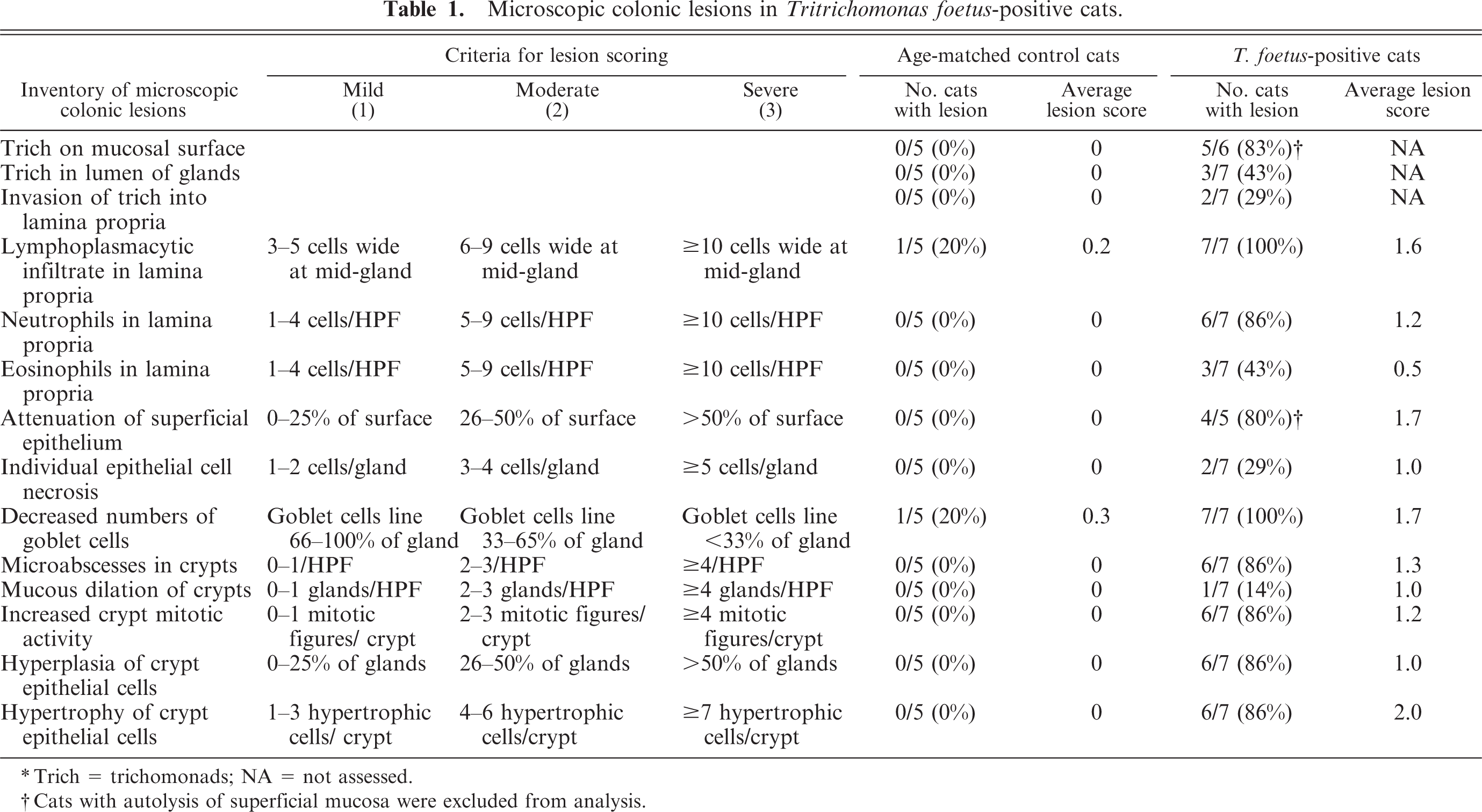
Histologic lesions are summarized in Table 1. All cats had mild to moderate expansion of the colonic lamina propria with an infiltrate of plasma cells and lesser numbers of lymphocytes (Fig. 1). Moderate numbers of neutrophils were observed in the lamina propria in six of seven (86%) cats. Eosinophils were not a prominent feature of the lamina propria infiltrate, were observed in only three of seven (43%) cats, and were generally present in low numbers. Crypt epithelial cell hypertrophy, hyperplasia and increased mitotic activity, loss of goblet cells, crypt microabscesses, and attenuation of the superficial colonic epithelium were consistent microscopic features, as each was identified in ≥80% of the cases (Figs. 2– 4). Mucus dilatation of colonic crypts was rarely observed. Lesions tended to be segmental, with 2–3 adjacent crypts exhibiting striking changes, whereas neighboring crypts were minimally affected.
Photomicrograph. Colon; cat No. 6, with enteric Tritrichomonas foetus infection. The lamina propria is moderately expanded by an infiltrate of plasma cells, lymphocytes (score = 1), and neutrophils (score = 2). There is moderate loss of goblet cells (score = 2) and hypertrophy of crypt epithelial cells (score = 1.5). HE stain. Bar = 100 µm.
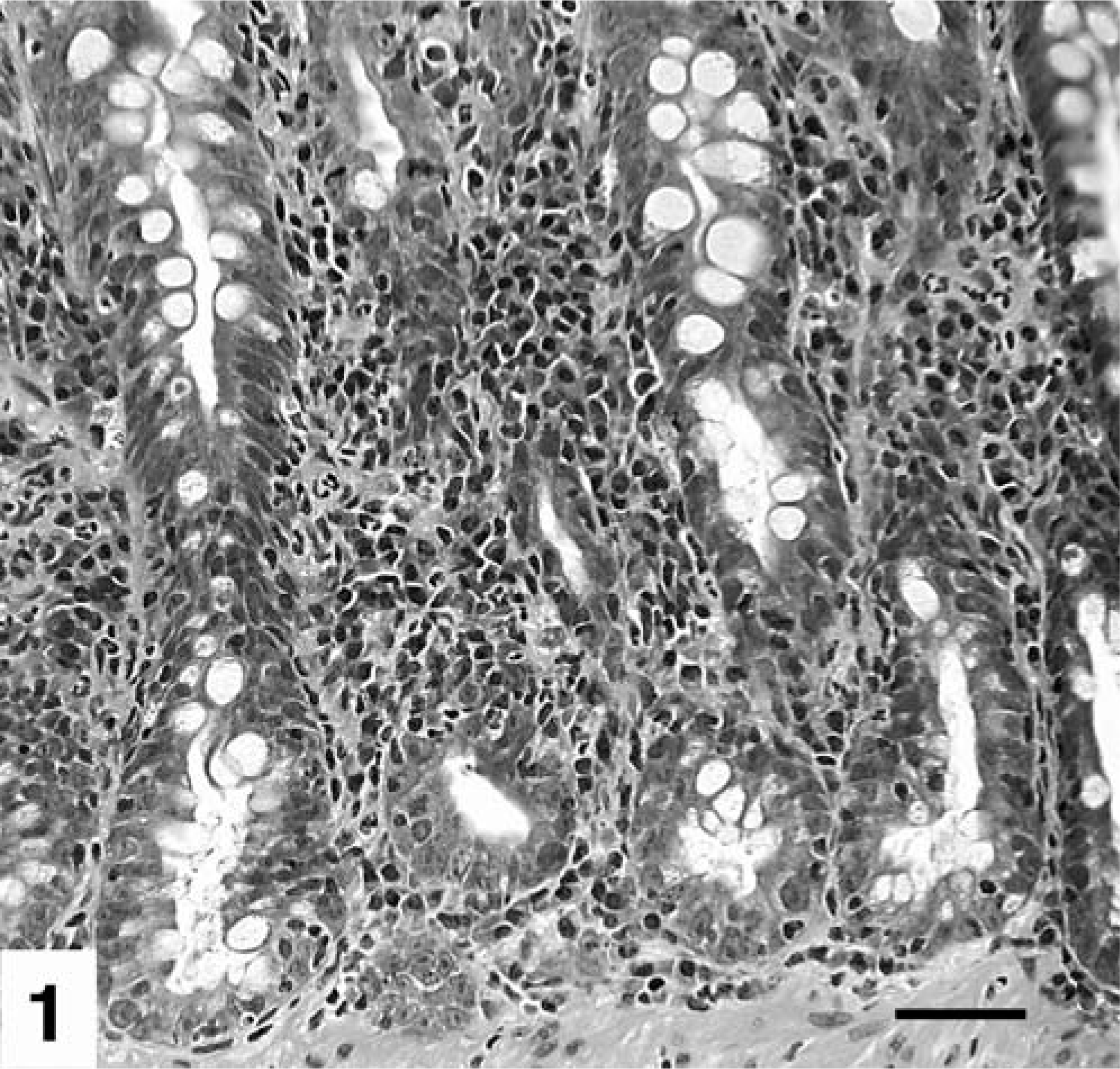
Transmission electron micrograph. Colonic mucosa; normal cat (A) and T. foetus-infected cat (B; cat No. 1). Notice attenuation of colonic epithelium and inflammatory subepithelial infiltrate. Colonic epithelial cell (e), intraepithelial lymphocyte (L), blood vessel containing erythrocytes and platelets (v), and plasma cell (p). Bar = 5 µm.
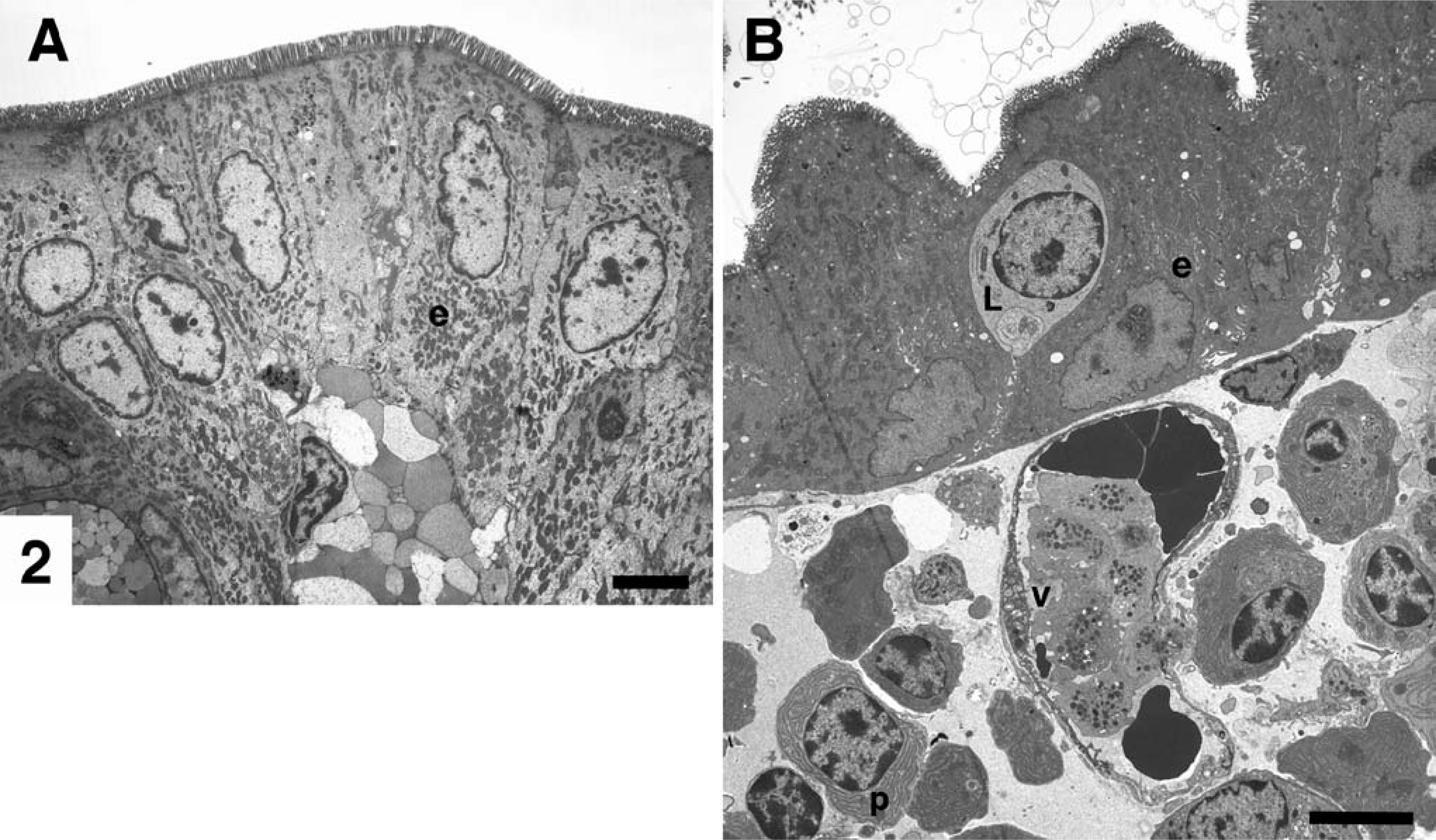
Photomicrograph. Colon; cat No. 4, with enteric T. foetus infection. The height of superficial colonic epithelial cells is attenuated, and there are numerous trichomonads, admixed with bacteria, in the lumen of the colon adjacent to the mucosa. HE. Bar = 10 µm.
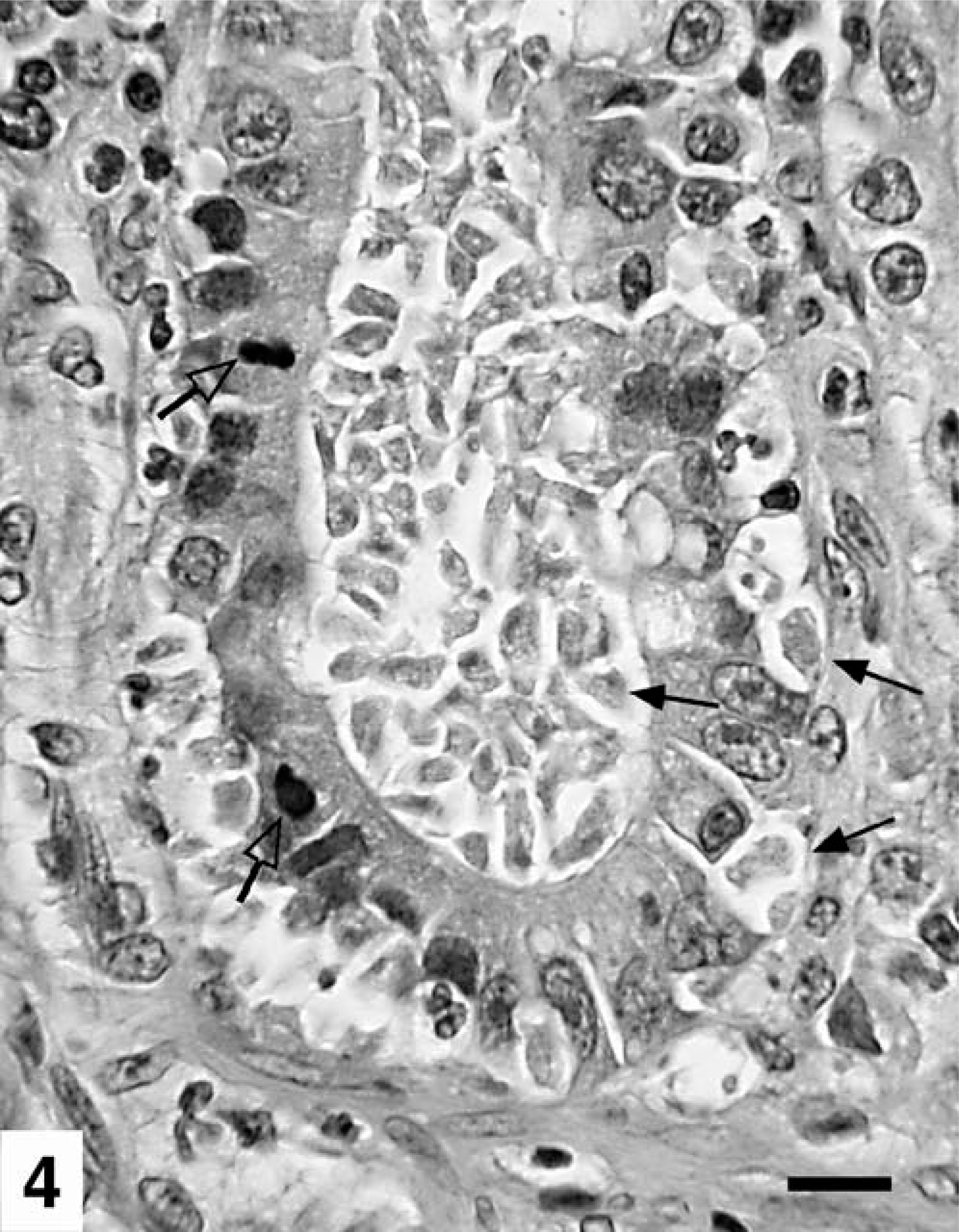
Photomicrograph. Colon; cat No. 6, with enteric T. foetus infection. Trichomonads are seen in the lumen of a colonic crypt, with invasion into the lamina propria (closed arrows). Notice crypt epithelial cell hypertrophy, increase in mitotic activity (open arrows), and absence of goblet cells. HE. Bar = 20 µm.
The diagnostic feature in all cats was the presence of colonic trichomonads (Figs. 3– 6). In tissue sections, trichomonads were elongate, teardrop to crescent-shaped organisms, 7–8 µm long by 4–5 µm wide, with eosinophilic cytoplasm and a moderately faint, hyperchromatic round-to-oval nucleus 1.0–1.5 µm in diameter typically located in the apical or widest portion of the cell. Flagella were not visible in HE-stained sections. Trichomonads were most consistently observed in close proximity to the surface of the colonic mucosa (83%) and less frequently in the lumen of colonic crypts (43%) (Figs. 3– 5). When trichomonads were compressed within the lumen of colonic crypts, they were often more elongate with flattened sides (Fig. 4).
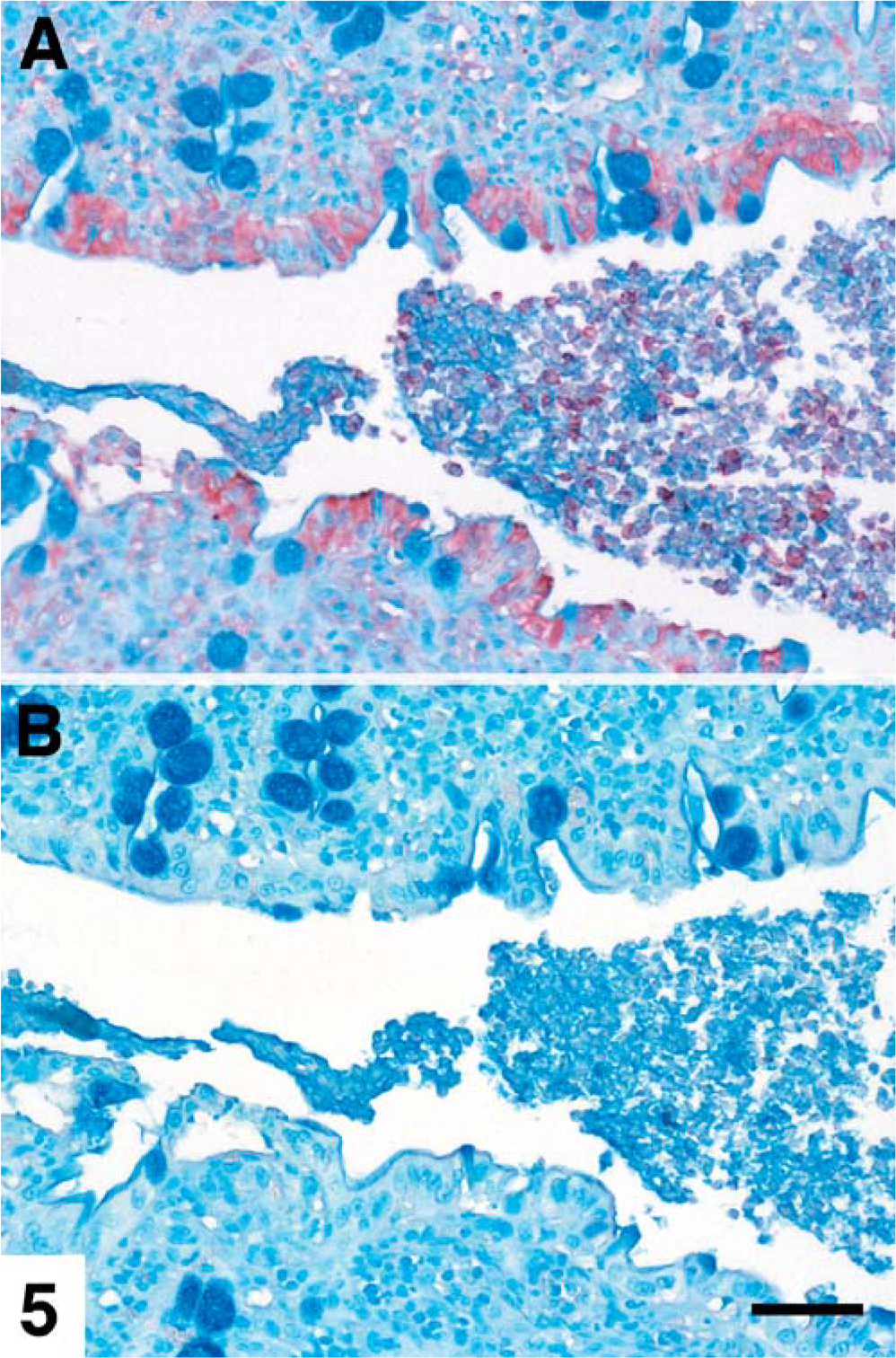
Photomicrograph. Colon; cat No. 4, with enteric T. foetus infection. The biopsy specimen was stained with (A) and without (B) antiT. foetus monoclonal antibody (TF1.15) 15 demonstrating AEC-labeled trichomonads in the colonic lumen and antigen uptake by colonic epithelium. Methyl green counterstain. Bar = 45 µm.
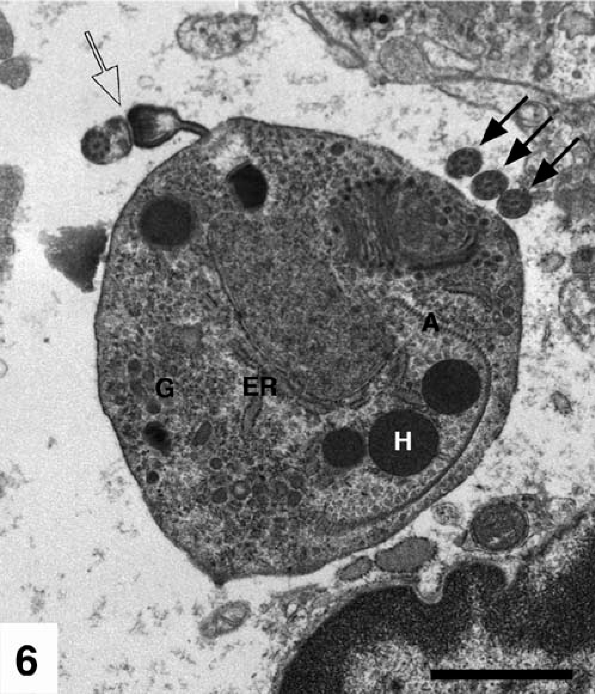
Transmission electron micrograph. Colonic lumen; trichomonad with three anterior flagella (closed arrows) and one recurrent flagellum (open arrow). G = glycogen granules, ER = rough endoplasmic reticulum, H = hydrogenosome, and A = and axostyle. Bar = 1 µm.
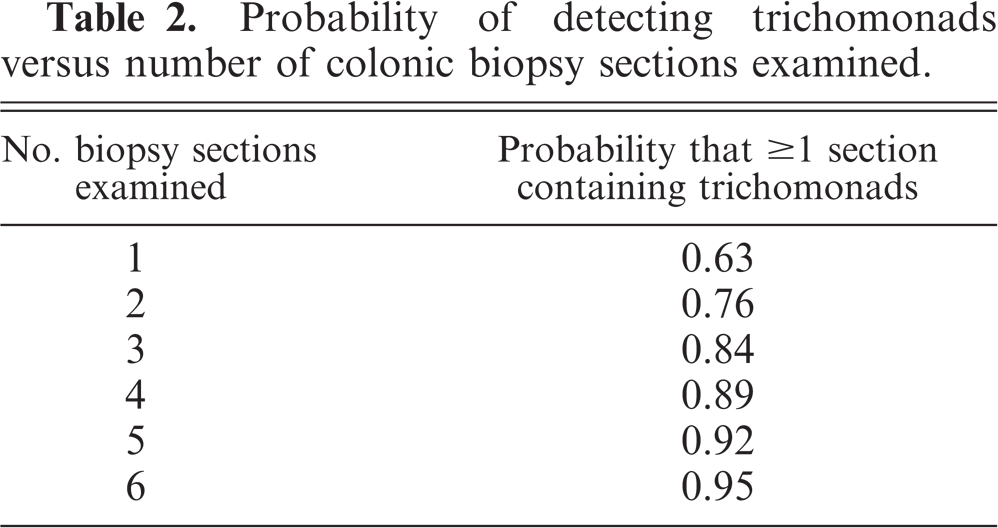
Trichomonads were identified in 24/43 (56%) sections of colon. The estimated probability of detecting trichomonads versus the number of biopsy sections examined was calculated (Table 2). In an infected animal, to have ≥95% confidence that trichomonads will be identified in at least one section, a minimum of six sections of colon would need to be examined.
Probability of detecting trichomonads versus number of colonic biopsy sections examined.
Trichomonads were not detected in all sections of colon from infected individuals. An evaluation of lesion severity versus the presence of the organism was assessed in two cats, in which surface material was evident in all sections of colon, and sections with and without trichomonads were identified. In these two cats, the average lesion severity scores for several parameters were higher in sections in which trichomonads were identified. The lamina propria infiltrate was more intense (2.0 vs. 1.25), the superficial epithelium was slightly more attenuated (1.5 vs. 1.25), and greater numbers of crypt microabscesses (2.0 vs. 1.0) were identified in sections of colon with organisms, compared with those without organisms.
In two of the seven cats, histologic lesions were more severe and were associated with the presence of trichomonads in the lamina propria and/or deeper layers of the colon. In one cat, trichomonads had infiltrated the colonic lamina propria, principally at the base of the crypts (Fig. 4). This was associated with severe and diffuse crypt changes consisting of crypt epithelial cell hypertrophy and hyperplasia accompanied by increased crypt mitotic activity. Macrophages were a prominent component of the lamina propria infiltrate, and a few AEC-labeled fragments, assumed to be those of trichomonads, were observed within the cytoplasm of large histiocytic cells in the lamina propria and submucosa. Crypt microabscesses were abundant, and loss of goblet cells was marked. The second cat had multifocal ulcerations, and marked transmural, suppurative-to-pyogranulomatous inflammation, with numerous trichomonads in the submucosa, tunica muscularis, and subserosal lymphatics. Multifocal chronic, granulating, necrotizing, and pyogranulomatous nodules were present on the serosal surface of the colon. Draining lymph nodes were hyperplastic, with multiple moderately sized foci of necrosis. In neither of these cases were trichomonads observed in sections of lymph node examined.
Trichomonads were not observed in any of the age-matched, negative-control cats. Of the five controls, four received a score of zero for each of the 14 graded categories (Table 1). Mild lesions were identified in the colon of one cat. These changes consisted of mild focal lymphoplasmacytic infiltrates in the lamina propria, which received a score of one in two of 10 fields, and decreased numbers of goblet cells, which received a score of one in three of the 10 randomly evaluated fields.
Discussion
Trichomonads are flagellated protozoans that reproduce by binary fission and undergo direct transmission from host to host. 6 Both commensal and pathogenic species of trichomonads exist. In domestic cats, T. foetus colonizes the colon, resulting in chronic large-bowel diarrhea. 8, 10, 12, 18 The infection is prevalent among young, densely housed cats. 10, 13 Trophozoites of T. foetus can be difficult to distinguish from those of Giardia sp., except that the former has a distinct undulating membrane, does not form cysts, and is refractory to treatment with antiprotozoal drugs such as metronidazole and fenbendazole. 10 Feline T. foetus infection is diagnosed by direct fecal smear examination for trichomonads, cultivation of feces using a commercially available system (In PouchTM TF), 11 or by extraction of DNA from feces and amplification of T. foetus rDNA by use of PCR analysis with species-specific primers. 9 One of the distinctive features of feline trichomonad-induced colitis is that affected cats are typically young. 10 At the time colon specimens were collected, all cats of this study were 1 year old or younger and ranged in age from 10 weeks to 12 months (mean, 6.7 ± 1.7 months).
To the authors' knowledge, this study is the first to describe the histologic lesions associated with naturally acquired T. foetus infection in the feline colon. The most distinctive microscopic feature is the presence of trichomonads in sections of colon. In tissue sections, trichomonads are elongate, teardrop- to crescent-shaped organisms, measuring approximately 7 by 5 µm. The organisms have eosinophilic cytoplasm and a faint, hyperchromatic round-to-oval nucleus, approximately 1.5 µm in diameter located in the apical portion of the protozoan. Trichomonads are generally present in large numbers, are most consistently identified in close association with the surface of the colonic mucosa, and are found to a lesser extent within the lumen of colonic crypts.
The presence of colonic trichomonads is consistently associated with a mild-to-moderate lymphoplasmacytic and neutrophilic colitis, with crypt epithelial cell hypertrophy, hyperplasia and increased mitotic activity, loss of goblet cells, crypt microabscesses, and attenuation of the superficial colonic mucosa. Eosinophils are not a prominent feature of the inflammatory infiltrate. Macrophages are identified in cats in which trichomonads have infiltrated into the lamina propria and beyond.
Trichomonads were detected in 56% of the 43 sections of colon examined. This figure may not accurately reflect the true prevalence of the organism in colonic sections from infected cats as trichomonads were identified less often in sections with little adherent mucus. As such, tissue collection and processing could impact the ability to detect the organism. When tissue is collected by biopsy or at necropsy, our findings suggest that examination of multiple sections of colon will increase the likelihood of detecting trichomonads in infected cats. To have ≥95% confidence that trichomonads will be present in at least one section, six sections of colon should be examined.
Experimental studies have fulfilled Koch's postulates regarding the ability of T. foetus to act as a primary enteric pathogen in cats. 12 However, the current understanding of factors mediating pathogenicity of trichomonads is limited. Identified mechanisms include alterations in host endogenous bacterial flora, adherence to the epithelium, and the elaboration of cytotoxins and enzymes. 2, 16, 19, 24, 25, 28, 29 With respect to the potential virulence of this organism in cats, of particular interest were the two cats (29%) of this study in which the organism was identified in the deeper layers of the colon. One cat had erosive and ulcerative lesions, and trichmonads were identified in all layers of the colon. At the time of necropsy, it was unclear whether the trichomonads were the cause of these lesions or whether the organism used pre-existing breaches in mucosal integrity to pass into the deeper layers of the colon. In the second cat, tissues were harvested immediately after euthanasia and the mucosa appeared intact; yet, there were numerous trichomonads in the submucosa. Findings in this animal support direct enteroinvasion. Compared with swine, where invasion beyond the mucosal surface has not been demonstrated and the pathogenic potential is uncertain, the ability of this organism to invade into the submucosa and beyond bears a distinct resemblance to enteroinvasive T. foetus in aborted bovine fetuses 22 and further supports the pathogenic potential of this organism in cats.
In young cats with large-bowel diarrhea that is unresponsive to antimicrobial therapy, underlying infection with T. foetus should be given strong consideration. The organism can be diagnosed by microscopic observation of motile trichomonads in saline-diluted fecal smears, cultivation of trichomonads from feces using In PouchTM TF media, 11 demonstration of T. foetus rDNA in feces by use of PCR analysis, 9 or by histologic evaluation of multiple sections of colon. If biopsies are to be evaluated, it is important to collect several specimens, as current data indicate that the organism is not necessarily present in all sections.
Footnotes
Acknowledgements
We thank Drs. Richard Brown, Sarah Cowan, Linden Craig, Mark Klopfenstein, Mac Law, Kristen Lowrimore, Martin Randell, Jenny Salpeter, and Jacqueline Simon for providing the histologic specimens, and the Laboratory for Advanced Electron and Light Optical Methods and Biomedical Communications, College of Veterinary Medicine, North Carolina State University. We also thank Dr. Richard Evans for his assistance with statistical analysis and Dr. Lynette Corbeil for her gift of TF1.15 antibody.
This study was funded by grants from Fort Dodge Animal Health (JLG) and the National Institutes of Health (DK 02868 to JLG).