
Abstract
Dermoid cysts are developmental abnormalities usually located at the dorsal body midline. Histologically, these lesions reduplicate the skin, with associated collagen bundles, adnexal structures, and are filled by keratin and hairs. While these cysts have well-recognized breed and anatomical predispositions in dogs, the information in feline patients is restricted to scattered case reports and anecdotal data. Through a multi-institutional retrospective case series, we aim to describe the clinical and demographic features of this condition in cats. We retrieved a total of 220 cases. The average age at the time of diagnosis was 5.5 years, with 59.5% (131/220) being males. Domestic short hairs were the most represented breed, accounting for 56.4% (124/220) of the cases. The average cyst diameter was 1.4 cm, and 99.5% (219/220) of the cases the cysts were cutaneous and subcutaneous with the most common anatomical location being the neck (55.9%; 123/220). One dermoid cyst was in the mesentery. In most cases, no associated inflammatory lesions were reported (66.4%; 146/220). The anatomical location of the dermoid cyst did not differ significantly among sexes (Chi-square test, P = .840), breeds (Chi-square test, P = .999), ages (Chi-square test, P = .627), or other histological findings related to the cyst (Chi-square test, P = .363).
Dermoid cyst (dermoid sinus) in veterinary pathology is a developmental abnormality identical to its counterpart in humans. 7 Dermoid cysts are solitary lesions usually located at the dorsal midline and represent an incomplete separation between the ectoderm and neuroectoderm during embryonal development. 21 These lesions can be cutaneous and extracutaneous.1,3–20 Dorsal cysts can extend through the subcutis reaching the spinal canal and dura mater.6,11,21 Histologically, these cysts mimic the haired skin, are filled with keratin and hair, and are lined by squamous epithelium with adnexal structures (sebaceous and apocrine glands, hair follicles) radiating from the cyst wall and surrounded by collagen bundles parallel to the cyst’s wall.7,6,21 They are considered uncommon lesions, more frequently diagnosed in dogs than cats.7,21 Although breed predispositions (Rhodesian ridgeback, boxer, and Kerry blue terrier) and common anatomical locations (dorsal midline, neck, and shoulder) have been identified in dogs,7,6 information in cats is scarce and reported mostly in scattered case reports.1,3–5,9,10,12,13,15–20
Our main purpose here is to elucidate and expand the demographic, anatomic, and histologic features of feline dermoid cysts through a comprehensive retrospective analysis of pathological submissions at 2 veterinary diagnostic institutions and to compare our findings with a literature review of this condition in cats.
Materials and Methods
The cases were collected retrospectively from the computerized diagnostic pathology reports of databases from 2 institutions: Athens Veterinary Diagnostic Laboratory and Antech Diagnostics, Mars Petcare Science & Diagnostics. The database search was done using the words “feline,” “cat,” “dermoid cyst,” and “dermoid sinus” of biopsy and autopsy submissions between 2010 and 2024 at Athens Veterinary Diagnostic Laboratory and from 2021 to 2024 at Antech Diagnostics, Mars Petcare Science & Diagnostics. The inclusion criteria for the case selection included a final diagnosis of a dermoid cyst or dermoid sinus upon the following diagnostic histological criteria: a cystic structure lined by squamous epithelium with keratohyalin granules; filled by keratin and hairs; and surrounded by collagen bundles parallel to the cyst wall, with small well-developed hair follicles (“folliculettes”) attached to and radiating from the cyst wall, sebaceous glands, and apocrine glands.7,6,21 We included all cases that fulfilled these criteria, regardless of the anatomical location. Relevant demographic information was retrieved from each case, when available, including age, sex, and breed. Clinical findings included the lesion size (diameter), and anatomical location reported in the original submission. Finally, the histological findings in association with the cyst including the presence of inflammation, rupture, or concurrent neoplastic and nonneoplastic lesions were used in the data analysis. The descriptive statistics for continuous data (age and cyst diameter), and frequencies and percentages for categorical data (sex, anatomic location and associated histological findings) were calculated through the IBM SPSS Statistics software package (International Business Machines Corp., Armonk, NY, USA). A McNemar’s chi-square test was performed to evaluate the statistical significance of the correlation between the categorical data. An alpha value of P ≤ .05 was considered statistically significant. The literature review was performed by a comprehensive search of Google, PubMed, CAB Direct, Web of Science, and Scopus, using search terms “cat,” “feline,” “dermoid cyst,” and “dermoid sinus.”
To better assess the rarity of dermoid cyst in cats, the computerized records of biopsy and autopsy submissions between 2010 and 2024 at Athens Veterinary Diagnostic Laboratory and from 2021 to 2024 at Antech Diagnostics, Mars Petcare Science & Diagnostics were analyzed for reports with diagnosis of “follicular cysts” and “infundibular cysts.”
Results
A total of 220 cases were retrieved after the database search (Supplemental Table S1). Twelve cases were retrieved from Athens Veterinary Diagnostic Laboratory between 2010 and 2024. At the same time frame a total of 46 cases of “infundibular cysts” were issued in this institution (DRR, personal communication). The remaining 208 cases were retrieved from Antech Diagnostics, Mars Petcare Science & Diagnostics between 2021 and 2024. During this time a total of 1811 diagnoses of follicular cysts were issued as follows: 48.2% (873/1811) follicular cysts without specific histological subtype, 51.2% (928/1811) infundibular cysts, 0.5% (9/1811) isthmus cysts (tricholemmal cysts), and 0.1% (1/1811) matrical cyst.
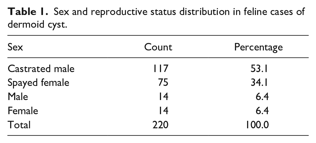
The age was available in 214 cases. The average age of affected cats was 5.5 years, ranging from 6 months to 18 years (±3.9 years standard deviation) (Supplemental Table S1). The sex distribution of the studied population (Table 1) was 59.5% males (131/220) from which 117 (53.1%) were castrated and 14 (6.4%) were intact males. Females were 40.5% (34.1%; 75/220 spayed and 6.4%; 14/220 intact females). Domestic short hair was the most commonly reported breed (56.4%; 124/220), followed by domestic long hair (8.6%; 19/220), domestic medium hair (6.8%; 15/220), Persian (2.3%; 5/220), and Russian blue and Siamese (1.8%; 4/220 each). Maine coon and Scottish fold were represented by 3 individuals each (1.4%). Two individuals were identified as “mixed breed” (0.9%). The remaining breeds (ragdoll, sphynx, Abyssinian, Bengal, Burmese, Manx, Oriental, and Ukrainian Levkoy) were represented by 1 or 2 individuals each. The breed was not specified in 14.1% (32/220) of the cases (Table 2).
Sex and reproductive status distribution in feline cases of dermoid cyst.
Distribution of frequency and percentage of cat breeds with dermoid cyst diagnosis.
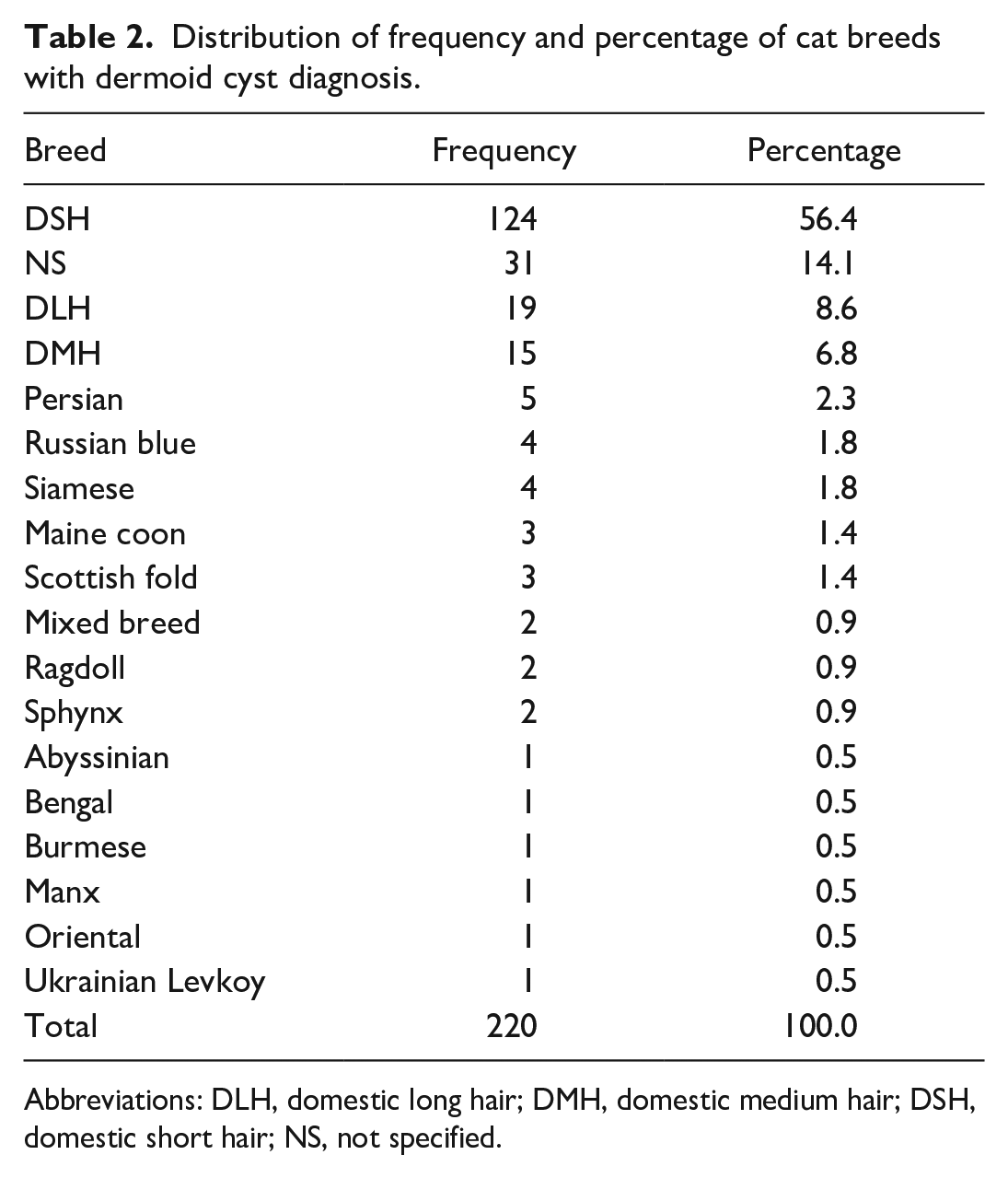
Abbreviations: DLH, domestic long hair; DMH, domestic medium hair; DSH, domestic short hair; NS, not specified.
Dermoid cysts were described in the skin and subcutaneous tissue in 99.5% (219/220) of the cases. There was only an extracutaneous dermoid cyst in the mesentery (case 216). In 11 cases (5%), the anatomic location in the skin was not specified. The most common anatomical region was the neck (55.9%; 123/220) (Fig. 1a, Supplemental Table S1). In the neck, 24.1% (53/220) were described in the ventral region, 15% (33/220) lateral right, 8.6% (19/220) lateral left, 2.3% (5/220) lateral without specification, and 2.3% (5/220) in dorsal neck. Only 8 (3.6%) cases were described as “neck” cysts without further specifications. A total of 12.2% (27/220) of the cases were located at the head (
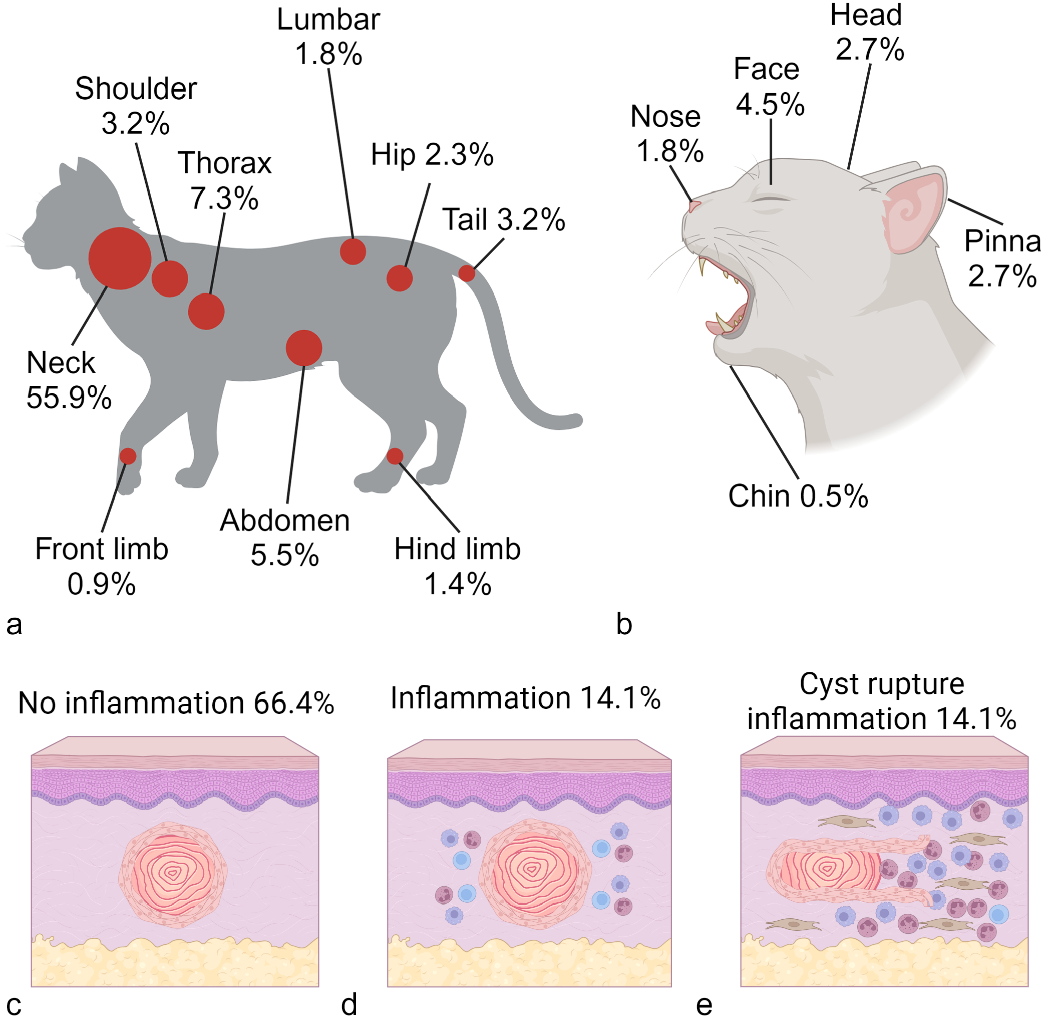
Feline dermoid cysts. (
The dermoid cyst size was recorded in 217 cases, with an average diameter of 1.4 cm, ranging from 0.25 to 6 cm (±0.9 cm standard deviation) (Supplemental Table S1). A 6-cm diameter dermoid cyst was removed from the left caudal thorax (case 114). Inflammatory changes were not reported in 66.4% (146/220) of the cases, whereas chronic stromal lymphoplasmacytic or mixed infiltrates were documented in 14.1% (31/220) of the cases; an equal percentage was recorded in cases with rupture of the cyst wall, marked pyogranulomatous inflammation, and proliferation of organizing granulation tissue (
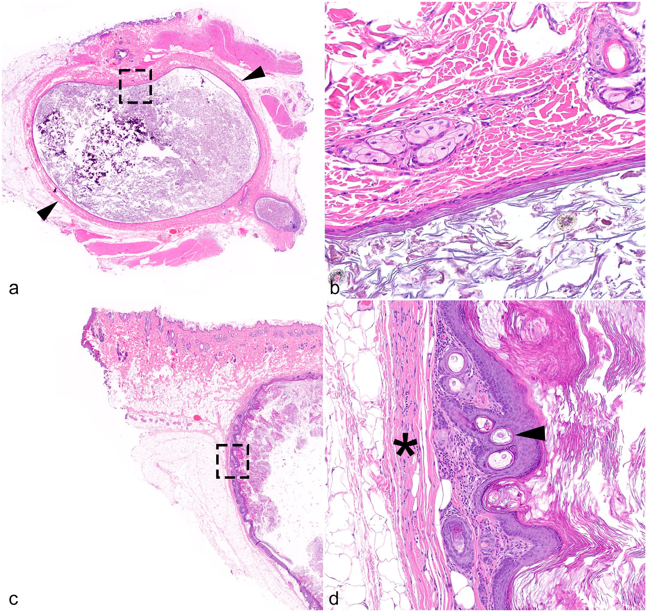
Histologic features of feline dermoid cyst. Hematoxylin and eosin. (
The anatomical location of the dermoid cyst did not differ significantly among sexes (Chi-square test, P = .840), breeds (Chi-square test, P = .999), ages (Chi-square test, P = .627), and other histological findings related to the cyst (Chi-square test, P = .363).
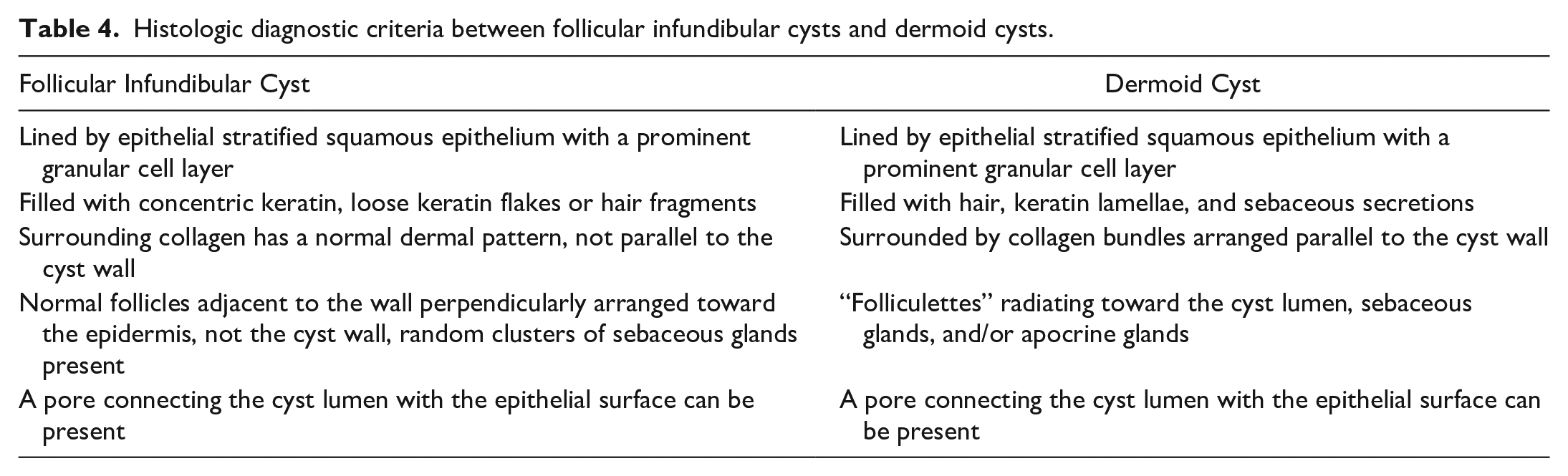
A total of 15 cases were retrieved after the literature review (Table 3). The main diagnostic criteria between infundibular cysts and dermoid cysts are summarized in Table 4.
Published cases of dermoid cysts in cats.
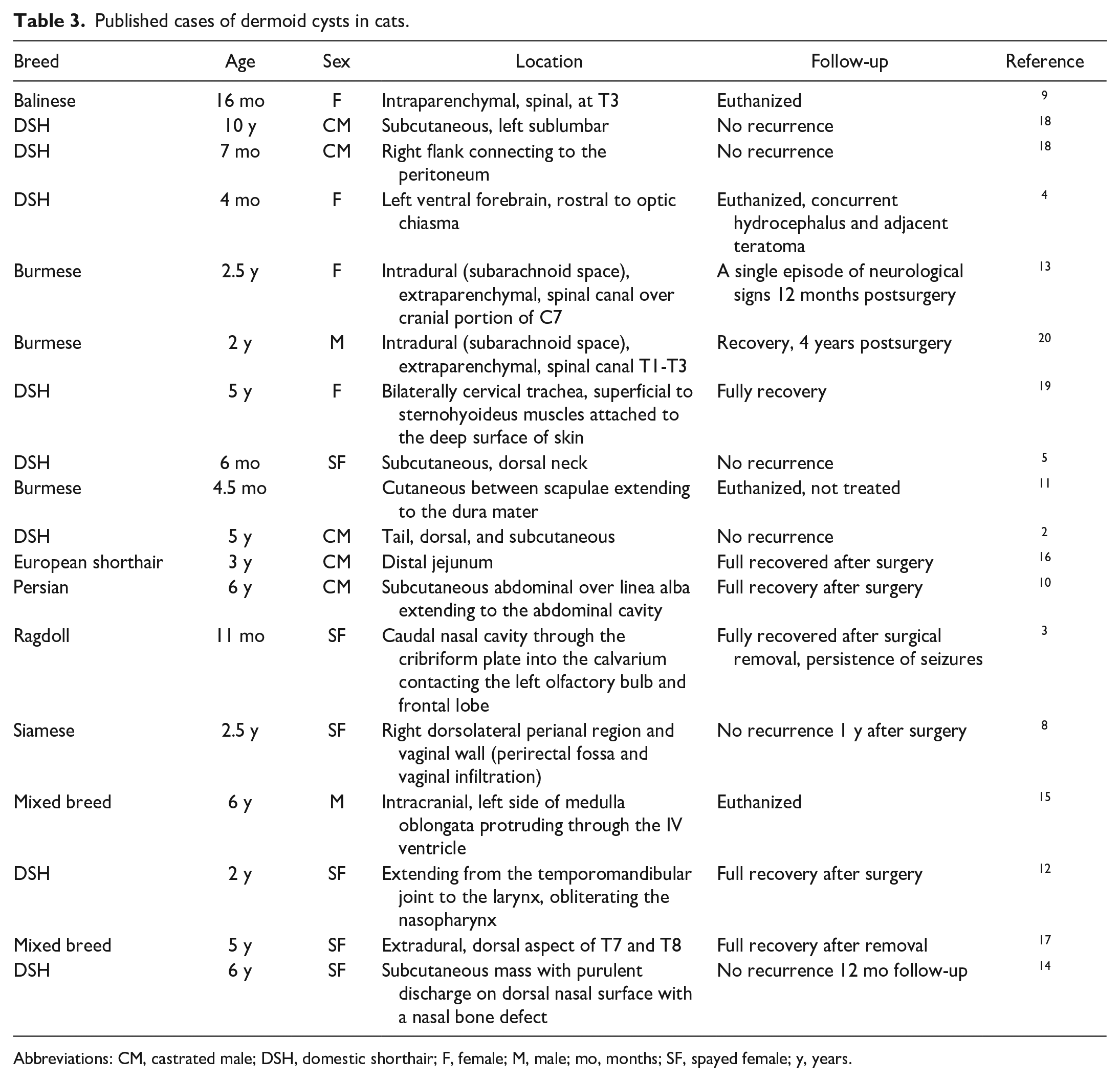
Abbreviations: CM, castrated male; DSH, domestic shorthair; F, female; M, male; mo, months; SF, spayed female; y, years.
Histologic diagnostic criteria between follicular infundibular cysts and dermoid cysts.
Discussion
Our results highlight important contrasts between the collected cases and the information available in the literature. Dermoid cysts are believed to affect younger dogs and cats since they represent a developmental abnormality. 21 Thus, these cysts are conventionally reported in young feline and canine patients (<2 years),6,21 with cases documented in cats as young as 4 months old. 4 The average age of the affected cats in our study was 5.5 years, suggesting that this lesion is either not noticeable or subclinical in most of the cases until the patients reached an older age. Sex predilection is not reported in cats, with almost 50% of the retrieved cases in the literature review as females.3–5,8,9,12–14,17,19 In our study, males were slightly more commonly affected than females (59.5% vs 40.5%), although this was not statistically significant. Breed predispositions are not determined in cats, 20 but the vast majority of our cases were domestic short hair cats (56.4%). Nevertheless, rather than a true breed predisposition, we suspect that domestic short hair cats are overrepresented in our studied population due to their abundance as pets. Dermoid cysts have been documented in several breeds besides domestic short hair cats, including Balinese, 8 Burmese,11,13,20 ragdoll, 3 European shorthair, 16 and Persian. 10 Our findings add to this list domestic longhair, domestic medium hair, Russian blue, Siamese, Maine coon, Scottish fold, Sphynx, Abyssinian, Bengal, Manx, Oriental, and Ukrainian Levkoy, suggesting that dermoid cysts can occur in a wide variety of breeds.
The skin and subcutis were the most common locations reported for our dermoid cysts, with 99.5% identified in these locations. The dorsal midline is the predilected site for the development of these lesions in dogs due to its pathogenesis. 6 However, in our study, only a few cases (8.2%) were specifically presented in the dorsal body midline, suggesting that cats do not have similar trends as dogs. They are reported to occur particularly in the lateral neck and shoulder of cats.7,20 Other cutaneous locations include the left sublumbar area, 18 bilateral cervical, 19 dorsal neck, 5 tail, 1 perirectal fossa, 8 and ventral abdomen. 10 The neck is the most common location for dermoid cysts in cats according to our results, with 55.9% reported in this region (24.1% in the ventral neck and 25.9% in the lateral neck). The shoulder was less frequently involved (3.2%) than the head (12.2%), thorax (7.3%), and abdomen (5.5%). Although more infrequent, in our study, these lesions were identified in other anatomical sites, including nose, chin, face, limbs, and pinna.
Extracutaneous locations reported for dermoid cysts in cats include the spinal cord (intradural and intraparenchymal, 9 intradural and extraparenchymal,11,13,20 and within the spinal canal 17 ), intracranial,4,15 pharyngeal, 12 nasal,3,14 and intestinal. 16 Rare case reports describe the extension of the cysts from subcutaneous tissues into the abdominal cavity and peritoneum.10,18 None of our cutaneous dermoid cysts extended to the internal anatomical structures. Only 1 case was noncutaneous and was in the mesentery. No associations between the location of the cysts and other factors like the age, sex, and breed of the cats were found in our study.
The main differential diagnoses for dermoid cysts are trichofolliculomas, infundibular cysts, and fibroadnexal hamartomas.6,21 The trichofolliculomas have more hair follicles radiating from the main cysts, and the epithelium is not exclusively a squamous epithelium with infundibular differentiation containing keratohyalin granules.7,20 Trichofolliculomas can be lined by epithelium similar to the isthmic and inferior segment of the hair follicle.7,20
The differentiation between dermoid cysts and infundibular cysts can be challenging. 7 Dermoid cysts have folliculettes and folliculosebaceous units radiating and perpendicularly oriented toward the cyst’s wall; infundibular cysts can have clusters of sebaceous glands adjacent to the wall, but the hair follicles are normally oriented toward the epidermal surface and not the cyst’s wall. 7 The concentric arranged collagen fibers at the periphery of the dermoid cysts is lacking in infundibular cysts. 7 Infundibular cysts were more common lesions than dermoid cysts. When comparing the diagnoses from the 2 different institutions within the same timeframe, the infundibular cyst to dermoid cysts ratio was 4:1 (46 infundibular cysts vs 12 dermoid cysts) in Athens Veterinary Diagnostic Laboratory and 4:1 (928 infundibular cysts vs 208 dermoid cysts) at Antech Diagnostics, Mars Petcare Science & Diagnostics. Fibroadnexal hamartomas might represent another differential diagnosis, particularly in older animals; however, the folliculosebaceous units in hamartomas are not oriented around a central cyst filled with keratin and hairs and are disorganized. 6
In cats, dermoid cysts are described clinically as small lesions, less than 2 cm in diameter, with a small pore connecting it with the surface.7,21 The presence of a pore connecting the lumen with the epithelial surface was evidenced in only 1.8% (4/220) of the cases in our study; thus, we consider this feature not common in this lesion. The average size of the cysts in our study was 1.4 cm; however, we documented a case that was 6 cm in diameter. The cysts are amenable to surgical excision, which is considered curative in most cases, and do not represent a life-threatening condition, unless lesions occur in the intracranial or spinal location, which typically leads to euthanasia of the patient.9,4,15 If the dermoid cyst is incompletely excised it might recur. 6 The rupture of the cyst will release keratin and hairs into the cutaneous or subcutaneous stroma, leading to a foreign-body inflammatory response with large infiltrates of macrophages and neutrophils.7,6 Inflammatory changes in association with the cysts were observed in 38.2% of our cases, with severe pyogranulomatous inflammation in association with the wall rupture in 14.1% of cases. In most cases (66.4%), secondary inflammatory changes were not reported.
In summary, in a cohort of 220 cats, dermoid cysts most commonly occurred in the ventral and lateral neck regions, with the domestic shorthair cat being the most represented breed. Affected animals were, on average, 5.5-years-old at the time of diagnosis. Most lesions lacked an inflammatory response and were less than 2 cm in diameter, often remaining unnoticed. A connecting pore with the epithelial surface was found to be an uncommon histological feature of dermoid cysts in cats. For diagnosis, the pathologist should evaluate specific features, such as hair folliculosebaceous units oriented perpendicularly to the cyst wall and surrounding collagen bundles aligned parallel to the cyst wall.
Supplemental Material
sj-pdf-1-vet-10.1177_03009858251317457 – Supplemental material for Feline dermoid cyst: Retrospective case series and literature review
Supplemental material, sj-pdf-1-vet-10.1177_03009858251317457 for Feline dermoid cyst: Retrospective case series and literature review by Daniel Felipe Barrantes Murillo, Daniel R. Rissi, Dominique J. Wiener and Tatiane Terumi Negrão Watanabe in Veterinary Pathology
Footnotes
Author Contributions
DFBM and TTNW designed and supervised the study. DFBM, DRR, and TTNW collected the data and performed the histopathological evaluations. DFBM performed the data analysis. DFBM, DRR, DJW, and TTNW wrote the manuscript. All the authors reviewed and critically edited the manuscript. All authors read and approved the final manuscript.
Declaration of Conflicting Interests
The author(s) declared the following potential conflicts of interest with respect to the research, authorship, and/or publication of this article: TTNW is employed at Antech Diagnostics, Mars Petcare Science & Diagnostics. The remaining authors declared no potential conflicts of interest concerning the research, authorship, and/or publication of this article.
Funding
The author(s) received no financial support for the research, authorship, and/or publication of this article.
Supplemental material for this article is available online.
References
Supplementary Material
Please find the following supplemental material available below.
For Open Access articles published under a Creative Commons License, all supplemental material carries the same license as the article it is associated with.
For non-Open Access articles published, all supplemental material carries a non-exclusive license, and permission requests for re-use of supplemental material or any part of supplemental material shall be sent directly to the copyright owner as specified in the copyright notice associated with the article.