
Abstract
Trichuris spp. infections can cause typhlitis or typhlocolitis in many species, but there are no published studies about its pathology in cats. Trichuris sp. infection in cats appears to be rare in most parts of the world but is frequent in some tropical and subtropical regions. The purpose of this study was to describe intestinal lesions associated with natural Trichuris sp. infections in cats of St. Kitts, West Indies. Comprehensive autopsies, histopathological assessment of small and large intestine, and total worm counts were performed in a cross-sectional study of 30 consecutive feline mortalities. Trichuris were found in 17 of 30 (57%; 95% confidence interval, 39%–74%) of the study cats with a median worm count of 11 (range, 1–170), indicating most cats had a low-intensity infection. Trichuris infection was associated with typhlitis but not consistency of feces or body condition score. In most cats examined, the typhlitis was categorized as mild (10/15, 67%) and, less frequently, moderate (2/15, 13%) or marked (3/15, 20%). The inflammatory infiltrate varied from predominantly eosinophilic (5/15, 33%) to neutrophilic (4/15, 27%), a mixture of eosinophilic and neutrophilic (2/15, 13%), a mixture of neutrophilic and lymphoplasmacytic (1/15, 7%), or a mixture of eosinophilic, neutrophilic, and lymphoplasmacytic (3/15, 20%). In some cats, surface erosions and catarrhal exudate were adjacent to adult worms. These findings are similar to those reported with low-intensity Trichuris infections in other species.
Observations of Trichuris eggs (proposed synonyms Trichuris felis, Trichuris campanula, Trichuris, order Trichinellida) in the feces of cats have been reported from around the world. While infection is considered rare in most locations, there are a few studies demonstrating considerably higher prevalence in certain tropical and subtropical areas. 2,8,15,18,27 In addition, it has been hypothesized that the distribution and prevalence of feline Trichuris might be increasing. 8
Trichuris in cats was first described in 1851 and originally described as 2 different species (Trichuris serrata von Linstow 1879 and Trichuris campanula von Linstow 1889). This taxonomic classification has since been challenged due to overlapping morphological characteristics and incomplete descriptions, and the name Trichuris felis has been suggested. 2 Trichuris of cats, as with other Trichuris spp. of a variety of domestic and wild animals, has a direct life cycle and parasitizes the intestine. 2 The host becomes infected by ingestion of eggs containing first-stage larvae (L1), which hatch and enter the intestinal mucosa. 12,14 Even though the mucosa of both the small and large intestine can be penetrated, the development of larvae to adults typically takes place in the mucosa of the large intestine. 12,14 In low-intensity infections, worms are typically only seen within the cecum and proximal colon, whereas individuals infected with a high number of worms can have adult Trichuris embedded throughout the mucosa of the large intestine and sometimes terminal ileum. 3 Adult worms have a characteristic morphology, with a thin anterior end, containing an esophagus equipped with a distinctive stichosome, and a thick posterior end. As worms approach maturity, the enlarging posterior portion breaks out of the mucosa and protrudes into the intestinal lumen, while the thinner anterior end remains within the intestinal mucosa. Eggs are shed in the feces and develop to the infective stage in the environment. The prepatent period varies between species and has been reported to be between 62 and 91 days in cats. 11
Experimental Trichuris infection in dogs and pigs, as well as natural infections in other domestic animals, can cause mucohemorrhagic typhlocolitis associated with chronic diarrhea and weight loss. 12,30 Little is known about the impact of Trichuris in cats, although it is typically considered of low clinical importance. 2 A parasitological survey of 50 stray cats in Argentina 27 mentioned that cats with Trichuris infections (median number of adult Trichuris 23.5, range 1–113) had grossly thickened ceca, pale nodules visible from the serosal surface and cecal mucosal petechiation at the site of worm implantation. However, there are no other studies of the pathology or clinical signs associated with Trichuris infection in cats. This study compared the presence of Trichuris as determined by total worm counts to the presence of intestinal inflammation determined by autopsy and histopathological grading, using feline mortalities in an area with previously demonstrated high prevalence of Trichuris infection. 15,18
Materials and Methods
The study population consisted of 30 cats, 14 consecutively submitted for autopsy to the pathology department of Ross University School of Veterinary Medicine (RUSVM) between January 1 and July 16, 2018, and for which owner consent was provided, and 16 roadside stray cat mortalities during the same period. Permission to collect roadside mortalities was obtained from the RUSVM Institutional Animal Care and Use Committee (IACUC). For the sake of generalizability, exclusion criteria were limited to ethical reasons (owners not giving consent to participate, for nonstray cats) and inability to provide good data (advanced stage of postmortem decomposition or ruptured gastrointestinal tract). Advanced stage of postmortem decomposition was defined before data collection as being after rigor mortis (flaccid skeletal musculature in the presence of signs of postmortem autolysis). After assessment of histological sections, the definition was extended to include cats in which the mucosa was completely lost in all large intestinal sections.
Age and body condition were assessed for each cat prior to autopsy. Body condition score (BCS) was recorded on a scale from 1 to 5 (1 = emaciated, 2 = below ideal, 3 = ideal, 4 = above ideal, 5 = obese). Age was estimated based on appearance and dentition and categorized into 5 groups (<6 weeks, 6 to 16 weeks, 16 weeks to 6 months, >6 months to 1 year, above 1 year).
A comprehensive autopsy was performed for each cat. Prior to collecting sections for histopathology, consistency of fecal material, if present, was recorded on a scale of 0 to 3 (0 = solid, 1 = semisolid, 2 = viscous, 3 = watery). Sections of stomach, duodenum, jejunum, ileum, cecum, proximal colon, distal colon, and rectum were fixed in 10% neutral-buffered formalin, routinely processed, and embedded in paraffin wax. Sections cut 4 µm thick were stained with hematoxylin and eosin and examined after all 30 cats were autopsied, using an Olympus BX51 microscope (Olympus corporation, Tokyo, Japan) with a standard field number of 22. Autolysis for each section was categorized as absent (if superficial epithelium was intact), mild (if there was loss of superficial epithelium), moderate (if there was loss of villi or superficial crypts), and marked (if there was loss of mucosa). Intestinal sections with absent to moderate autolysis were evaluated for mucosal fibrosis, crypt dilation/distention and crypt hyperplasia, and lamina propria cellular infiltrates following the histopathological standards for the diagnosis of gastrointestinal inflammation issued by the World Small Animal Veterinary Association group. 4 Furthermore, small intestinal sections with absent to mild autolysis were evaluated for villous stunting, intraepithelial lymphocytes, and lacteal dilation using the same guidelines. 4 Parameters evaluated without the use of the guidelines included presence of globular leukocytes, surface mucus admixed with neutrophils, lymphoid aggregates/hyperplasia, crypt elongation, and crypt herniation. Lamina propria lymphocytes, plasma cells, eosinophils, and neutrophils for each anatomical section were classified as normal (0), mildly (1), moderately (2), or markedly (3) increased based on number (per high-powered field). The guidelines for duodenum were used for all sections of small intestine, and the guidelines for colon were used for all sections of large intestine (ie, cecum and colon). When classifying intestinal sections, evaluation focused on areas of the section least obscured by autolysis and most representative of the overall pathology observed across the section.
Stomach, small intestine, and large intestine were soaked in saline for a minimum of 3 hours. The saline was poured over a 100-µm sieve to collect detached Trichuris and the gastrointestinal tract washed over a 100-µm sieve while gently scraping the mucosa with the contents of both sieve collections backwashed and fixed in 5% to 10% formalin. Total adult Trichuris worm counts were performed for all cases in a blinded manner. Trichuris in samples collected for histopathology were recorded separately and added to the total worm count. Additional nematodes (other than Trichuris) were also counted and the presence of other helminths (eg, cestodes and trematodes) recorded. The number of cestodes per host was not recorded because the methods used for nematode collection made scolex collection challenging. For helminths that were predominately (Platynosomum fastosum) or exclusively (Strongyloides sp.) seen in tissue section, the number of helminths per individual was not quantified.
Helminth infections were reported as prevalence with 95% confidence intervals, along with the numerator and denominator from which they were derived. For the parasites where quantification was possible, median and range of number of worms were chosen as measures of central tendency and dispersion rather than mean and standard deviation, as they are more robust for parasite population data, which tend to be overdispersed, resulting in outliers. 20 The association between Trichuris infection and stray status, age group, body condition score, feces score, presence of gastritis, enteritis, typhlitis, cecal lymphoid hyperplasia, and colonic lymphoid hyperplasia were examined with a nondirectional Fisher’s exact probability test using an online calculator. 21 For parameters where multiple levels were included (age group, body condition score, and feces score), a Freeman-Halton extension of the Fisher’s exact probability test was used. 22 To account for multiple testing, the significance level was adjusted from .05 to .005 with a Bonferroni correction.
All procedures were performed in accordance with local laws and regulations and following an approved RUSVM IACUC protocol.
Results
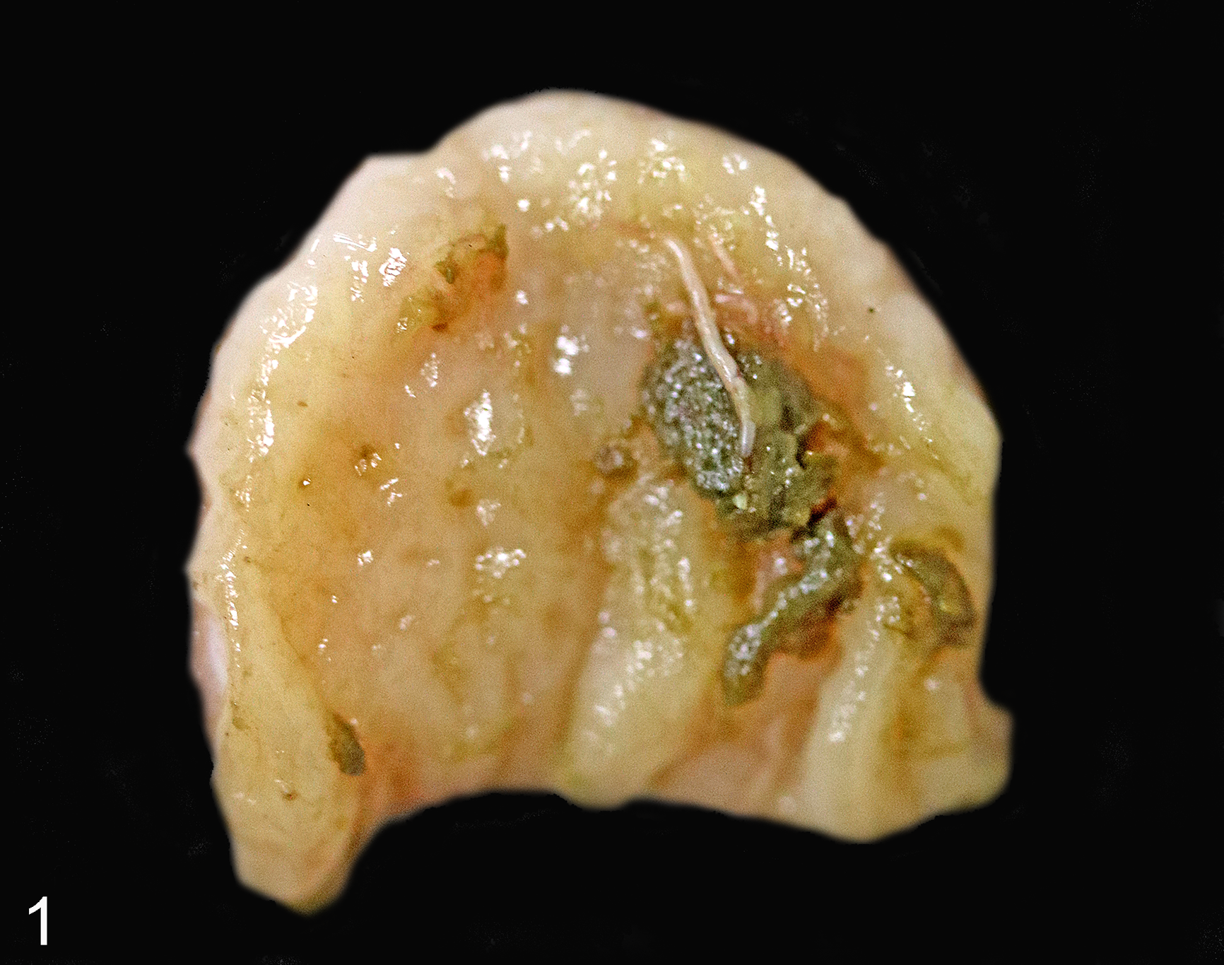
The study population consisted mostly of young cats, and the majority of cats were infected with Trichuris (Table 1). Almost all study cats (28/30, 93%; 95% confidence interval [CI], 84%–102%) had gastrointestinal helminth infections, and most of these (23/28, 82%; 95% CI, 68%–96%) had mixed infections with 2 to 6 different helminth species (Table 1). Trichuris sp. infection was present in 17 of 30 (57%; 95% CI, 39%–74%) of cats examined. Median number of adult Trichuris per cat was 11 (range, 1–170), with only 1 cat having more than 100. This cat had adult Trichuris in both the small and the large intestine and was the only cat in which adult Trichuris were observed grossly in locations other than the cecum. The cat was in adequate body condition (BCS 3/5) and had formed feces (feces score 0/3), and the large intestinal mucosa appeared normal on gross examination. The thick, posterior part of adult Trichuris could sometimes be observed grossly within the cecum but was often completely or partly obscured by feces (Fig. 1). Grossly appreciable typhlitis was seen in only 1 cat (with 62 adult Trichuris sp.), which had hemorrhagic cecal content but also hemorrhagic enteritis attributed to heavy Ancylostoma tubaeforme infection. Cecal mucosal lymphoid tissue was grossly evident in 19 of 30 (63%) study cats, including 10 that were not infected with Trichuris, and appeared as thickening and pale tan discoloration of the cecal wall, sometimes with coalescing flat mural nodules 1 to 2 mm in diameter.
Prevalence, Intensity, and Anatomic Location of Intestinal Helminth Infections (Based on Total Worm Counts and/or in Tissue Section) in 30 Cats.
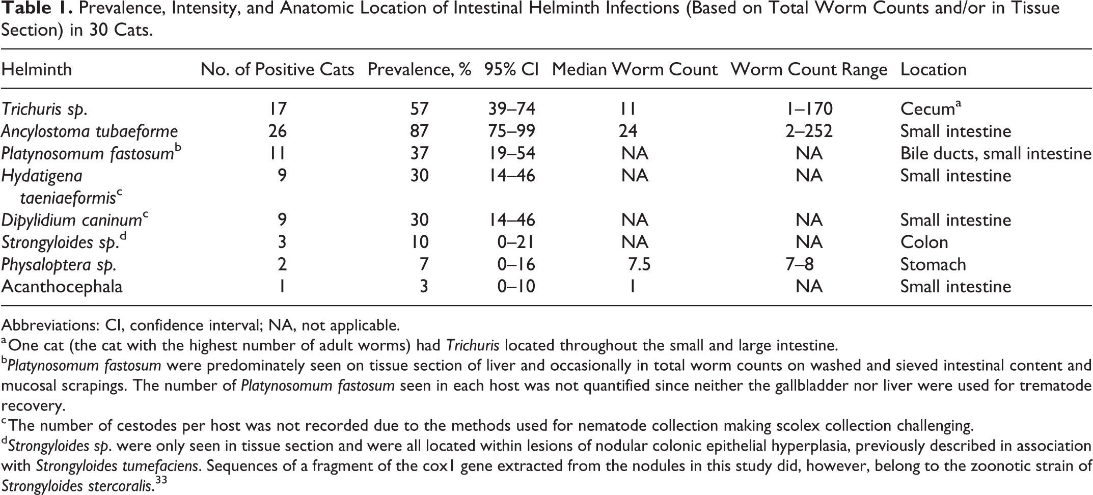
Abbreviations: CI, confidence interval; NA, not applicable.
a One cat (the cat with the highest number of adult worms) had Trichuris located throughout the small and large intestine.
b Platynosomum fastosum were predominately seen on tissue section of liver and occasionally in total worm counts on washed and sieved intestinal content and mucosal scrapings. The number of Platynosomum fastosum seen in each host was not quantified since neither the gallbladder nor liver were used for trematode recovery.
c The number of cestodes per host was not recorded due to the methods used for nematode collection making scolex collection challenging.
d Strongyloides sp. were only seen in tissue section and were all located within lesions of nodular colonic epithelial hyperplasia, previously described in association with Strongyloides tumefaciens. Sequences of a fragment of the cox1 gene extracted from the nodules in this study did, however, belong to the zoonotic strain of Strongyloides stercoralis. 33
There was no apparent difference in the distribution of body condition score or consistency of feces among Trichuris sp. positive and negative cats (Table 2). All study cats had histologically apparent inflammation in at least 1 part of the gastrointestinal tract, most commonly the small intestine. Only typhlitis, seen in 15 of 27 (56%) cats, was associated with Trichuris infection (P = .002) (Table 2). Typhlitis was mild (10/15, 67%), moderate (2/15, 13%), and marked (3/15, 20%) and was categorized as eosinophilic (5/15, 33%), neutrophilic (4/15, 27%), a mixture of eosinophilic and neutrophilic (2/15, 13%), a mixture of neutrophilic and lymphoplasmacytic (1/15, 7%), or a mixture of eosinophilic, neutrophilic, and lymphoplasmacytic (3/15, 20%). The cat with the highest infection intensity (170 adult worms) had a moderate eosinophilic and mild neutrophilic and lymphoplasmacytic typhlitis. The 2 other cats with moderate typhlitis and the 2 cats with marked typhlitis all had Trichuris counts below 30. The majority of the cats with moderate or marked typhlitis (4/5, 80%) were in good body condition (BCS 3/5) and had solid feces (feces score 0/3), whereas only 1 had a body condition score below ideal (BCS 2/5) and mucoid feces (feces score 2/3). Other histologic cecal lesions in cats with typhlitis included surface mucus admixed with neutrophils (ie, catarrhal exudate) in 4 of 15 (27%) cats (Fig. 3) and mild (11/15, 73%) to moderate (2/15, 13%) crypt distention. Catarrhal exudate was not histologically evident in cats without Trichuris typhlitis, but mild crypt distention was seen in 10 of 12 (83%) of these cats. Mucosal fibrosis, crypt hyperplasia, or crypt elongation was not present in any cecal sections examined. Adult Trichuris, characterized by coelomyarian musculature, bacillary bands, and stichosomes, were seen in the cecal histological sections of 10 of 17 (59%) infected cats (Figs. 2, 3). In most of these (7/10, 70%), Trichuris were limited to the lumen and/or mucosa, whereas in the remaining 3 of 10 (30%), Trichuris were also within submucosal lymphoid tissue (Fig. 3) surrounded by a thin rim of necrotic debris. Due to suboptimal fixation of the lamina epithelialis, the presence of mucosal erosions was often not appreciable, but of the 6 cases with optimal fixation of the epithelium, superficial erosions adjacent to Trichuris were seen histologically in all 3 cats with Trichuris and none of the 3 cats without Trichuris (Fig. 2).
Association Between Trichuris Status and Other Measured Parameters in 30 Cats.
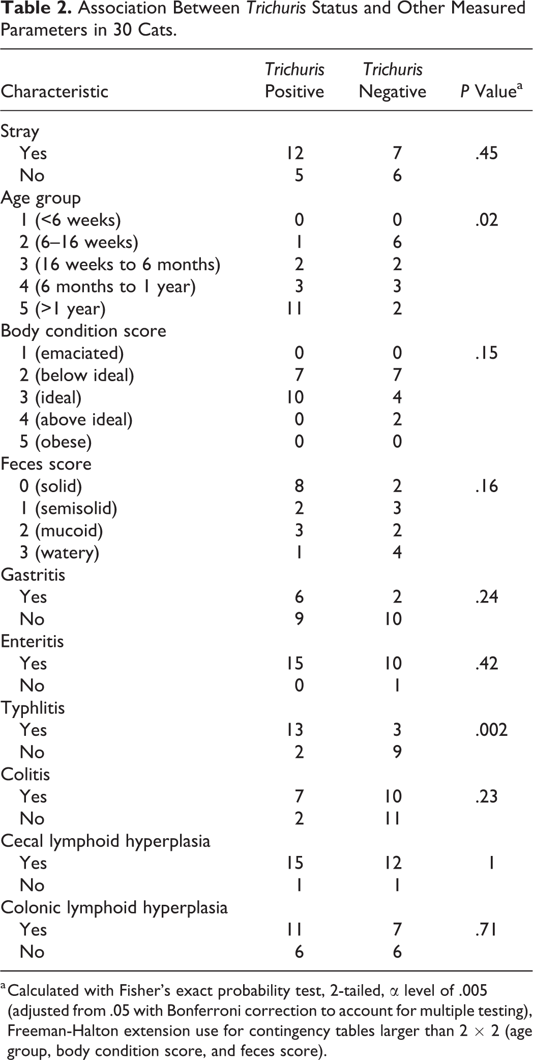
a Calculated with Fisher’s exact probability test, 2-tailed, α level of .005 (adjusted from .05 with Bonferroni correction to account for multiple testing), Freeman-Halton extension use for contingency tables larger than 2 × 2 (age group, body condition score, and feces score).
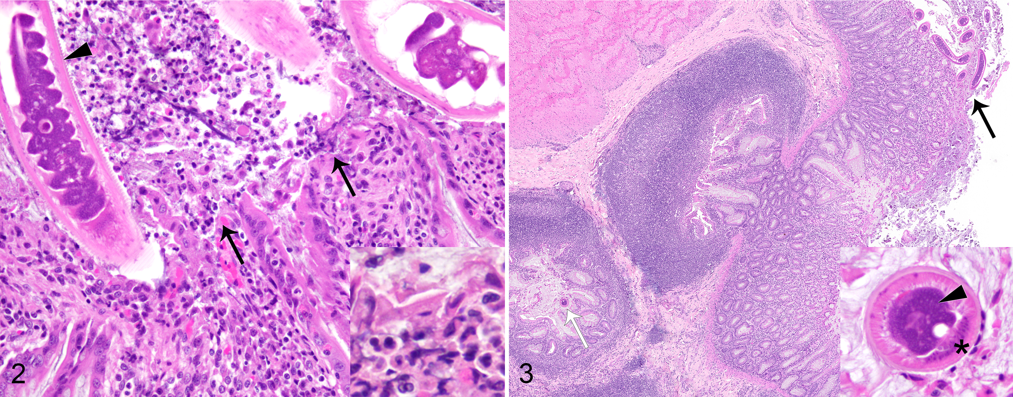
Trichuris adults are present in the lumen and within the mucosa. There is moderate neutrophilic and lymphoplasmacytic typhlitis. The Trichuris adult in the lumen is surrounded by catarrhal exudate (inset: neutrophils, necrotic cell debris, and mucus), and the mucosa adjacent to the Trichuris adult is eroded (arrows). Hematoxylin and eosin (HE).
Discussion
This is the first report of histologic lesions associated with Trichuris sp. infections in cats. In this study, Trichuris was consistently localized to the cecum and was associated with typhlitis, similar to several other species. 3,10,16
Cats in this study did not show gross lesions as a result of Trichuris infection, other than the presence of adult worms. This is in contrast to other host species where muco-hemorrhagic colitis and typhlocolitis are known to occur with Trichuris infection and are indicated by the presence of thickened, red, and edematous mucosa and hemorrhagic/mucoid large intestinal content. 7,25,28,31 However, these changes are typically seen with high-intensity infections (hundreds to thousands of adult worms), 5,7,9,16,25,28,29,31,32 and the absence of them may be due to the low-infection intensity in the present study where most cats (29/30) were infected by <100 worms. Trichuris-infected cats are reported to have thickened ceca with pale cecal nodules visible from the serosal side. 27 Pale cecal nodules visible from the serosal side (albeit most commonly coalescing, giving the cecal wall a diffuse white thickened appearance) were frequent in the current study but not associated with Trichuris infection. Histologically, these nodules consisted of lymphoid tissue, a normal component of the cecal wall. Prominent cecal lymphoid tissue also occurs in experimental Trichuris infections in dogs, 13,26 but since the dog studies lacked control groups, it is possible that the conditions coexisted and were not associated.
In the study presented here, as in most other reports from humans and domestic animals, 3,12,30 adult Trichuris and lesions were predominantly seen in the mucosa. However, in 3 cats, adult Trichuris were seen within submucosal lymphoid tissue where they were surrounded by a rim of necrotic tissue. This also occurred in a minority of experimentally infected dogs and pigs. 10,13,26 Although associated lesions are typically mild, it demonstrates the ability of adult Trichuris to penetrate deeper layers of the intestinal wall. Trichuris located in submucosa and tunica muscularis, surrounded by areas of fibrosis, granulomatous inflammation, and sometimes abscessation, occurs rarely in naturally infected dogs and cattle (with hundreds to thousands adult Trichuris). 16,29,32 These animals either die of the infection 16,29 or are euthanized due to being unresponsive to treatment. 32 Similar lesions also occur in humans with a high number of adult worms and secondary perforated bowel. 1,6
Despite the lack of gross lesions, most Trichuris-infected cats had microscopically evident mild eosinophilic and/or neutrophilic typhlitis. Lesions predominantly involved the mucosa, and, in addition to the inflammatory infiltrate in the lamina propria, catarrhal exudate and surface erosions adjacent to adult worms were seen in some cases. These findings are similar to what has previously been reported in pigs, dogs, and humans, 3,10,13,16,17,19,24 suggesting that the impact of infection on the feline host is probably similar to that observed in other species. The surface erosions seen in cats is this study were relatively mild and not associated with crypt elongation or crypt hyperplasia, a feature noted in pigs and humans with Trichuris infection. 3,23
In the present study, the severity of typhlitis did not seem related to the number of adult worms present. Four cats with moderate to severe typhlitis had Trichuris counts below 30, indicating that significant lesions can be observed in some cats with low-intensity infection. Possible contributors to typhlitis (such as bacterial pathogens) were not investigated and may have contributed to lesion severity in these cases. Trichuris were not grossly or histologically observed in 4 of 13 (31%) cats with typhlitis, which were later determined to have Trichuris after saline soaking and washing of the mucosa and intestinal contents. Trichuris infection should thus not be ruled out based on absence of parasite sections in cecal biopsies.
Although not statistically significant, Trichuris infection appeared to be less common in the youngest age group in our study (cats estimated to be below 16 weeks old) while most older cats were infected. This likely reflects the prepatent period (>60 days) and the increased opportunity for exposure over time.
In conclusion, this study demonstrated an association between feline Trichuris infection and typhlitis, which was most commonly mild. The study did not show an association between Trichuris infection and fecal consistency or body condition. Further study of the association between Trichuris status and clinical disease measurements, in addition to those assessed postmortem, is warranted to better elucidate its impact on the feline host.
Supplemental Material
Supplemental Material, Typhlitis_associated_with_natural_Trichuris_sp._infection_in_cats_-_supplemental_data - Typhlitis Associated With Natural Trichuris sp. Infection in Cats
Supplemental Material, Typhlitis_associated_with_natural_Trichuris_sp._infection_in_cats_-_supplemental_data for Typhlitis Associated With Natural Trichuris sp. Infection in Cats by Judit M. Wulcan, Jennifer K. Ketzis and Michelle M. Dennis in Veterinary Pathology
Footnotes
Acknowledgements
Terje Magnusson and Cyndie Demming assisted with cat collection. Samantha Zayas, Maurice Matthew, Candita Chapman, and Randel Thompson assisted with autopsies. David Hilchie assisted with histological processing.
Declaration of Conflicting Interests
The author(s) declared no potential conflicts of interest with respect to the research, authorship, and/or publication of this article.
Funding
The author(s) disclosed receipt of the following financial support for the research, authorship, and/or publication of this article: This research was funded by the Integrative Mammalian Research Center, Ross University School of Veterinary Medicine, St. Kitts, West Indies.
Supplemental Material
Supplemental material for this article is available online.
References
Supplementary Material
Please find the following supplemental material available below.
For Open Access articles published under a Creative Commons License, all supplemental material carries the same license as the article it is associated with.
For non-Open Access articles published, all supplemental material carries a non-exclusive license, and permission requests for re-use of supplemental material or any part of supplemental material shall be sent directly to the copyright owner as specified in the copyright notice associated with the article.