
Abstract
Hepatocellular carcinoma (HCC) with metastasis to the spleen in a Holstein cow was studied by histopathologic and immunohistochemical methods. The tumor was characterized by a pseudoglandular (acinar) pattern with an associated fibrous stroma. Individual cells often had a “hepatoid” appearance but were interspersed with scattered cells exhibiting a clear, periodic acid-Schiff (PAS)-positive cytoplasm and small eccentric nuclei. This pattern was present in nodules found in both liver and spleen. Moreover, hepatoid tumor cells were positive for alpha-fetoprotein. Immunohistochemical studies suggest that myofibroblasts were responsible for the production of fibrous septa surrounding the pseudoglandular structures of bovine HCC. In summary, our histologic and immunohistochemical findings support a diagnosis of primary HCC with splenic metastasis. Furthermore, the associated stromal response appears to be of a myofibroblast origin. The primary etiology of bovine HCC and the significance of the intralesional, PAS-positive clear cells remain undetermined.
Hepatocellular carcinoma (HCC) has been reported in various species of animals including dogs, cats, sheep, pigs, fowl, woodchucks, and trout. 6 There are relatively few reports on bovine HCC. 3, 5 Incidence data and age distribution of HCC in cattle is not known because most cases were obtained at the time of slaughter.
This report represents the gross, histopathologic, and immunohistochemical findings of a bovine HCC with metastasis to the spleen, found in a Holstein cow during a routine postmortem inspection at an abattoir. The cow was of normal appearance at the time of slaughter, without any preexisting medical conditions. We also describe the myofibroblasts' involvement in the production of a fibrous stroma of bovine HCC.
A 4-year-old Holstein cow was slaughtered. Postmortem inspection of the carcass revealed small white-gray nodules, ranging in size from several millimeters to 1 cm in diameter, randomly scattered on the liver surface. Also, there was peritonitis. However, the lungs appeared grossly normal. The nodules were nonencapsulated and poorly demarcated from the surrounding liver tissue. The spleen was enlarged, with hemorrhagic lymphoid follicles. The cut surface of the spleen revealed hemorrhaging and bulging lymphoid follicles, 0.3–0.5 mm in diameter.
Representative sections of the liver and spleen were fixed immediately in 10% neutral-buffered formalin, processed routinely, and embedded in paraffin. Tissue sections were cut to 4 μm thickness and stained with hematoxylin and eosin, periodic acid–Schiff (PAS), and Azan stain. For immunohistochemical studies, the primary antibodies used were: anti–α-smooth muscle actin (α-SMA) at a dilution of 1 : 800 (clone 1A4, Sigma Chemical Co., St. Louis, MO), anti-CD68 (EBM11) at a dilution of 1 : 80, alpha-fetoprotein (AFP) at a dilution of 1 : 800 (Dako, Carpenteria, CA), and anti-cytokeratin 18 (CK18) at a dilution of 1 : 100 (Novo-castra Laboratories Ltd., Newcastle Upon Tyne, UK). The antigen-antibody complex was observed by the labeled streptavidin-biotin method using a Histostain-plus bulk kit (Zymed Laboratories Inc., San Francisco, CA), with 3,3′-diaminobenzidine. Immunohistochemically stained sections were counterstained with Mayer's hematoxylin.
Microscopically, nodules in the liver were composed of ovoid to polygonal “hepatoid” tumor cells with pseudoglandular structures, and the nests or clusters of these cells were embedded in the fibrous stroma. Moderately differentiated tumor cells resembled normal hepatocytes. They were more rounded, with eosinophilic cytoplasm, large oval nuclei and nucleoli. The glandular structures were irregular in size and shape. Lymphatic invasion by tumor cells was observed in the portal regions. A few tumor cells in the pseudoglandular structure had clear cytoplasm with a small eccentric nucleus (Fig. 1). Scattered necrotic areas and infiltration of mono-nuclear cells and lymphocytes in the stroma were also present. Accumulation of bile pigment or mucin was not observed within the pseudoglandular regions. In the spleen, hemorrhage and macrophages filled with hemosiderin were present around the follicular areas, and lymphocytes within white pulp regions were replaced by tumor cells. The shape and size of these cells were morphologically similar with the tumor cells found in the liver. Scattered cells with clear cytoplasm were also observed. The clear cells found in liver and spleen had strong PAS staining, with many purple-colored granules found in the cytoplasm (Fig. 2). This reaction suggests that most of these granules consisted of glycogen deposits. Collagen fibers and α-SMA–positive cells were found in the fibrous stroma that surrounded the pseudoglandular tumor cell clusters in the liver (Fig. 3). A positive reaction for CD68, however, was rarely seen in the clusters although it was observed diffusely in the Kupffer cells of normal hepatic tissue. Moreover, tumor cells stained positively for CK18 and AFP (Fig. 4) in both the organs. It is believed that the collective data presented support a diagnosis of primary HCC with splenic metastasis.
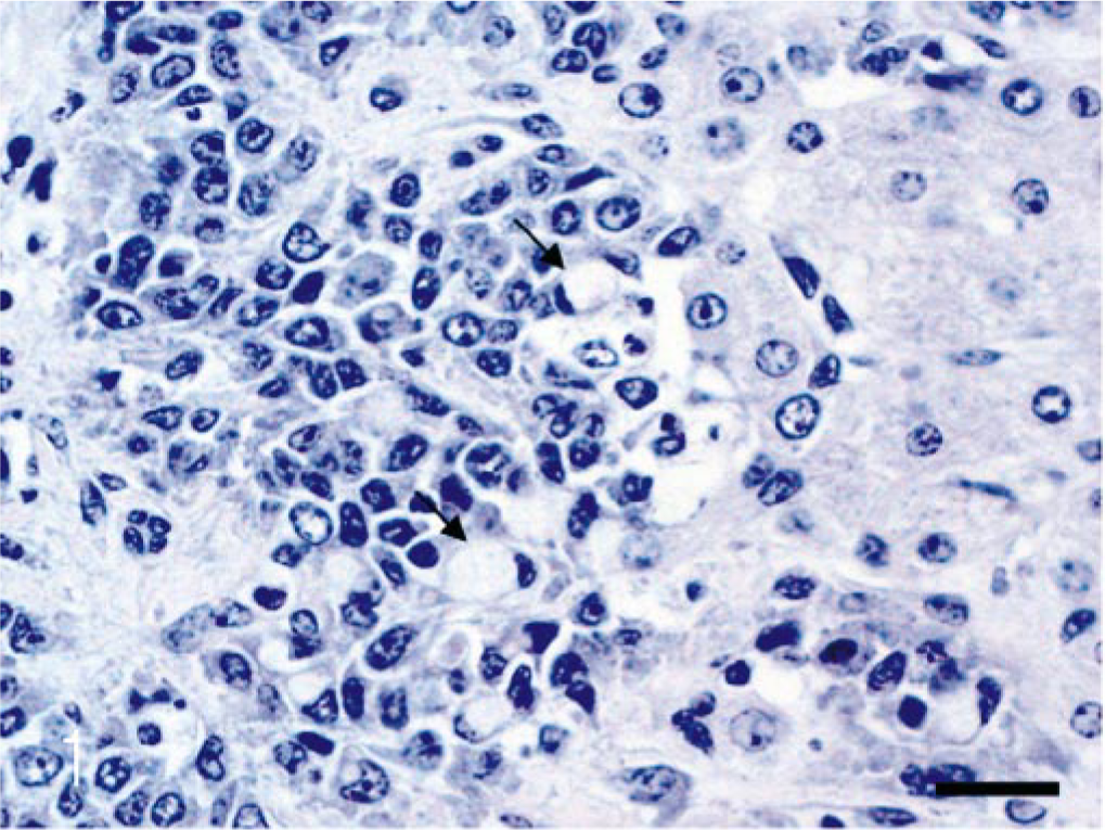
Liver; cow. Tumor cells (arrow) in a pseudoglandular structure had vacuolated cytoplasm and small eccentric nucleus. HE. Bar = 25 μm.
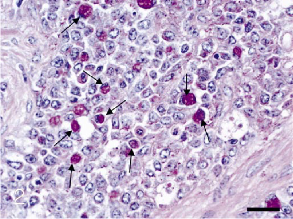
Liver; cow. Some cells (arrows) with vacuolated cytoplasm in pseudoglandular structures in the liver showing PAS-positive reaction. PAS. Bar = 25 μm.
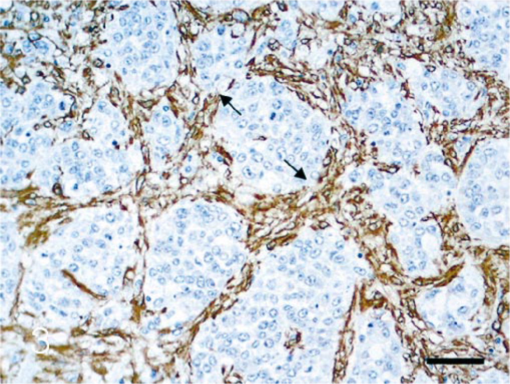
Liver; cow. α–SMA–positive cells (arrow) were detected in the fibrous stroma of HCC. Immunohistochemistry; DAB, hematoxylin counterstain. Bar = 50 μm.
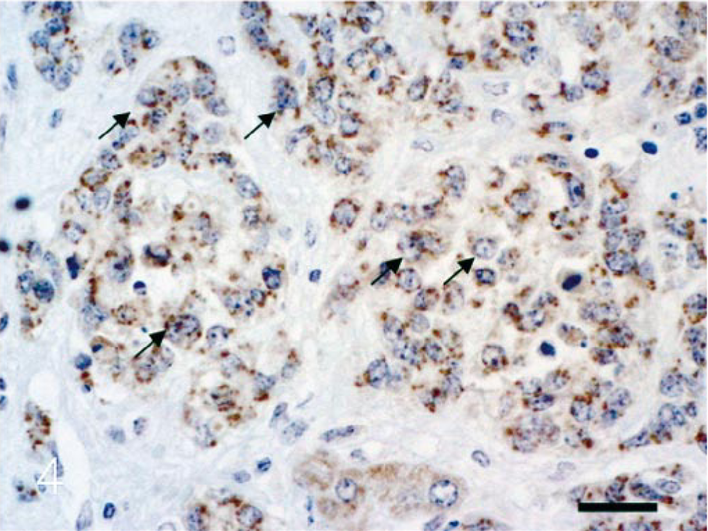
Liver; cow. AFP were detected in tumor cells (arrow) enclosed fibrous stroma. Immunohistochemistry; DAB, hematoxylin counterstain. Bar = 25 μm.
HCC progressively invades the adjacent hepatic tissue, and this attack tends to occur in clusters of neoplastic cells and rarely in individual neoplastic cells. 6 Metastasis of HCC occurs primarily in the lungs and regional lymph nodes through blood vessels and lymphatics, and a vascular invasion is more common than lymphatic invasion. 6 It has been also reported to metastasize in the spleen, kidneys, bones, and pancreas. 6 In our case, metastasis might occur through lymphatics because tumor cells were observed in the lymph vessels of the hepatic portal regions and in several follicles of the spleen. It is often difficult to distinguish HCC with a pseudoglandular pattern from cholangiocarcinoma. Intraluminal mucin accumulation in tubular/glandular structures was a useful histologic feature in cholangiocellular carcinoma. 9, 11 AFP and CK18 are also known as tumor markers of HCC. 3, 4, 8
Currently, it has been reported and accepted that myofibroblasts play an important role in hepatic fibrosis for the production of collagen fibers resulting in fibrous septa or capsules. Specifically, myofibroblasts can express α-SMA, and α-SMA–positive spindle cells were detected in the areas of fibrosis, within the neoplastic regions. The formation of collagenous fibrous capsules or septa around HCC has been frequently observed in humans. 2, 6, 7 Recent evidence suggested that stromal collagen fibers in human HCC were produced by activated hepatic stellate cells (myofibroblasts) and that these cells were mostly α-SMA positive. 2, 7 In our study, collagen fibers and α-SMA–positive myofibroblasts were observed within the newly formed fibrous septa surrounding the pseudoglandular structure of bovine HCC, indicating that α-SMA–positive myofibroblasts produced collagen fibers as in humans. Some reports showed that there was a marked reduction in the number of Kupffer cells in poorly or moderately differentiated HCC. 3, 10 We used anti-CD68 antibody to delineate Kupffer cells in bovine liver tissue. 1 Our study revealed a marked decrease of Kupffer cells within the HCC, when compared with their distribution in adjacent nonneoplastic liver tissue.
The findings of this study support a diagnosis of HCC based on morphologic features (collagenous septa formation surrounding HCC) and immunohistochemical characteristics (positive reaction for AFP and CK18) as well as general histologic patterns (ovoid to polygonal hepatoid cells). It is interesting to note that tumor cells of both liver and spleen were scattered cells with clear cytoplasm and small eccentric nuclei. These cells showed positive PAS cytoplasmic staining and they were found along with more typical hepatoid cells. The significance of these clear cells, however, remains unclear.
In summary, our histologic and immunohistochemical findings support a diagnosis of primary HCC with splenic metastasis. Furthermore, the associated stromal response appears to be of a myofibroblast origin. The primary etiology of bovine HCC and the significance of the intralesional, PAS-positive clear cells, remain undetermined.
Footnotes
Acknowledgements
We wish to thank the veterinary meat inspectors, who collected the material. The work presented in this paper was supported by the Brain Korea 21 Project in 2004.