
Abstract
Between January 2002 and November 2003, 50% (n = 395) of short-toed larks (Calandrella rufescens) and 28% (n = 139) of Berthelot's pipits (Anthus berthelotti) examined on the islands of Fuerteventura and Lanzarote, Canary Islands, had gross lesions compatible with avian pox. However, Spanish sparrows (Passer hispaniolensis, n = 128) and trumpeter finches (Bucanetes githagineus, n = 228), which inhabit the same steppe habitats associated with goat husbandry, did not have poxlike lesions. Histopathology and electron microscopy confirmed poxvirus in the lesions, whereas serology using standard, fowl poxvirus- and pigeon poxvirus-based diagnostic agar gel immunodiffusion techniques was negative, likely because of the limited (74.6% pipit; 74.9% lark) similarity between the viruses in our species and fowlpox virus on which the serologic tests rely. on the basis of polymerase chain reaction analyses, the virus isolated from dried lesions of C. rufescens has 80.5% similarity with the virus isolated from A. berthelotti and 91.3% similarity with canarypox, whereas A. berthelotti poxvirus has only 80% similarity with canarypox. We have two distinct and possibly new avian poxviruses. Both poultry and the wild birds on the farms were heavily infested by fleas, which may have acted as vectors in transmission of poxvirus. Disease prevalence in these Canary Island passerines is higher than that described in song birds in Hawaii that are now threatened, endangered, or extinct. Environmental and biological factors contributing to increased disease susceptibility of these isolated populations must be investigated.
Keywords
Introduction
Avian pox is a well-recognized infectious disease in birds caused by a family of viruses collectively known as avipoxviruses. These viruses are antigenically and immunologically distinguishable from each other, but cross-relationships complicate strain identification, 26 and the actual number of species or strains of avian poxvirus are not known. Although fowl-, pigeon-, quail-, canary-, turkey-, and juncopox are among some of the identified species, studies in domestic poultry with fowlpox virus (FPV) have been responsible for most of the information currently available. 23 , 26 There is evidence of considerable heterogeneity among species of avian poxviruses. 26 , 30 Serology has revealed cross-reactivity among several of the viral species, 20 whereas genomic and antigenic characterization have been only moderately useful for identifying differences among strains. 26 A great range of pathogenicity is seen among various species exposed to different types of avian poxvirus. Host specificity appears to be particularly strong in wild birds regarding both pathogenicity and protectivity against other strains of the virus. 3 , 5 , 10 , 11 , 16 A recently identified condorpox virus isolated from an Andean condor (Vultur gryphus) caused an aggressive, diphtheritic form of the disease in the condor but produced small, localized cutaneous lesions in inoculated chickens, which subsequently received no related cross-protection when challenged with FPV. 15
Regardless of the strain of virus or species of bird, the associated pathology is very similar. The most common cutaneous form of avian pox involves the unfeathered parts of the body: legs, feet, face at the base of the beak, and eyelids. Lesions consist of epithelial hyperplasia of the epidermis resulting in proliferative, wartlike projections. With the diphtheritic form, caseous, necrotic lesions develop in the mucous membranes of the upper respiratory tract, mouth and pharynx. Although very convincing on gross examination, presence of the typical skin lesions is not sufficient to definitively diagnose avian poxvirus infection because nutritional deficiencies or mycotoxins 26 and other agents such as papilloma virus or scaly leg mites 21 could produce similar lesions. Light microscopic evaluation of affected tissues can confirm the presence of the typical large, solid or ringlike, eosinophilic intra-cytoplasmic inclusions known as Bollinger bodies. Histopathologic changes include hypertrophy and hyperplasia of epithelial cells resembling a papilloma. Virus isolation and propagation on chorioallantoic membranes of chicken embryo is one method of confirming poxvirus. 29 An alternate means of reaching a confirmatory diagnosis is through electron microscopic (EM) examination of affected tissues.
Although the worldwide distribution of avian poxvirus is well known, 2 , 26 the effect of poxvirus on populations of wild birds is less well studied. 27 , 30 Fuerteventura and Lanzarote are two small, primarily steppe habitat, desert islands in the Canary Islands. Here, we have initiated studies into the ecology and conservation of several species of passerines. In the course of banding birds, a high proportion was found to have proliferative lesions on the legs and occasionally the face. In this article, we describe the pattern of disease and the species-specific susceptibility to clinical pox infection and we attempt to identify the causative agent. We introduce the need for an epidemiologic investigation of this problem.
Materials and Methods
Study sites and species
This study was conducted on the islands of Fuerteventura and Lanzarote, Canary Islands. All collections were made around farmyards in the desert, steppe habitats, where the principal livestock is dairy goats, although domestic sheep, chickens, pigeons, peafowl, ducks, camels, and donkeys are also present in varying numbers. The habitat of both islands is dry, with annual precipitation being approximately 111 mm in Fuerteventura and 162 mm in Lanzarote. 18 Birds congregate in large flocks in and around the farms for feeding on grain provided to goats and poultry, as well as on worms living in the manure. The most common species in these habitats are the Spanish sparrow Passer hispaniolensis (with a circum-Mediterranean distribution), the short-toed lark Calandrella rufescens polatzeki and the trumpeter finch Bucanetes githagineus amantum (both subspecies endemic to the Canary Islands), and the Berthelot's pipit Anthus berthelotti (endemic to the Canary, Savage, and Madeira islands). 18 The Canarian stone chat Saxicola dacotiae is endemic to Fuerteventura but rarely uses the farms or surrounding flat steppes that we sampled, preferring rocky habitats. 12 All five species were caught and examined in Fuerteventura, but only Berthelot's pipits were captured in Lanzarote.
Field techniques
Bird captures were made during several periods between January 2002 and November 2003, in both the breeding and nonbreeding seasons although relatively few birds (17–20% of total) were caught in the nonbreeding season. Birds were captured using either 3- to 5-m-wide spring-released nets baited with ground corn or individual, spring-loaded traps baited with mealworms. Each captured bird was examined closely for evidence of abnormalities, which primary consisted of proliferative lesions on the legs or face. If the lesions were in a location and of a size that permitted sampling with minimal risk to the bird, a small portion was excised with a sterile scalpel, placed into an Eppendorph vial, and frozen at −20 C for later analysis in the Department of Veterinary Pathology at the University of Saskatchewan, SK, Canada. Lesions were preserved in 10% neutral-buffered formalin for histopathologic assessment. The number of pox-like hyperplastic lesions on legs, feet, face, or eyelids (or all) was recorded, as well as the number of nails and toes that were missing in each bird. In birds with pox-compatible lesions, the severity of infections were ranked following scoring methods used by Van Riper et al. 29 as light (one lesion), moderate (two lesions), or heavy (three or more lesions, or one lesion on the head). Birds with missing digits or toenails were placed into a fourth category because these could represent healed poxvirus infections but other etiologies such as trauma could not be ruled out.
Pathologic evaluations and virus identification
Serologic samples were prepared from 128 birds representing all four wild species and included 15 samples from domestic chickens and pigeons from the farms where the wild birds were being captured. Of the 128 serum samples from these birds, 52 were from birds with lesions resembling pox infections. Frozen sera were sent to the Animal Disease Diagnostic Laboratory (Ohio Department of Agriculture, Reynoldsburg, OH). The diagnostic agar gel immunodiffusion (AGID) technique was used to determine the presence of cross-reacting antibodies to avian poxvirus in the sera from birds from the Canary Islands. This diagnostic assay is designed to detect avian poxviruses that cross-react with FPV and pigeonpox virus strains that commonly affect domestic birds (Avian Pox Kit, Charles River Laboratories, Wilmington, MA). Because of the conservative nature of this technique, reactions are classified into three categories of response: positive, weakly positive, or negative.
Formalin-fixed tissues were routinely prepared and stained with hematoxylin and eosin for histopathology. From two birds with histopathologic findings consistent with poxvirus infection, tissue samples were prepared for electron microscopy. One-millimeter sections were excised from the middle of lesions, fixed in 5% glutaraldehyde, subsequently fixed in 1% osmium tetroxide, dehydrated, and embedded into Epon resin. Ultrathin sections were stained with lead citrate and uranyl acetate to allow examination with transmission electron microscopy. Fresh dried lesions collected from live birds were prepared for polymerase chain reaction (PCR) to attempt to identify the species or strain of virus that is involved in this disease outbreak. Nucleic acid extraction of skin lesions for viral amplification was conducted using a PCR thermacycler (PTC200, M.J. Research, Reno, NV) following a standard extraction method using sodium dodecyl sulfate (SDS), SDS lysis buffer, and proteinase K digestion followed by phenol and chloroform extraction. DNA was recovered by ethanol precipitation. Primers used were from an FPV strain HP44, FPV-F1 and FPV-R1. 9 Sequencing was done in one direction only, FPV-F1 CAGCAGGTGCTAAA-CAACAA, CGGTAGCTTAACGCCGAATA.
Results
A total of 893 birds were captured and examined for poxlike lesions. There was a remarkable species specificity in the likelihood of developing clinical poxvirus infection. Spanish sparrows (n = 128), trumpeter finches (n = 228), and Canary island stonechats (n = 3) did not have clinical signs of poxvirus infection. Sample size for the last species is not representative enough to indicate its poxvirus status. However, more than 100 adult stonechats were recently trapped and examined in the course of ecological studies, and none of them showed signs of poxvirus infection. 14 In contrast with these findings, 50% (n = 395) of short-toed larks and 28% (n = 139) of Berthelot's pipits had lesions typical of avian pox (Table 1). Prevalence was significantly higher in larks than in pipits (chi-square test, χ 2 = 18.97, P < 0.001). Berthelot's pipits were similarly affected in Fuerteventura (29%, n = 79) and Lanzarote (26%, n = 60) islands. Prevalence of larks with lesions was fairly similar in the breeding (49%) and nonbreeding (55%) seasons, whereas pipits were more than twice as likely to have pox lesions in the nonbreeding season (56 versus 22%). Of the 395 juvenile and adult larks examined, 35.4% of the young-of-the-year had poxlike lesions compared with 56.7% of the adults. Of the 139 pipits captured from Fuerteventura and Lanzarote, 27 and 26%, respectively, of the young-of-the-year had gross lesions, whereas 33.3% of the adults had lesions. Among affected birds, nine C. rufescens and one A. berthelotti had lesions involving the eyelid, cere, or commissures of the mouth (Fig. 1a, b). Five of them had concurrent lesions on the legs or feet. Most of the affected birds had one to two lesions on their feet, although some of them had up to eight distinct foci or multiple, large coalescent lesions, occasionally with missing nails and toes, possibly secondary to the infection (Table 1). Severity of infection did not differ between larks and pipits (chi-square test, χ 2 = 0.63, P = 0.73). Birds showing only missing nails or digits may represent those with healed poxlike lesions, although other etiologies cannot be ruled out. However, 75 of 80 birds with missing nails and all the 13 birds with missing toes also had proliferative poxlike lesions. Therefore, missing nails and toes could be cautiously interpreted as cases of heavy infection with poxvirus. Both the birds that died subsequent to being handled had concomitant pox infections, one of which involved the mucous membranes of the oral cavity. This was the only bird that showed the more virulent, diphtheritic form of poxvirus infection.
Numbers and percentage of birds with proliferative skin lesions compared with total number captured in two native passerines species with poxlike lesions. Lesions are ranked as mild (1 lesion), moderate (2 lesions), or severe (>2 lesions). Missing nails and digits are recorded also.
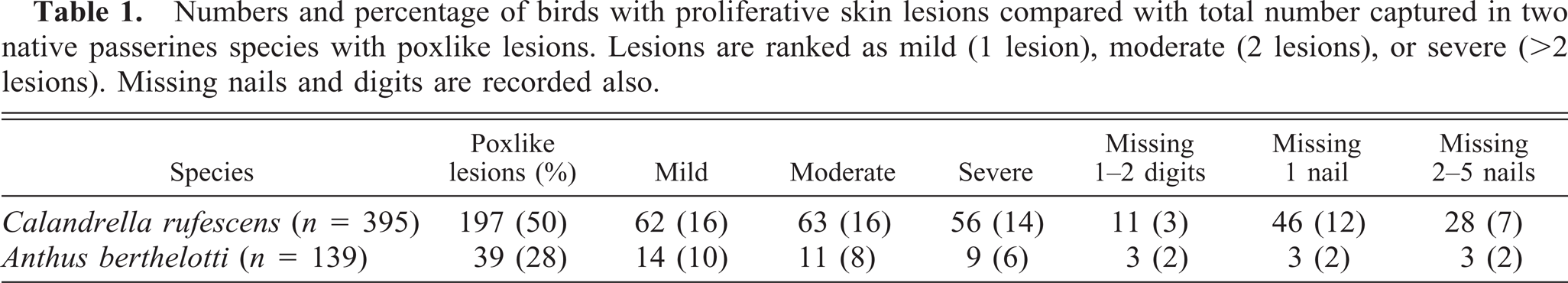
Short-toed lark, Calandrella rufescens, with poxvirus lesion proliferating from the conjunctiva on the lower eyelid (a), and on the feet and toes (b).
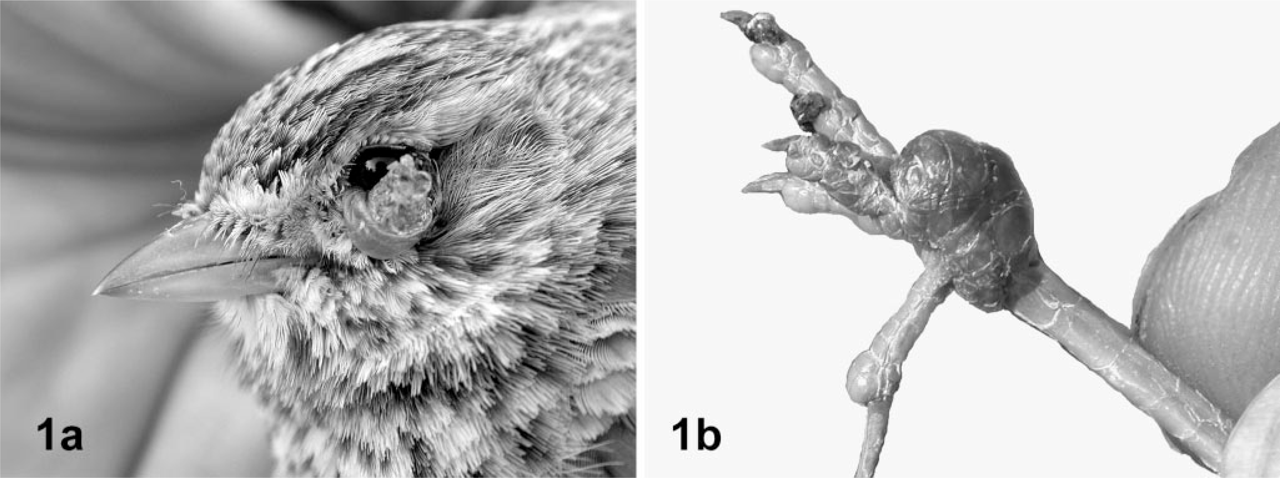
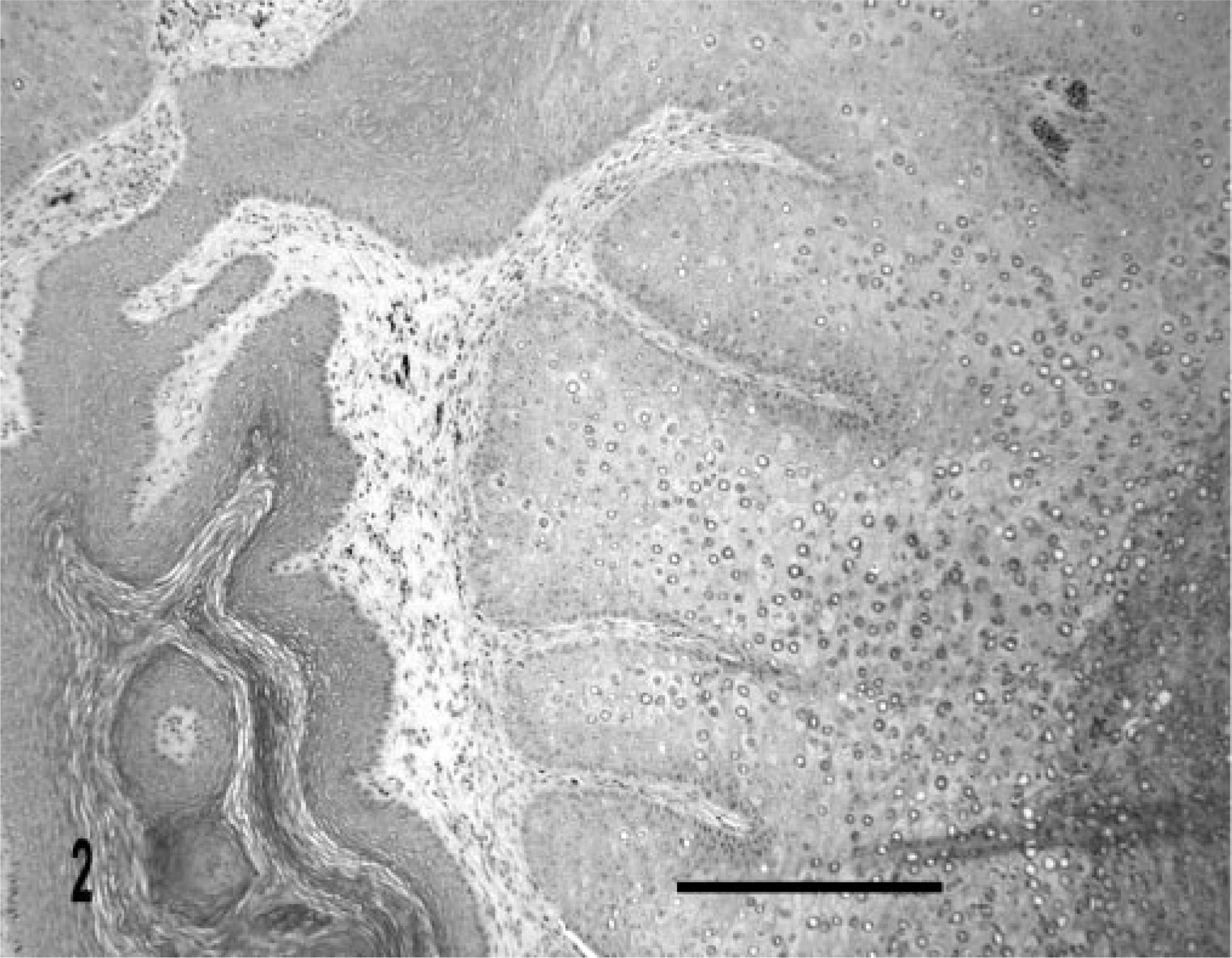
Serology for antibodies to avian poxvirus was unrewarding, yielding only three positives of 128 serologic samples, of which 52 birds had physical evidence of pox infection. Histopathologic examination of lesions from 10 of these birds showed similar pathology in all but one bird. Affected areas had marked hypertrophy and hyperplasia of the epidermis, with ballooning degeneration of epithelial cells. In the majority of cases, a heterophil-rich inflammatory cell population surrounded lesions consisting largely of large eosinophilic inclusions with solid or ring structures distending the cytoplasm of epithelial cells (Figs. 2, 3). Infiltrating bacterial colonies were visible in lesions in which there was ulceration of the keratinized superficial epidermis. On transmission electron microscopy, the inclusions consisted of numerous, dumbbell-shaped bodies typical of pox virions (Figs. 4, 5). Multiple granulomas without the typical inclusions were seen in the bird without eosinophilic inclusions.
Leg tissue; Berthelot's pipit, Anthus berthelotti. Affected limb has a proliferative lesion heavily infected with poxviral inclusions. HE. Bar 250 µm.
Leg tissue; Berthelot's pipit, Anthus berthelotti. Solid and ringlike poxviral inclusions are visible in greatly distended epithelial cells. HE. Bar = 50 µm.
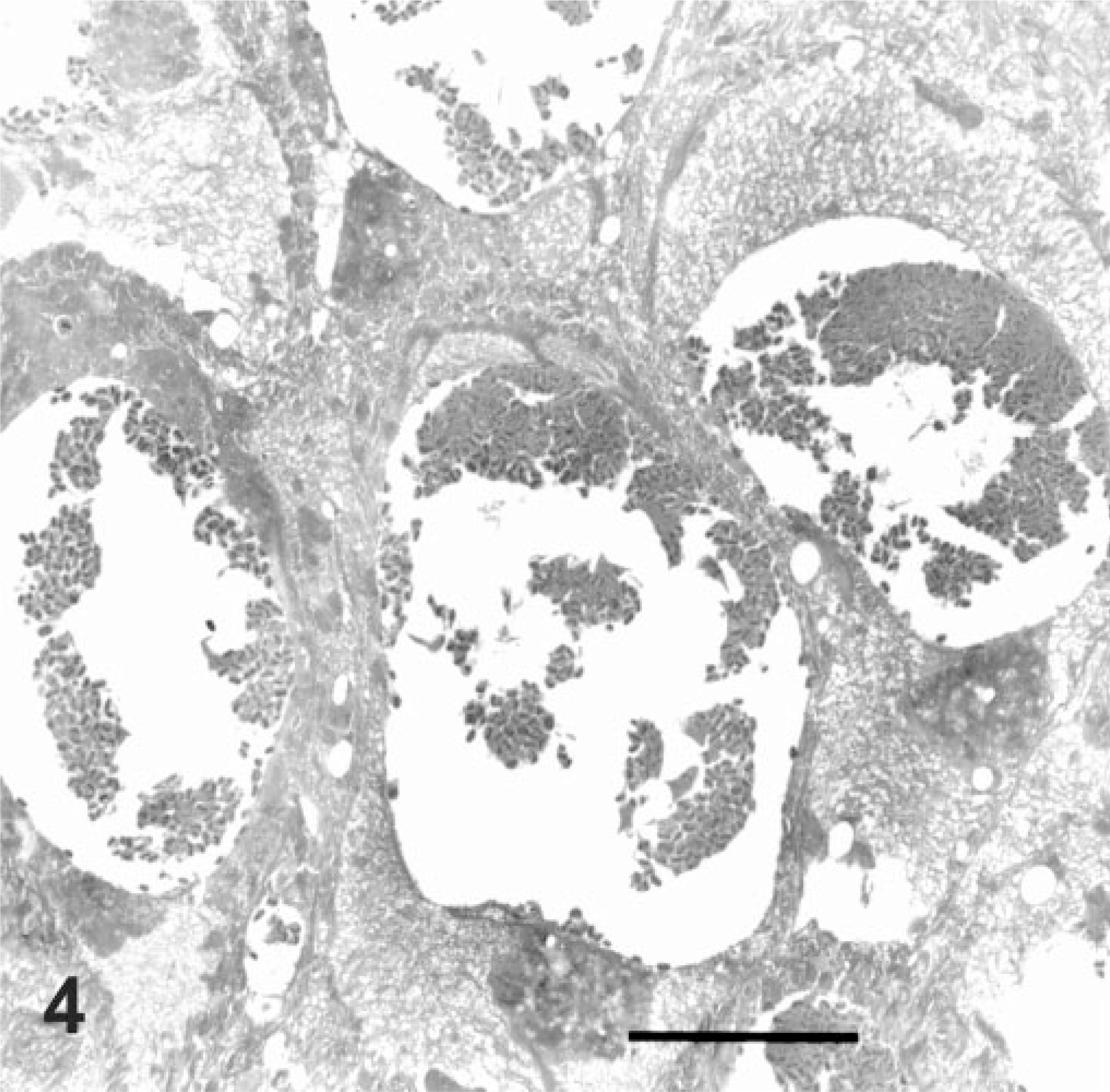
Foot tissue; short-toed lark, Calandrella rufescens. Electron micrograph shows epithelial cells with pox viral inclusions. Bar = 5 µm.
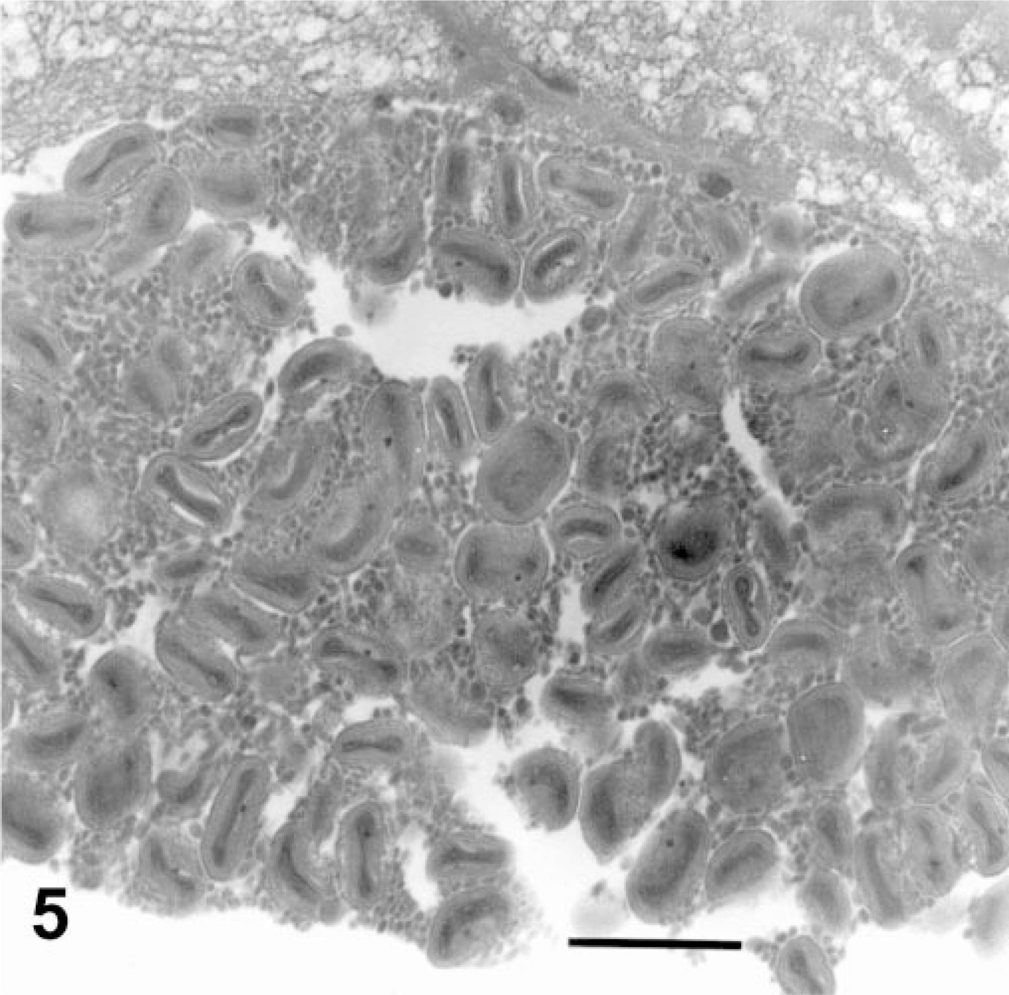
Foot tissue; short-toed lark, Calandrella rufescens. Electron micrograph shows typical dumbbell shaped poxviral particles within epithelial cell inclusions. Bar = 500 nm.
A primer set designed from the 4b core protein gene of an FPV strain amplified a 575-bp product in the virus extracted from both larks and pipits. Virus from C. rufescens had 91.3% similarity with canarypox virus, 80.5% similarity with virus from A. berthelotti, and 74.9% similarity with FPV. Virus isolated from A. berthelotti had only 79.2% similarity to canarypox virus and 74.6% similarity to FPV.
Discussion
Avian poxvirus infections have been described since the middle of the 19th century, 2 , 26 affecting more than 232 species in 23 orders of birds. Because the genomic sequences of only a small number of isolates are known, determining the species of poxvirus involved with naturally occurring disease remains elusive. Serologic tests such as the AGID are helpful for thoroughly studied poxviruses of domestic species, but the test suffers from limitations. Because of cross-reactions, one cannot differentiate different species or strains of poxvirus. 23 Yet, lack of cross-reaction with other strains of the virus yields negative results, despite histopathologic evidence of avian poxvirus infection, as was the situation in this case. The diagnostic AGID assay recognizes both clearly and weakly positive samples, presumably because of the unpredictable cross-reactivity among different strains of the various identified avipoxviruses. Our study shows no detectable cross-reactivity between the two newly described poxviruses infecting the passerines from the Canary Islands and the FPV and pigeonpox virus-based AGID kits.
Histopathology provides sound evidence of poxvirus in individuals clinically affected with poxlike lesions, 9 out of 10 of our samples and 20 of 22 samples examined by Van Riper et al. (2002) 29 being confirmed using this method. Viruses isolated from lesions can be further identified with EM revealing the well-recognized dumbbell shape (hypothesized to be an artifact of fixation) 26 or the more recently described brick-shaped 8 poxvirus elementary bodies. DNA sequencing using PCR for the viruses in the larks and pipits was hoped to help shed light on the lack of agreement between the clinically obvious poxlike lesions and the poor concordance with the AGID assay. These results are not surprising now that we have determined that the two viruses from the affected species show less than 75% similarity with FPV, the avianpox virus used in the AGID assay kit.
The discovery of the high prevalence of pox infection in short-toed larks and Berthelot's pipits on the islands of Fuerteventura and Lanzarote was unexpected. Poxvirus infection in the Canary Islands has been only recently reported for two white-tailed laurel-pigeons (Columba junoniae), an endemic species living in laurel forests. 19 Our study is the first recognition of an epizootic of poxvirus infection in these passerine species, 2 despite considerable ornithological research occurring on these islands. 7 , 12 , 14 , 17 , 19 Of note are 1) the high prevalence in these populations (28–50%), which is in stark contrast to modal avian pox prevalence in continental regions of Europe (0.5–1.3%) 29 and to the low prevalence of pox virus in short-toed larks inhabiting continental Spain (only two birds infected of 236 examined in several populations, prevalence = 0.84%, J. L. Tella et al., unpublished) and 2) that in both affected species, disease is more prevalent in adults than juveniles. Levels of pox infection in the Canary Island species are even higher than those reported in native Hawaiian birds (16.8–34.9%), which are being studied 27 , 28 , 29 because of the concern that poxvirus is one factor responsible for the decline in populations of native birds. Introduced species of poultry and wild birds, as well as mosquito vectors, have a well-defined temporal relationship with avian pox epizootics in Hawaii, where native birds are far more likely to suffer from avian poxvirus than are the introduced species. 29 In our study, however, both infected and uninfected species are native. 18 Therefore, susceptibility to infection should be related to other factors such as age, species-specific resistance to avianpox, or changes in habitat use, or all.
In contrast to the findings of Buenestado et al., 4 in our study, adults were more likely to have pox lesions than young-of-the-year. Regarding habitat use, feeding on goat farms seems to be a recent behavior for larks and pipits. This behavior of short-toed larks has never been recorded in their other native areas in continental Europe and Central Asia, where the species breed and forage exclusively in natural and seminatural steppe-like habitats (J. L. Tella, D. Serrano, unpublished data from Spain and Kazakhstan). 6 , 22 Unlike the larks, Spanish sparrows, and to a lesser degree, trumpeter finches, do commonly frequent areas associated with human habitation and farming activities in the Canary Islands. By foraging on the farms, larks may experience novel contact with poxvirus and vectors not commonly found in remote habitats. Supporting this hypothesis is the fact that poultry on the farms were so severely parasitized by fleas that some of their combs looked black instead of red due to the density of the infestation. The same species of flea was commonly found on wild birds captured on these farms. Twenty of 180 larks inspected for ectoparasites had fleas attached to the skin near the beak (mean of nine fleas per infected bird, range 1–32). These are unusual findings in wild birds. Regarding other potential hematophagous parasites, only four birds had hippoboscid flies, which was lower than the prevalence of infestation reported in other avian populations. 24 , 25 Mosquitoes are not abundant in Fuerteventura and Lanzarote because of the scarcity of pools of water in these desert habitats. Annual rainfall has been low (64–155 mm, average 111 m) over the last decade, 13 so a recent increase in the abundance of mosquitoes as potential vectors contributing to the poxvirus outbreak is not likely. Therefore, perhaps we are dealing with naive and/or displaced, stressed populations, which are susceptible to pox infection, whereas the sparrows and finches have long been exposed to various strains of avianpox viruses through contact with domestic livestock. Further research could elucidate whether this or other hypotheses may explain the differences in species susceptibility.
The conservation implications of this level of avian pox in the Canary Islands are considerable. Of the species we have studied, all except the sparrow are included in different national and international catalogues of threatened bird species. 18 Subspecies of other globally endangered birds, the houbara bustard (Chlamydotis undulata fuerteventurae), the stone curlew (Burhinus oedicnemus insularum), the spectacled warbler (Sylvia conspicillata orbicularis), the great grey shrike (Lanius meridionalis koenigi), and the Canarian Egyptian vulture (Neophron percnopterus majorensis), 7 , 18 share the same habitats. Subspecies on the mainland have been diagnosed with poxvirus infections, 1 , 2 indicating their susceptibility. It will be essential to investigate environmental, immunologic, and other biological factors contributing to the remarkable, species-specific susceptibility to poxvirus in these isolated populations to predict the effect on population stability and to reverse this alarming trend in disease occurrence.
Footnotes
Acknowledgements
We would like to thank Antonio Domingo and Fernando Peña for giving us access to their farms for trapping the birds, C. J. Palacios, M. Vögeli, J. C. Illera, and L. Gangoso for their help in the field, Y. Zhang from Ohio State Animal Disease Diagnostic Laboratory for the serology, and G. Appleyard and the PCR lab of Prairie Diagnostic Service, University of Saskatchewan, for PCR analysis. This work was conducted without financial support.