
Abstract
The American crow (Corvus brachyrhynchos) is a common urban and rural inhabitant of the Northeast and Midwest United States that is commonly infected with West Nile virus (WNV). The current study was initiated to determine non–WNV-associated causes of mortality in the American crow. All animals (40/40) tested negative for WNV infection via polymerase chain reaction and had no evidence of infection based on immunohistochemistry. Common gross necropsy findings included external trauma (6/40), hepatosplenomegaly (6/40), poxviral dermatitis (5/40), and pneumonia (3/40). Common histologic findings included endoparasitism (32/40), multifocal hepatic and splenic necrosis (7/40), pigment accumulation in the spleen (5/40), and disseminated bacterial infection (3/40). The most significant and debilitating diseases included fungal pneumonia and poxvirus-associated lesions. The present report increases the knowledge of diseases present in the American crow population.
Keywords
The American crow (Corvus brachyrhynchos) is ubiquitous throughout most of North America, with large populations found in both suburban areas and rural settings. Like many corvids, crows have a complex social structure and live in extended family groups, with individuals representing multiple generations maintaining discrete territories. 13 This tight-knit social structure can aid in the dissemination of disease and, indeed, in 1999, crow populations were the first to be devastated by the introduction of West Nile virus (WNV) to North America. 3 West Nile virus (family Flaviviridae, genus Flavivirus) is arthropod-borne, circulates between reservoir hosts and Culex species of mosquito, and can cause fatal neurologic disease in dead-end hosts (i.e., Equidae and some species of birds). 5
Although there have been several pathologic and immunohistochemical studies of WNV infection in the American crow, data are lacking with regard to other potential infectious and noninfectious causes of mortality circulating in the crow population. 9,14 American crows are known to be susceptible to pulmonary aspergillosis as well as disseminated bacterial infections from Pasteurella multocida. 11,15 Experimental data suggest that American crows are susceptible to Mycobacterium avium; however, there are no known natural cases reported in the veterinary literature. 2 Sporadically, other diseases have been reported, including avipoxvirus infection and filariasis. 1,4 Noninfectious causes of mortality in the American crow are similarly absent from the veterinary literature except for a single report of nutritional secondary hyperparathyroidism in a cohort of fledgling birds. 10 Because of the relative lack of information regarding natural disease occurrence, the current study was undertaken to assess causes of mortality in a defined population of American crows that has been part of a long-term ecological study.
Between November 1, 2006, and May 1, 2008, 40 American crows were presented to the Cornell University Department of Biomedical Sciences, Section of Anatomic Pathology (Ithaca, NY) for necropsy and subsequent histologic examination. Some birds that were submitted for examination had recorded ages based on banding as nestlings and known hatch dates. All submitted animals were subjected to a complete necropsy in which a written gross necropsy report was developed for each animal. All organs except for bone were sampled and fixed in 10% neutral buffered formalin. Representative tissue sections were embedded in paraffin, sectioned at 4 μm, mounted on glass slides, stained with hematoxylin and eosin using standard histological technique, and examined by light microscopy. Because of the presence of bacteria, fungi, and pigments of unknown origin, paraffin-embedded sections of select tissues were further processed for Gram stain, Gomori methionine silver stain, Fontana–Masson stain, and Prussian blue stain.
Prior to necropsy, all birds tested negative for WNV by reverse transcription polymerase chain reaction on oralpharyngeal swabs. Briefly, nucleic acid extraction and amplification were performed using the probes and protocol previously described with amplification using SmartCycler a technology. Reactions were performed using 2 μl of RNA in a 25-μl reaction. Amplification was done in a 2-stage reaction with reverse transcription at 42°C for 900 sec, denaturation at 95°C for 600 sec, followed by 39 cycles of 95°C for 15 sec, 50°C for 10 sec, and 60°C for 100 sec. 6 Immunohistochemistry was also employed on sections of cardiac muscle from all birds. Briefly, sections of cardiac muscle were deparaffinized and rehydrated through graded alcohol concentrations to water and subsequently blocked with 3% hydrogen peroxide. Slides were pretreated with pronase for 5 min at room temperature. b All steps were followed by a Tris-buffered saline (TBS) wash. Prior to application of primary antibody, all slides were treated with Zymed protein block for 10 min. c Sections were incubated with antimouse WNV (1:75). d Antibody was incubated for 30 min followed by incubation with secondary antibody biotinylated goat antimouse. c A streptavidin–peroxidase conjugate was then applied for 10 min, and the slides were rinsed in TBS for 5 min followed by treatment with 3,3'-diaminobenzidine chromogen for 2 min. c All slides were counterstained with hematoxylin, washed in tap water, and rinsed in distilled water. Negative control slides were processed with primary antibody and substitution with mouse immunoglobulin G. b
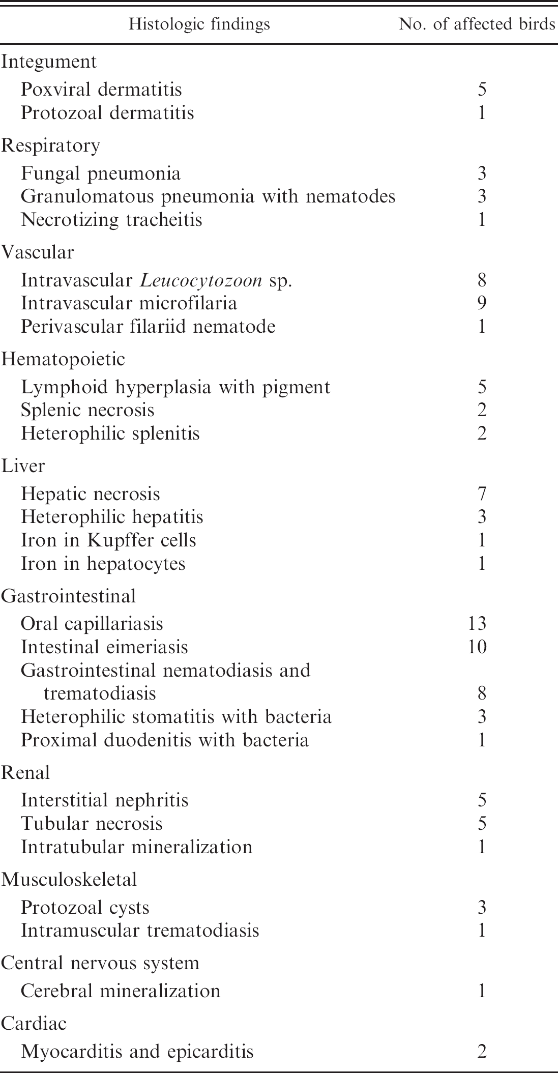
Gross necropsy findings in American crows (Corvus brachyrhynchos) in the current study.
Of the 40 birds examined, 10 were males, 14 were females, and 16 had undetermined sexes. Of the birds with recorded ages, 7 were juvenile (<1 year old), 4 were young adult (between 1 and 2 years old), and 4 were adult. The average weight of the males was 0.40 kg (range: 0.25–0.52 kg), and the average weight for the females was 0.42 g (range: 0.37–0.56 g). Of the 40 birds, 6 had marked autolysis, 12 had mild to moderate autolysis, and 22 had minimal to mild autolysis. None of the birds examined tested positive for WNV via immunohistochemistry.
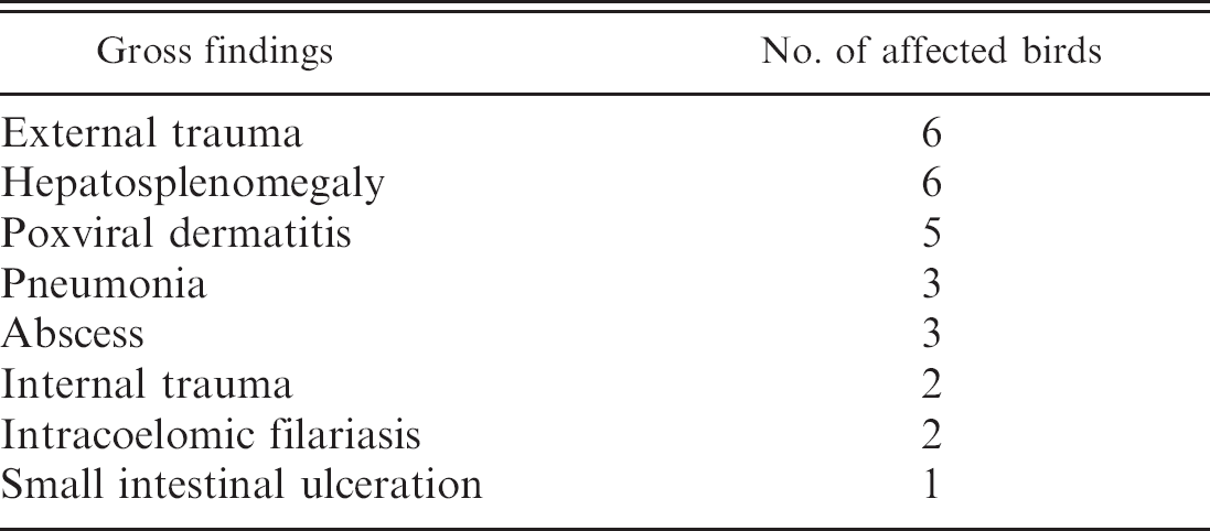
The gross findings are presented in Table 1. Six of the birds had external trauma. One bird had a full-thickness, mid-diaphyseal fracture of the left femur, 3 birds had multiple rib fractures, and the 2 remaining birds had gunshot wounds to the thorax. The 3 birds with rib fractures also had a hemocoelom. Five of the birds had singular to multiple, irregular, proliferative masses present on both legs (3 birds) or around the oral cavity (2 birds). On section, the masses were homogenous, off-white–tan, speckled with areas of necrosis and hemorrhage, and confluent with the adjacent skin. The 2 oral masses were confined to the outer portion of the mouth and did not invade into the oral mucosa (Fig. 1).
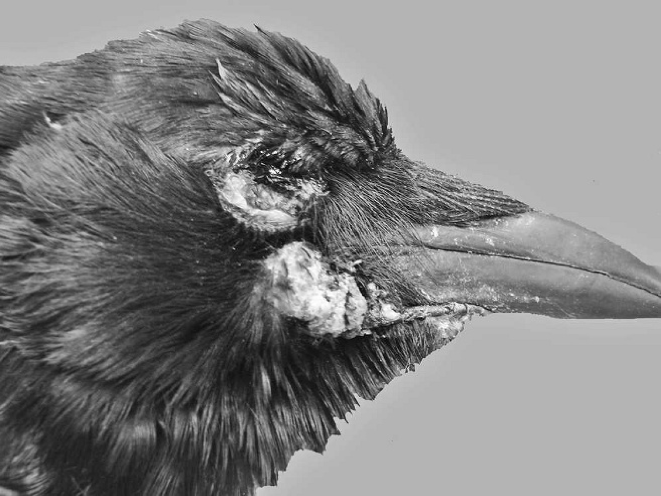
Three birds had severe pneumonia, which was characterized by dozens of round, variably sized, white to tan foci scattered randomly throughout the lung lobes. On section, these foci exuded necrotic cellular debris that occasionally contained small chalky specks (mineral). Of the 3 birds, 2 also had concurrent thickening and proliferation of the air sacs (Fig. 2).
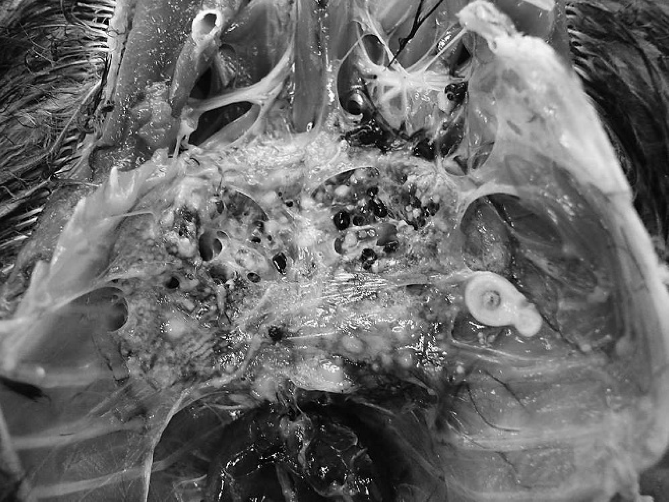
Three birds had marked hepatosplenomegaly with myriad, variably sized white foci scattered randomly throughout the hepatic and splenic parenchyma. The foci were homogenous on section and contained a small amount of white, necrotic debris. Three other birds had marked splenomegaly; however, the 3 birds lacked the characteristic miliary pattern and instead were diffusely meaty and mottled red–brown (lymphoid hyperplasia). Other gross findings included left pectoral muscle abscess (1 bird); liver abscess (1 bird); pharyngeal abscess (1 bird); hepatic capsular fracture and hemorrhage (1 bird); proximal small intestinal ulceration (1 bird); facial hemorrhage (1 bird); and intracoelomic filariasis (Diplotriaena tricuspis; 2 birds; Fig. 3).
Head, American crow (Corvus brachyrhynchos). Arising adjacent to the commissure of the mouth is a large proliferative mass consistent with poxvirus infection.
Lungs, American crow (Corvus brachyrhynchos). The lungs have been obliterated by multiple yellow–tan masses consistent with multiple fungal granulomas.
Coelom, American crow (Corvus brachyrhynchos). Within the coelom is a tightly coiled adult filariid nematode.
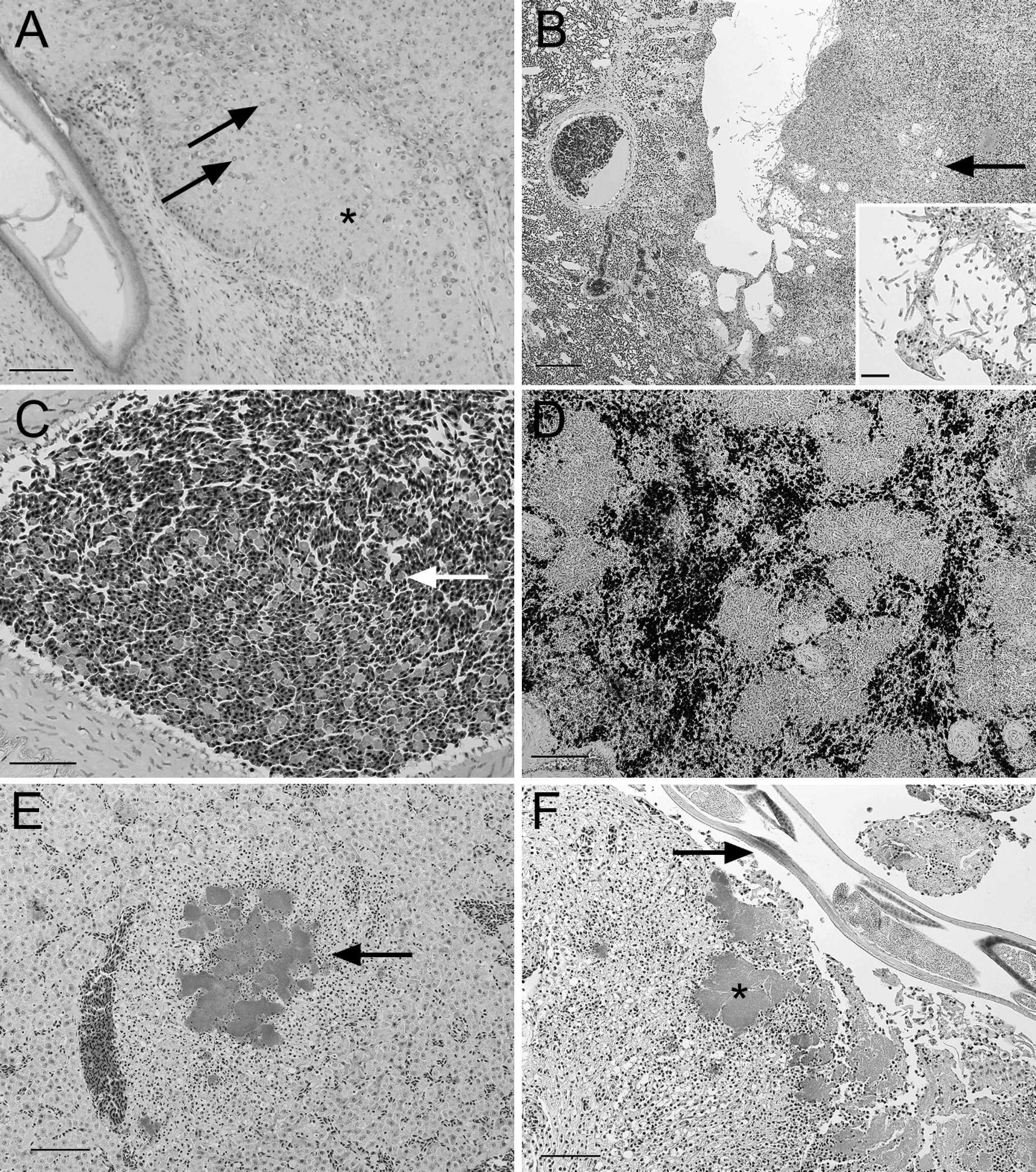
The histologic findings are presented in Table 2. Only 5 birds did not have any histologic findings. The only skin lesions that were detected were in those birds noted to have proliferative lesions consistent with poxvirus infection. Briefly, all sections examined were composed of markedly proliferative epidermis in which the keratinocytes were markedly enlarged, had areas of ballooning degeneration, and contained singular to variable numbers of deeply eosinophilic, round, 4–8-μm, intracytoplasmic inclusion bodies (Fig. 4A). One of these birds also had concurrent protozoal dermatitis that was characterized by moderate numbers of lymphocytes and macrophages infiltrating the dermis, of which the latter often contained small numbers of round, 2–4-μm, intracytoplasmic protozoal organisms.
Histologic lung lesions were present in 7 of 40 birds. A total of 3 of the 7 birds had multifocal to coalescing, granulomatous pneumonia centered on a focus of liquefactive necrosis that contained variable numbers of fungal hyphae that had parallel, septate walls and occasional right-angle branching (Fig. 4B, inset). Two birds had multifocal foci of bacterial embolization and vasculitis. In the 2 birds, the affected vessels were surrounded and infiltrated by a low number of heterophils, with occasional lymphocytes and macrophages. The airways were within normal limits in these birds. The final 2 birds had focal granulomatous pneumonia that was centered on degenerate metazoan parasites. Because of the state of autolysis, more precise identification could not be performed; however, these organisms had a thick cuticle, coelomyarial musculature, and a pseudocelom. In addition, 1 bird also had necrotizing heterophilic tracheitis.
Intravascular parasitism was noted in 18 of 40 birds. A total of 8 of 18 birds had small to large numbers of intravascular, intramonocytic parasites (consistent with Leucocytozoon sp.; Fig. 4C), whereas 9 of the 18 birds had small to moderate numbers of microfilaria within the pulmonary and, rarely, hepatic blood vessels. Finally, 1 bird had a tangential cross-section of a large nematode embedded in the periaortic connective tissue. This nematode had a prominent cuticle, coelomyarial musculature, and a pseudocelom.
Splenic changes were present in 11 of 40 birds. Of the 11 birds, 2 birds had multifocal necrotizing and heterophilic splenitis with intralesional coccal bacteria (Gram negative); 2 birds had multifocal, random splenic necrosis without concurrent inflammation or bacteria; and 5 birds had marked lymphoid hyperplasia with concurrent proliferation of variably sized aggregates of macrophages that contained abundant, intracytoplasmic, brown–black, granular pigment (Fig. 4D), which did not stain with Prussian blue or Fontana–Masson.
Histologic findings in American crows (Corvus brachyrhynchos) in the current study.
Liver lesions were present in 14 of 40 birds. Of the 14 birds, 7 birds had multifocal, random hepatic necrosis without inflammation, and 3 birds had multifocal suppurative hepatitis with intralesional coccal bacterial (Fig. 4E), of which 2 also had concurrent lesions in the spleen and the oral cavity. Last, 2 birds had focal abscesses in the liver, 1 had increased iron retention in the Kupffer cells, and 1 had iron storage in the hepatocytes.
The oral cavity and small intestine frequently had microscopic disease, even though there were no lesions detected grossly. The bird with gross ulceration in the proximal duodenum had a locally extensive necrosuppurative duodenitis with intralesional coccal bacteria. A total of 13 of 40 birds had parasitism in the oral cavity consistent with Capillaria sp. Of the 13 birds, 3 also had concurrent necrotizing and heterophilic stomatitis with intralesional coccal bacteria (Fig. 4F). These bacteria were similar in morphology to those noted in the spleen and liver. Although the small intestinal sections examined were often severely autolyzed, 10 of 40 birds had intraepithelial schizonts and gamonts consistent with Eimeria sp. A total of 7 of 40 birds had intraluminal nematode parasites, and 1 of 40 had intraluminal trematodes parasites.
Histologic changes in the kidney were detected in 11 of 40 birds. Of the 11 birds, 5 of these cases consisted of focal to multifocal aggregates of heterophils, lymphocytes, and macrophages that surrounded colonies of coccal bacteria (2/5) or were intermixed with protozoal cysts (3/5). In these areas, the renal tubules were often filled with irregular casts of degenerate and necrotic epithelial cells and occasional degenerate leukocytes. Five birds also had multifocal renal tubular necrosis characterized by loss of cell cohesiveness, increased cytoplasmic eosinophilia, and nuclear dissolution. One bird had a focal deposit of tubular crystals. Other, less common histologic findings included protozoal cysts within cardiac and skeletal muscle (3/40), encysted trematode within the skeletal muscle (1/40), focal cerebral mineralization (1/40), and heterophilic myocarditis and epicarditis (2/40). The skeletal system was not analyzed for the current study.
Since the discovery of WNV in the United States in 1999, it has been well documented that the virus can have a significant effect on population dynamics within the American crow population. 3 In this population, the first recognized cases of WNV were in 2002. 3 By 2003, WNV resulted in the deaths of about 37% of the study population. 3 Because of the drastic changes in population dynamics that diseases can cause, the current prospective study was undertaken with the goal of determining other diseases circulating within the population that could lead to increased morbidity and mortality, and thus alter population dynamics. All of the birds described in the current study were negative for WNV via polymerase chain reaction and immunohistochemistry, and although both tests can miss positive cases because of sample degradation or lack of the appropriate histologic section, the combined negative tests significantly decrease the likelihood that WNV-positive animals were included in the present study. 9 It has previously been shown that the disease status in this population was strongly affected by inbreeding, with highly inbred birds more likely to succumb to the effects of disease. 12 In the present population, there is a very high survival rate, with normal yearly mortality for nonjuveniles less than 20%, juveniles at 50%, and only 8% for breeders. 7,8 For a passerine species, these mortality numbers are quite low and certainly indicate that the currently population is not being devastated by diseases other than WNV.
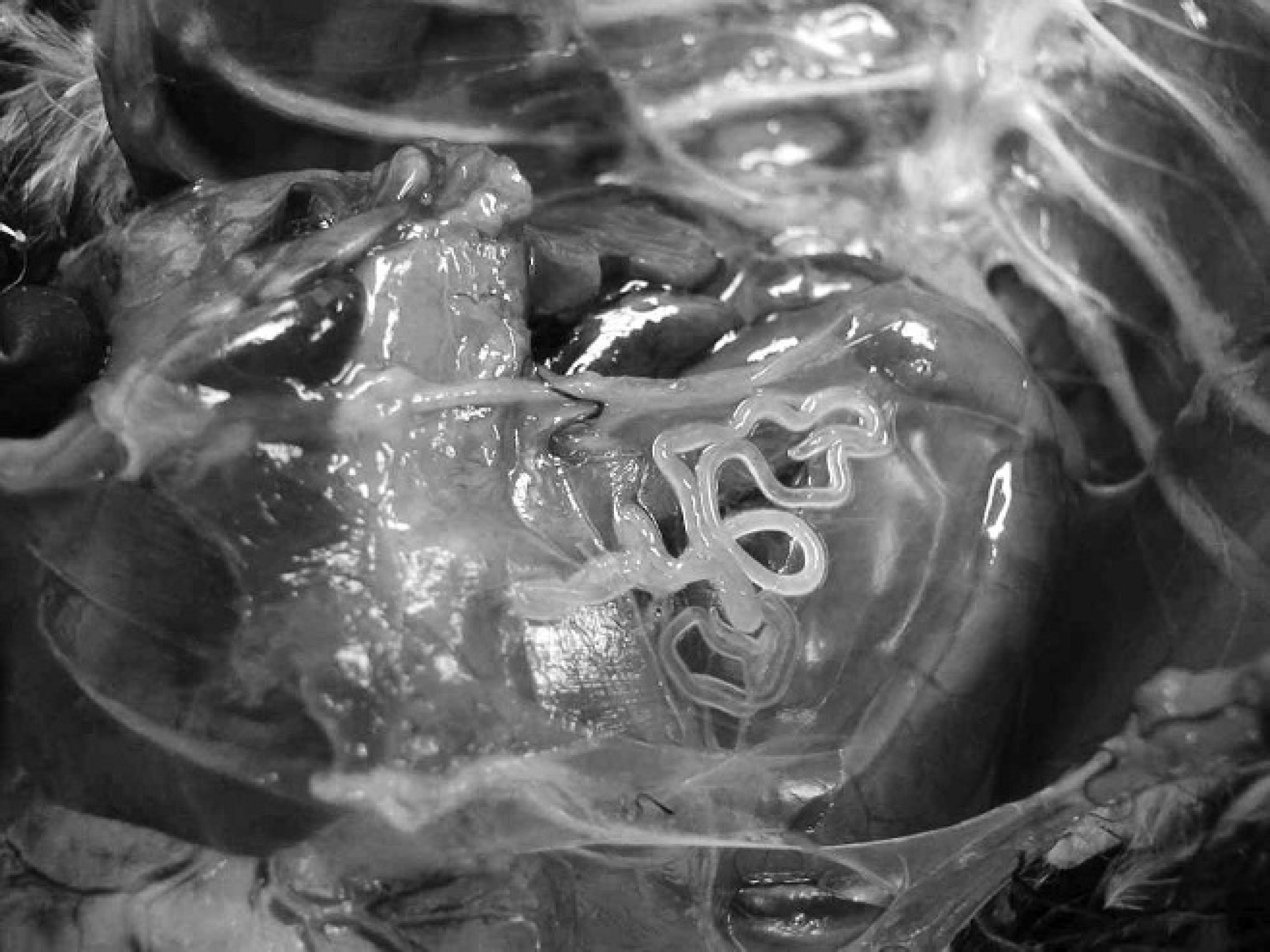
American crow (Corvus brachyrhynchos).
Poxvirus has only been described once in the American crow, and in the current study it was seen in 12.5% of the birds necropsied. 4 Poxviruses are large DNA viruses that replicate within the cytoplasm, forming characteristic intracytoplasmic inclusion bodies. 4 Animals commonly develop lesions around the legs and the mouth because these are sites that are predisposed to microabrasions. These microabrasions allow for the virus to enter into the epidermis and propagate. Interestingly, none of the cases of poxvirus described here developed within the oral cavity (i.e., wet pox). Whether this is a species-specific difference in the American crow is not known, and further laboratory tests are ongoing to culture and determine the molecular characteristics of this poxvirus.
Similar to a single previous report, a low prevalence of fungal pneumonia was present in this population (7.5% of birds examined). 15 Although fungal culture was not performed in any of these cases, the histomorphology was most consistent with Aspergillus sp. Based on the extent of the pneumonia with the concurrent invasion into the air sac in 2 of the 3 cases, the fungal pneumonia was directly the cause of death in these birds. The 3 cases of fungal pneumonia were seen in late summer and fall, corresponding to the warmest and wettest parts of the year, and therefore may have played a role in increasing exposure of these birds to environments rich in fungi. Two birds also had granulomatous pneumonia with intralesional nematode parasites. Although the state of degeneration made it impossible to identify the species of these parasites, they were consistent with Chandlerella chitwoodae, which is relatively common in the American crow in the Northeast (A. L. Glaser, personal communication, 2009). The significance of microfilaria in the lung, liver, and kidney vessels is not known; however, the presence of a large, periaortic filariid nematode in 1 bird with microfilariasis is likely causal. A filariid parasite (Splendidofilaria caperata) is known to infect crows in the northeast and might be the cause in this case. 1 Additionally, the presence of a coelomic filariid, Diplotriaena tricuspis, in several of the birds may also have contributed to the microfilariid burden in several of these birds.
Bacterial disease was present in 7 of 40 birds (17.5% prevalence). A total of 3 of 40 birds had a necrotizing stomatitis with intralesional, Gram-negative bacteria. In these same birds, there was also dissemination to the liver, lung, spleen, small intestine, and kidney. It is presumed that the primary site of infection was the oral cavity; however, culture was not available for any of the 3 cases, and therefore the exact causative agent is not known. Intriguingly, all 3 of the birds with necrotizing oral stomatitis also had concurrent oral capillariasis. Although Capillaria sp. was present in 13 of 40 birds examined, it was especially prolific in the 3 birds with bacterial infection. It is possible that the localized inflammation caused by the parasites allowed for bacterial invasion and replication, which rapidly outpaced the immune response and led to dissemination. Focal bacterial infections were also noted in 3 birds with pectoral, hepatic, and pharyngeal abscesses, and although dissemination did not occur, the sheer size of the abscesses certainly would have led to cachexia.
Protozoan parasitism was very common in all birds examined. Intramonocytic protozoal organisms were detected in 8 of 40 birds (20% prevalence). Based on the morphology, these protozoa were consistent with Leucocytozoon sp. This is a common, seasonal parasite that is transmitted by hematophagous arthropods. The entire life cycle is not known, but it is usually thought of as an incidental finding in birds. However, in one of the cases described here, the infection was extremely robust and coincided with poxviral dermatitis, potentially indicating a broad immunosuppressive state in this bird. Protozoal cysts and free protozoal zoites were also seen in the muscle and skin of 3 birds. The cysts in the muscle were consistent with Sarcocystis sp. as described in a variety of avian species.
Multifocal renal inflammation and necrosis were also common in the crow population in the present study. This change most commonly represented dissemination from bacterial disease; however, 5 cases were devoid of bacteria or other infectious organisms. In these 5 cases, the direct cause of the necrosis and inflammation is not clearly known, although it is possible that the birds could have ingested nephrotoxic material.
Additionally, 5 birds had large amounts of pigment noted within the splenic macrophages. The origin of the pigment is purely speculative, because it did not stain for melanin via Fontana–Masson or for hemosiderin via Prussian blue. It is still possible that either of these endogenous pigments could represent the storage material but has since lost its staining characteristics. None of the 5 birds with the splenic pigment accumulation had concurrent pigment accumulation in the liver, and the presence of pigment always coincided with lymphoid hyperplasia.
Although the current study systematically examined a cohort of birds for evidence of gross and histologic disease, there were several aspects of the investigation that require further investigation. There was an inability to determine the microbiologic characteristics of the causative bacteria and fungi. In addition, the bones of the birds were not studied on a histologic level, and therefore the incidence of bone disease is not known in this population. Age analysis was only able to be performed on a small cohort of the birds studied. However, among the few birds for which accurate age data were obtained, there was an increase in severe, debilitating disease (i.e., fungal pneumonia, pox viral dermatitis, and cavitary abscesses) in younger birds.
In conclusion, the marked variation in disease manifestations in the present study was surprising and illustrates the multitude of infectious and noninfectious diseases present within the American crow population. Although several of the processes reported here are unlikely to have caused primary disease and are considered incidental, many others directly led to ill thrift and ultimately death of the bird and would have a negative impact on a population (e.g., poxviral dermatitis, fungal pneumonia).
Footnotes
a.
Cepheid, Sunnyvale, CA.
b.
Dako North American Inc., Carpinteria, CA.
c.
Zymed Laboratories Inc., South San Francisco, CA.
d.
7H2 clone, BioReliance Corp., Rockville, MD.