
Abstract
A 7-year-old Holstein cow developed a large cystic mass in the region between the atlantoaxis and larynx. The mass extended to the synovium in the atlanto-occipital joint. Many villous projections were present on the inner surface of the tumor tissue, and irregular clefts were formed in the inside. Two cell types, epithelioid-like synovioblasts and spindle cells, were present. Immunohistochemical analysis showed that the cells stained positively for cytokeratin (AE1/AE3) and vimentin. Both cells had similar fine structures ultra-structurally. Vacuoles present in the cytoplasm were full of an acid mucous substance. The tumor was diagnosed as a well-differentiated biphasic synovial sarcoma. This is the first report of a rare case of synovial sarcoma, from the viewpoint of its origin.
Synovial tumors generally occur in the joints of extremities and are common in dogs, cats, 1,6–8 and humans 2 but rarely occur in large animals. To date, only three cases of synovial tumors have been reported in cattle, all of which occurred in the joint of the fore or hind extremities. 3,5,10 We discovered a very rare case of synovial sarcoma that developed in the atlanto-occipital joint of a cow. Histopathologic, immunohistochemical, and ultrastructural findings are detailed in this report.
A 7-year-old pregnant Holstein cow developed swelling in the submandibular region and was presented at the large-animal hospital of Kitasato University. On clinical examination by palpation and echography, the swelling was found to be due to a cystic mass in the dorsal region of the pharyngolarynx. Cytologic examination was not performed. No functional disturbance of swallowing and breathing due to the developed cystic mass was observed. The cow was sacrificed 3 weeks after a Caesarean operation and was submitted for pathologic investigation of the cystic mass.
The ovoid cystic mass was located between the atlanto-axial vertebrae and pharyngolarynx. Its size was measured as 24 × 14 × 13 cm. The mass was encapsulated by thin connective and muscular tissue. Sticky yellowish fluid, resembling articular fluid, spurted out at incision. Blood clots and fibrinous debris were present inside the cyst. The inner surface of the cyst was smooth and white-yellowish in color, and many soft polypoid projections of various sizes were present. The cyst extended to the atlanto-occipital joint by the peduncle. A tentative diagnosis of synovial tumor was made. The tissue of the peduncle was harder in comparison with the tissue of the cyst wall. After separation of the cyst from the atlanto-occipital joint, further examination revealed that one third of the articular surfaces of both the occipital condylus and fovea articularis cranialis of the atlas was eroded (Fig. 1) and the tumor tissue had invaded the bone marrow from the eroded surface. The tumor did not infiltrate inside the dura mater. In addition, metastases were not observed in the accessory lymph nodes or organs.
Synovial sarcoma, atlanto-occipital joint; cow. Tumor in fovea articularis cranialis of the atlas. Soft tumor tissue (asterisk) resembling synovium proliferates on the surface. One third of the articular surface is eroded (arrow).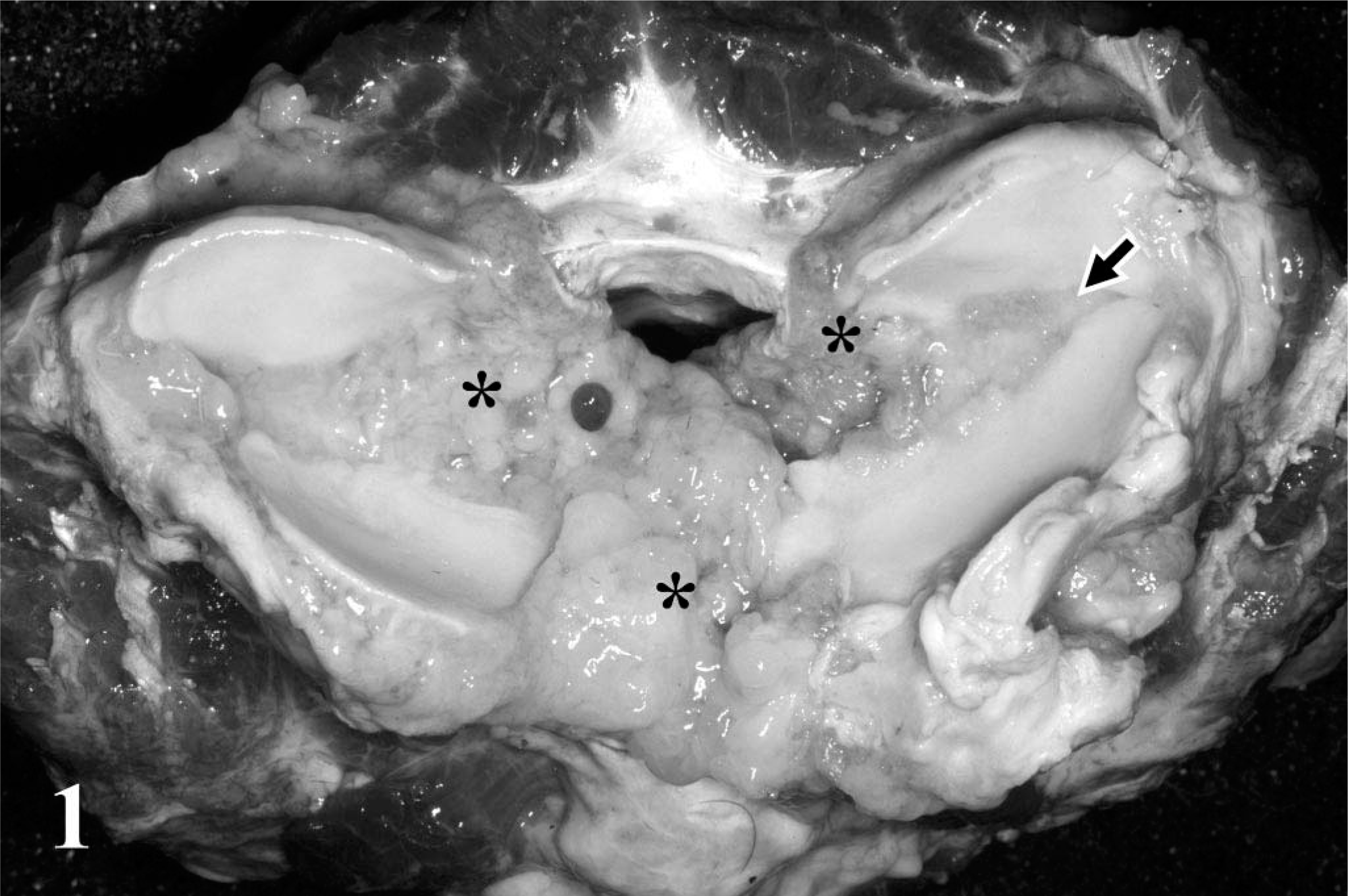
Samples for histopathology were fixed in 10% neutral buffered formalin and embedded in paraffin, and thin sections were stained with hematoxylin-eosin (HE). Selected sections were also stained with periodic acid–Schiff (PAS), toluidine blue (pH 4.1), alcian blue (pH 2.6), and Masson trichrome. Immunohistochemical examination was performed using HISTOFINE simple stain MAX PO (Nichirei, Tokyo, Japan) for cytokeratin (AE1/AE3) (Nichirei), vimentin (Nichirei), and S100 protein (Nichirei) on formalin-fixed and paraffin-embedded sections. As a control, synovial tissue from the left stifle joint of the same cow was compared with the tumor. Small tissue blocks were fixed in 0.5% glutaraldehyde, postfixed in 1% osmium tetraoxide, and routinely processed for electron microscopy.
The histologic structure of the tumor tissue resembled that of normal synovium (Fig. 2). Many villous projections of variable size and length were formed on the inner surface, and irregular clefts were observed (Fig. 2, arrow).
Synovial sarcoma, atlanto-occipital joint; cow. Tumor in the inner surface of the cystic mass. Many villous projections are formed on the surface. Arrow indicates a cleft in the tumor tissue. HE. Bar = 200 µm.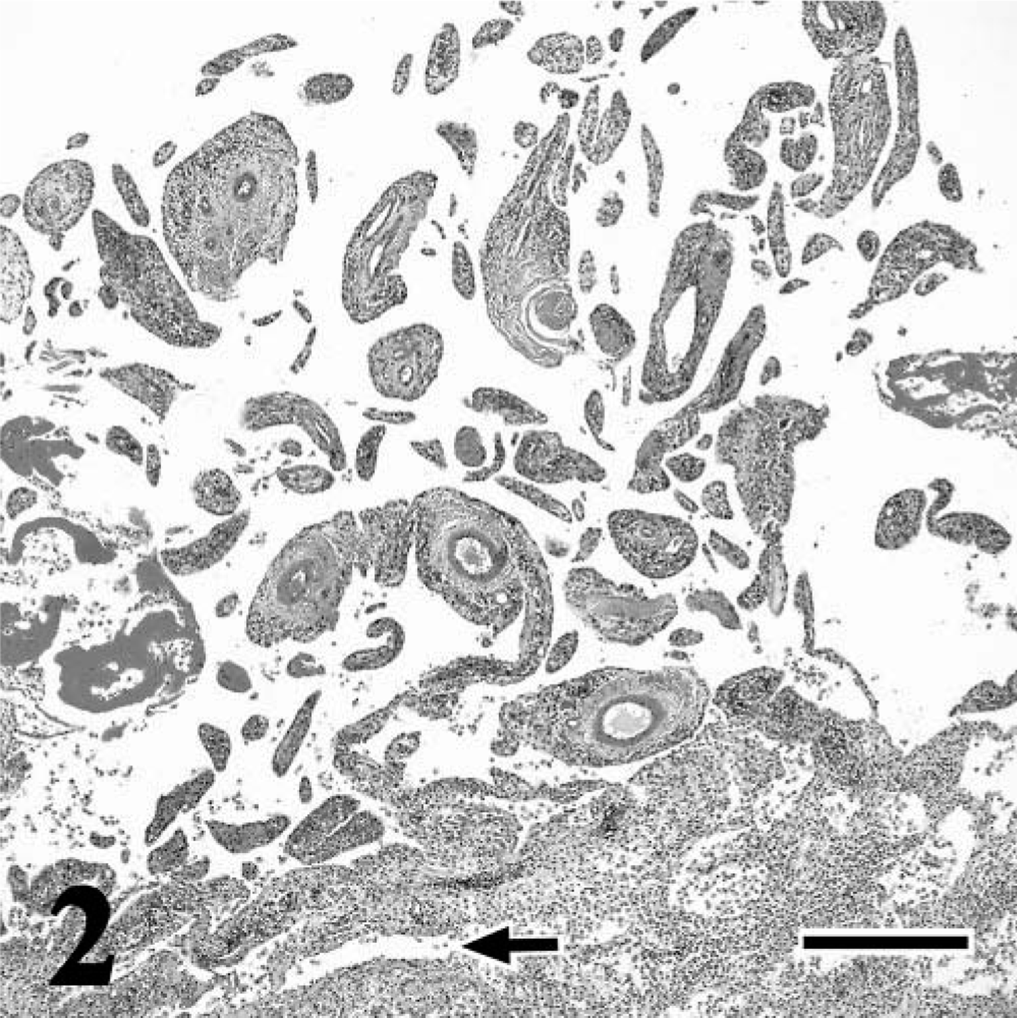
Two cell components, the epithelioid-like synovioblast and the spindle cell, were present, but no multinucleated giant cells were observed. Cells covering the villi and the surface layer of the cyst wall were epithelioid-like synovioblasts, characterized as round or polyhedral in shape with a foamy cytoplasm and small hyperchromatic nucleus. The foamy cytoplasm was positively stained with PAS, and the presence of a metachromatic acid mucinous substance was demonstrated by staining with toluidine blue (pH 4.1) and alcian blue (pH 2.6). Neither a mitotic figure nor an atypic morphology was observed. Cell density was low, and inter-cellular connections were very loose: a few fine collagen and reticular fibers surrounded the cell bodies. Synovioblastic cells were also present in the deeper part of the cyst wall (Fig. 3). In the transitional region between the cyst wall and the deeper connective tissue, tumor cells decreased in size and changed shape from polygonal to spindular, similar to fibroblasts (Fig. 4).
Synovial sarcoma, atlanto-occipital joint; cow. Tumor cells in the deeper part of the cystic mass. Synovioblastic cells are apparently derived from the spindle cells in the interstitium (arrow). HE. Bar = 20 µm.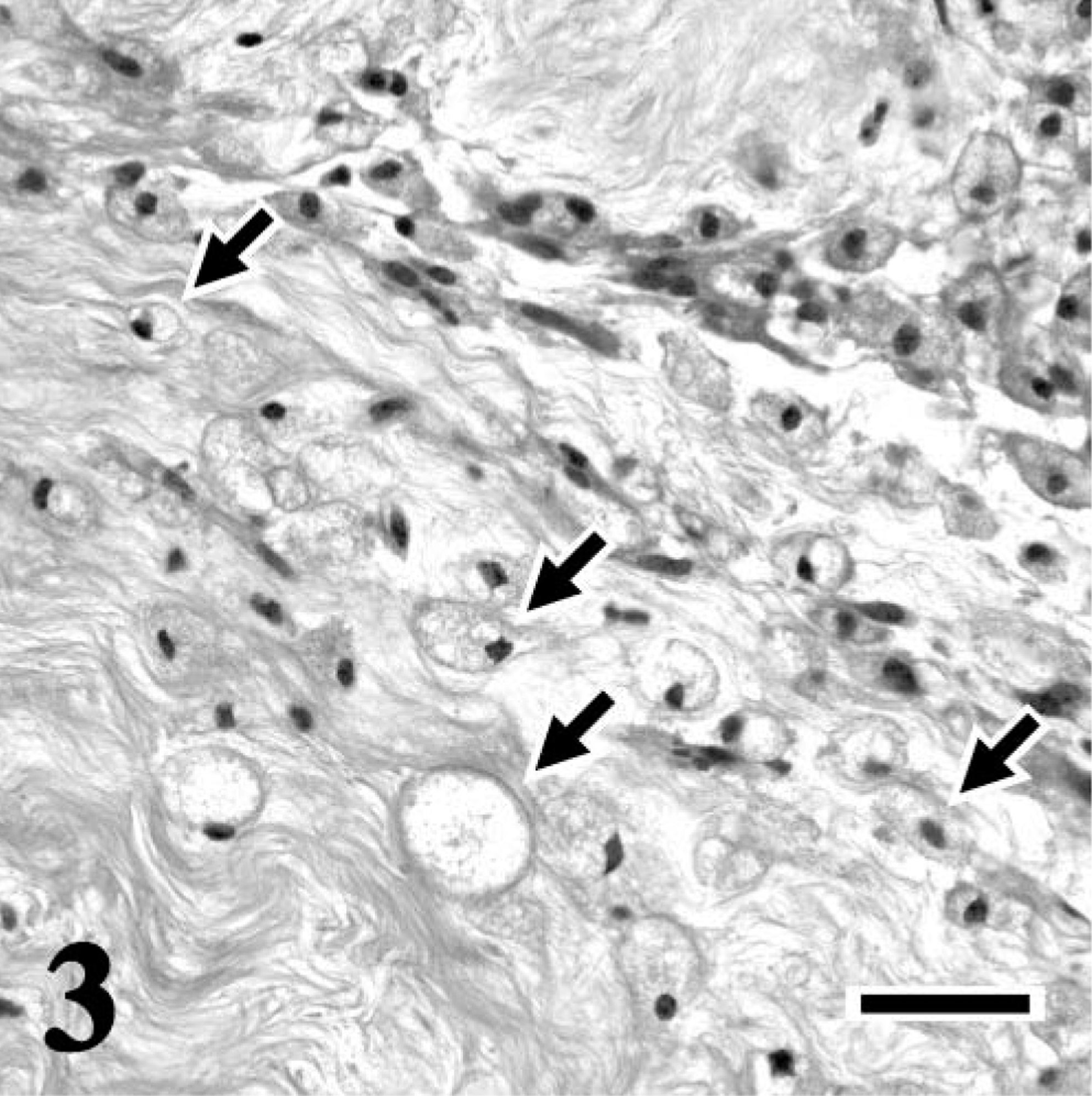
Synovial sarcoma, atlanto-occipital joint; cow. Tumor tissue in the bone marrow of atlas. Both synovioblastic (S) and fibroblastic (F) tumor cells proliferate. HE. Bar = 100 µm.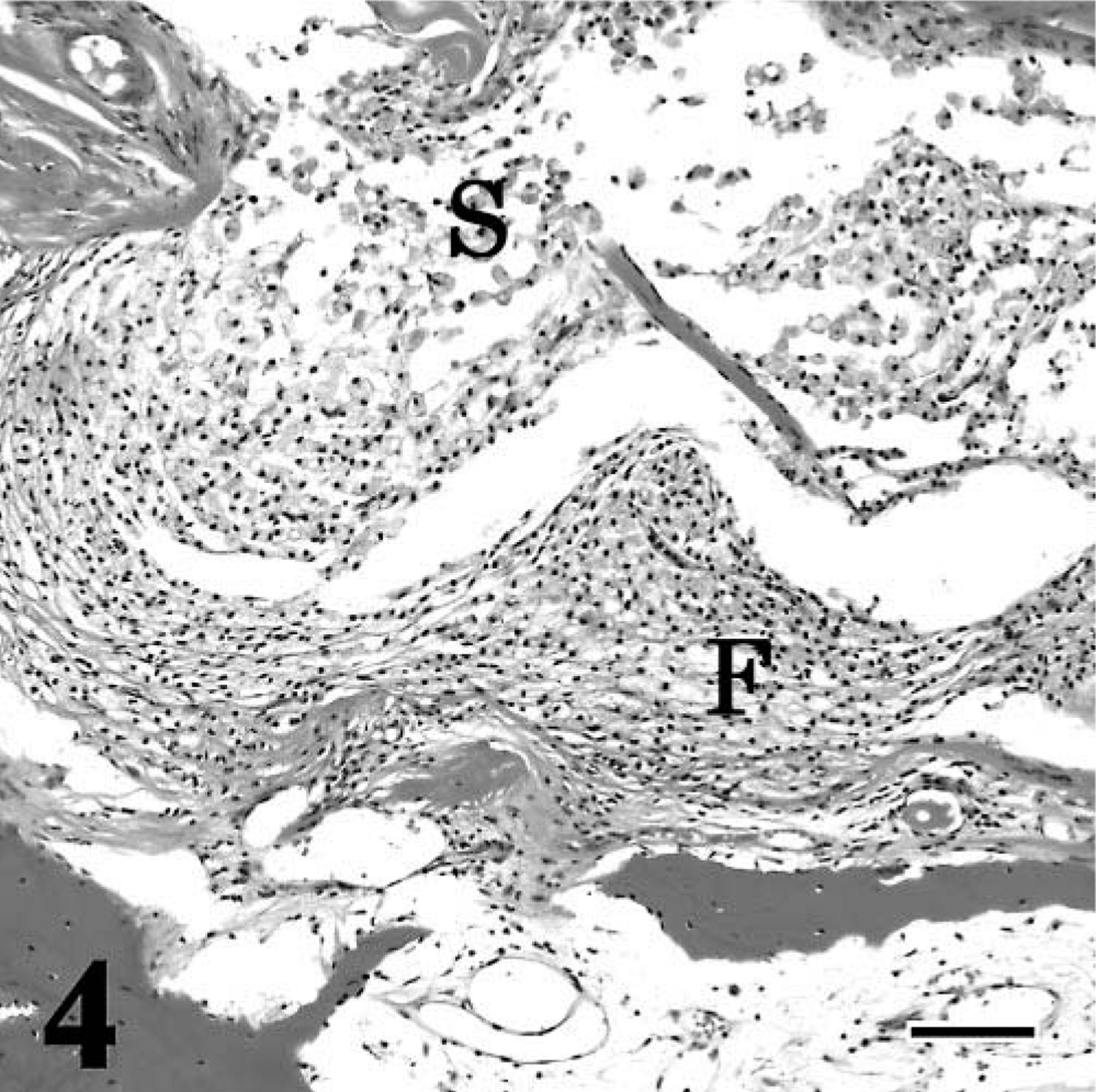
Epithelioid-like synovioblast and fibroblast-like spindle cells were observed in the peduncle of the tumor mass and bone marrow tissue; however, the cell density of the peduncle and bone marrow was higher than that of the tumor tissue in the cyst wall. The inner surface layer of the tumor tissue was composed of characteristic synovioblasts, but small spindle cells with indistinct cellular boundaries and eosinophilic cytoplasm were predominant (Fig. 3).
Neoplastic synovioblasts and spindle cells in the surface layer of the cyst wall stained positively for cytokeratin (AE1/AE3) (Fig. 5) and vimentin (Fig. 6) but stained negatively for S100 protein. The cells of normal synovium from the stifle joint stained positively for vimentin only.
Synovial sarcoma, atlanto-occipital joint; cow. Polyhedral synovioblastic cells (arrow) are positive for cytokeratin (AE1/AE3). Cells in the surface layer of the cyst wall (arrowhead) are also positive. HISTOFINE simple stain and counterstain with hematoxylin. Bar = 20 µm.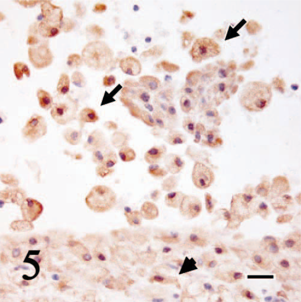
Synovial sarcoma, atlanto-occipital joint; cow. Both synovioblastic (arrow) and fibroblastic (arrowhead) tumor cells are strongly positive for vimentin. HISTOFINE simple stain and counterstain with hematoxylin. Bar = 20 µm.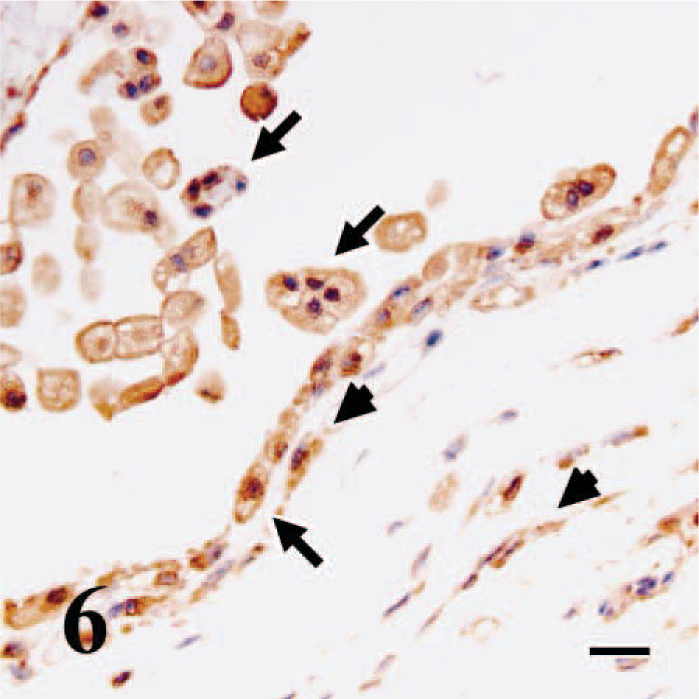
Electron microscopy revealed that neoplastic synovioblasts and spindle cells had a similar morphology (Fig. 7). Neoplastic synovioblasts had a concentrated round nucleus and large cytoplasm containing many vacuoles of variable sizes. The content of some vacuoles was either scant, finely granular, or fibrillar. Few organelles, such as mitochondria, rough and smooth endoplasmic reticulum, and Golgi, were present in the cytoplasm. Neither desmosome nor basal membrane was observed. Spindle cells had an irregular-shaped dark nucleus and compact cytoplasm. Transitional cells, between spindle cells and epithelioid synovioblasts, were also observed and found to have similar vacuoles to the neoplastic synovioblast and spindle cells in their cytoplasm. In the surrounding interstitium, collagen bundles were rich and microvillous projections of spindle cells were scattered.
Synovial sarcoma, atlanto-occipital joint; cow. Tumor cells in the deeper part of the cystic mass. Two components of synovioblastic (arrow) and fibroblastic cells (arrowhead) are observed. They have multiple large vacuoles in their cytoplasm. Fine collagenous fibers are distributed in the surrounding interstitium (asterisk). Uranyl and lead stain. Bar = 5 µm.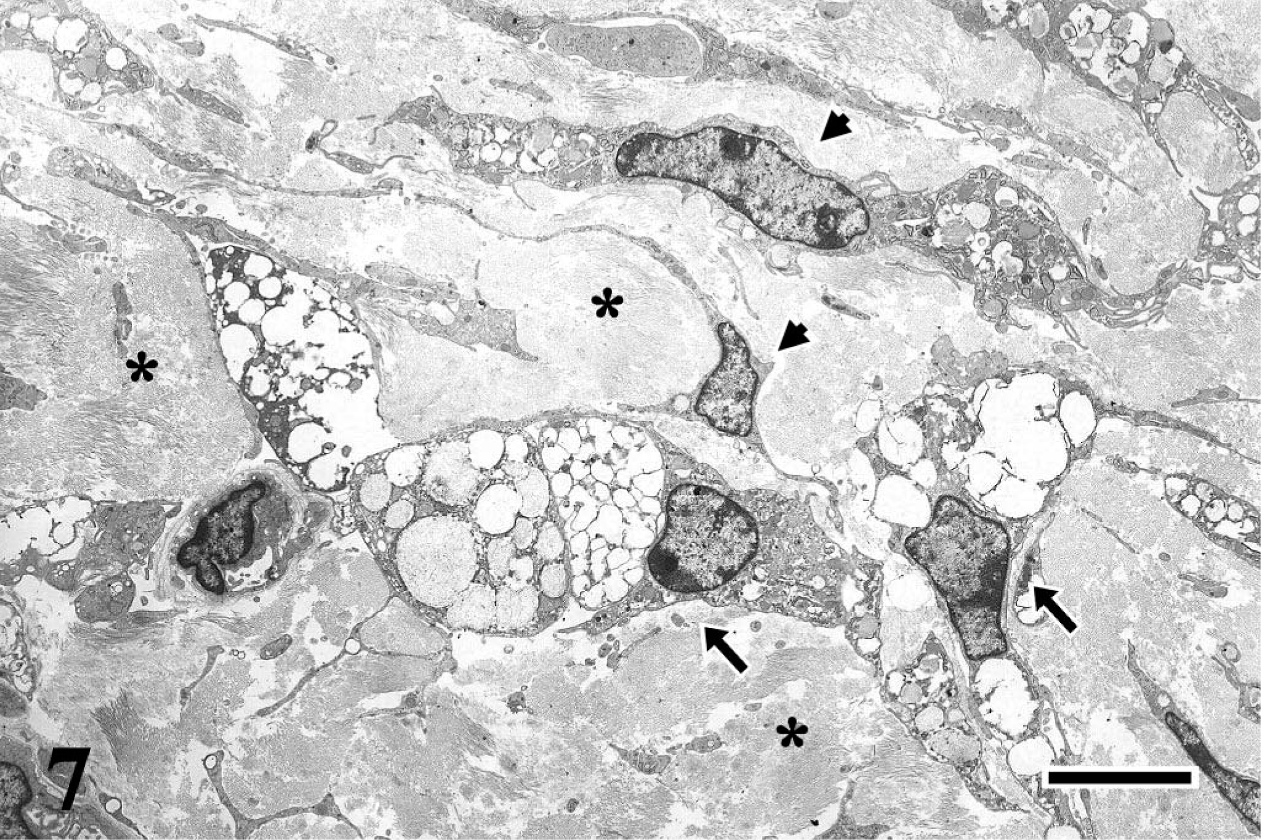
The submandibular cystic tumor in the 7-year-old Holstein cow was diagnosed as synovial sarcoma. Although removal of the mass was impossible, diagnosis of the synovial sarcoma could have been achieved if cytologic examination was performed before necropsy because of the presence of two neoplastic cell types, an epithelioid type and a fibro-blastic type. Transitional morphology between polygonal synovioblast and spindle cell was shown ultrastructurally.
Synovial sarcomas are morphologically classified into four subtypes: biphasic type, characterized by the presence of epithelial and mesenchymal cells, monophasic spindle cell type, monophasic epithelial cell type, and poorly differentiated cell type. 7 Immunohistochemical positive reactions for cytokeratin and vimentin and a negative reaction for S100 protein were helpful in the differential diagnosis from other types of sarcoma and tumors associated with the joint, such as neuroectodermal tumor, peripheral nerve sheath tumor, giant-cell tumor, or fibrosarcoma. Because malignant morphology such as nuclear atypism or frequent mitosis was not observed, the tumor was classified as a well-differentiated biphasic subtype. 6,7,9 The case reported in this study clearly filled two requirements of true synovial sarcoma: synovial differentiation and intra-articular location, as mentioned in a letter written by Fairley. 4 Tumor proliferation resulted in development of the large cystic mass outside the joint, which led to invasive proliferation of the bone marrow. Evidence of a metastatic tumor was not microscopically observed, which made the tumor reported in this study different from previous cases of synovial sarcoma in cattle. 3,5,10 All previous cases showed metastatic tumors in the lung and lymph node.
The most interesting fact in the case discussed in this study is its rare location. The tumor apparently developed from the synovium of the atlanto-occipital joint. There are previous reports of synovial sarcoma occurring in the connective tissue of the pharyngolarynx and other sites in humans, but its origin is not known. 8 Synovial sarcoma of the apophyseal joint of a cervical vertebra in dog has been reported, 6 but there have been no previous reports concerning synovial sarcoma in the vertebral joint of humans and animals.