
Abstract
The aim of this study was to correlate nuclear morphometric features with animal and human World Health Organization International Histological Classifications in canine seminomas. Twenty-three canine seminomas were classified, according to Animal World Health Organization International Histological Classification as intratubular, intratubular with signs of invasion, or diffuse and according to Human World Health Organization International Histological Classification criteria as spermatocytic and typical. The morphonuclear characteristics of tumors were quantitatively evaluated by means of digital cell image analyses of hematoxylin and eosin-stained nuclei. In particular, the mean nuclear area, mean nuclear perimeter, mean nuclear form factor, and their respective standard deviations were calculated. The relationship between the different variables and the tumor histologic types was assessed. On the basis of animal and human classification systems, statistically significant differences were observed only between intratubular seminomas with signs of invasion and the other two types and between spermatocytic and typical seminomas, respectively. In humans, it is well known that typical seminomas are more common and aggressive than spermatocytic ones. In our study, the canine seminomas classified as typical showed significantly larger and more variable nuclear area and perimeter than spermatocytic seminomas. These results support the opinion that most canine seminomas correspond to human spermatocytic seminomas and could explain the benign behavior of canine seminomas, which derive from a more differentiated type of germ cell.
Keywords
Spontaneous testicular tumors are common in aged dogs (mean age is 10 years), less common in aged bulls and stallions, and rare in other species. The principal canine testicular tumors consist of seminomas, Sertoli cell tumors, and Leydig cell tumors, and they occur with about equal frequency. The seminomas are usually solitary and unilateral and are more common in the right than in the left testicle; they can coexist with Sertoli cell and Leydig cell tumors. 9 Despite their malignant histologic appearance, in dogs, metastasis occurs in only a small percentage of the cases. 6 In contrast, human seminomas are the most common testicular neoplasms (comprising 40–50% of all testicular germ cell tumors) and have a greater tendency to metastasize. 5 In this respect, it is well known that the nuclear grading, expressing the degree of nuclear differentiation of neoplastic cells, is an important factor in determining the clinical outcome in patients with neoplastic disease. 1 The aim of this study was to correlate nuclear morphometric features of canine seminomas classified according to animal and human World Health Organization International Histological Classifications (WHO) criteria and to evaluate the diagnostic and prognostic usefulness of nuclear morphometry in these tumors.
Materials and Methods
Twenty-three spontaneously occurring canine seminomas were examined. Several tissue blocks from each tumor were formalin fixed and paraffin wax embedded, and sections (4 µm) were stained with hematoxylin and eosin (HE). Nuclear morphometric analysis was performed on HE-stained sections by means of an automated image analyzer (Sistema Mono, Immagini e Computer, Milan, Italy) connected to a Nikon Eclipse E-400 microscope (Tokyo, Japan), with the Image Pro Plus Program (Media Cybernetics, Inc., Silver Spring, MD). For each specimen, 10 images of cell fields, including areas with high and low cellularity, were captured by operator, who moved the microscopic field across the specimen from the surface to its margin. Areas of inflammatory and necrotic changes were excluded. Images were stored in the digital memory and displayed on the monitor screen. A total of 100 randomly sampled nuclei (avoiding nuclei of lymphocytes) from selected areas were measured by outlining their profiles with a computer “mouse.” In each case, the mean nuclear area (MNA; µm2), the mean nuclear perimeter (MNP; µm), the mean nuclear form factor (perimeter 2 /4Π area) (FF), and their standard deviations (SDNA, SDNP, and SDF, respectively) were calculated.
Associations between MNA, SDNA, MNP, SDNP, FF, and SDF and tumor histologic types were assessed by one-way analysis of variance followed by Student's t-test. The level of significance used was P < 0.01.
Results
The 23 tumors were classified according to the animal WHO criteria 10 as intratubular without signs of invasion (five), intratubular with signs of invasion (five), and diffuse (13) and according to the human WHO criteria 8 as spermatocytic (18) and typical (five corresponding to intratubular with signs of invasion). Two experienced pathologists performed this classification independently. The WHO histological classification of canine seminomas is based on intratubular (with or without signs of invasion) and diffuse types. In the intratubular type, atrophic seminiferous tubular epithelium is replaced by large, round neoplastic cells that fill the tubules (Fig. 1). The intratubular type with signs of invasion results from the extension of neoplastic cells from tubules into interstitium (Fig. 1). In the diffuse type the neoplastic cells form solid sheets, cords, or lobules 10 (Fig. 1). On the contrary, the WHO histological classification of human seminomas is based on typical or classic and spermatocytic types. Usually, the typical type shows a diffuse proliferation of a single morphologic type of neoplastic germ cells (inset Fig. 1) arranged in irregular lobules, cords, or strands. Rarely, the tumor is exclusively intratubular. The spermatocytic type is composed of a diffuse proliferation of polymorphic neoplastic germ cells, usually of three morphologic types: large, medium, and small cells. The nuclei of the large cells have a filamentous or “spireme” pattern of chromatin similar to that seen in spermatocytes in meiotic prophase (inset Fig. 1). Spermatocytic seminomas frequently show an intratubular growth pattern 7 . Nuclear morphometric results were then compared with histopathologic types.
Testis; dog. Intratubular seminoma according to animal WHO classification. HE. Bar = 20 µm.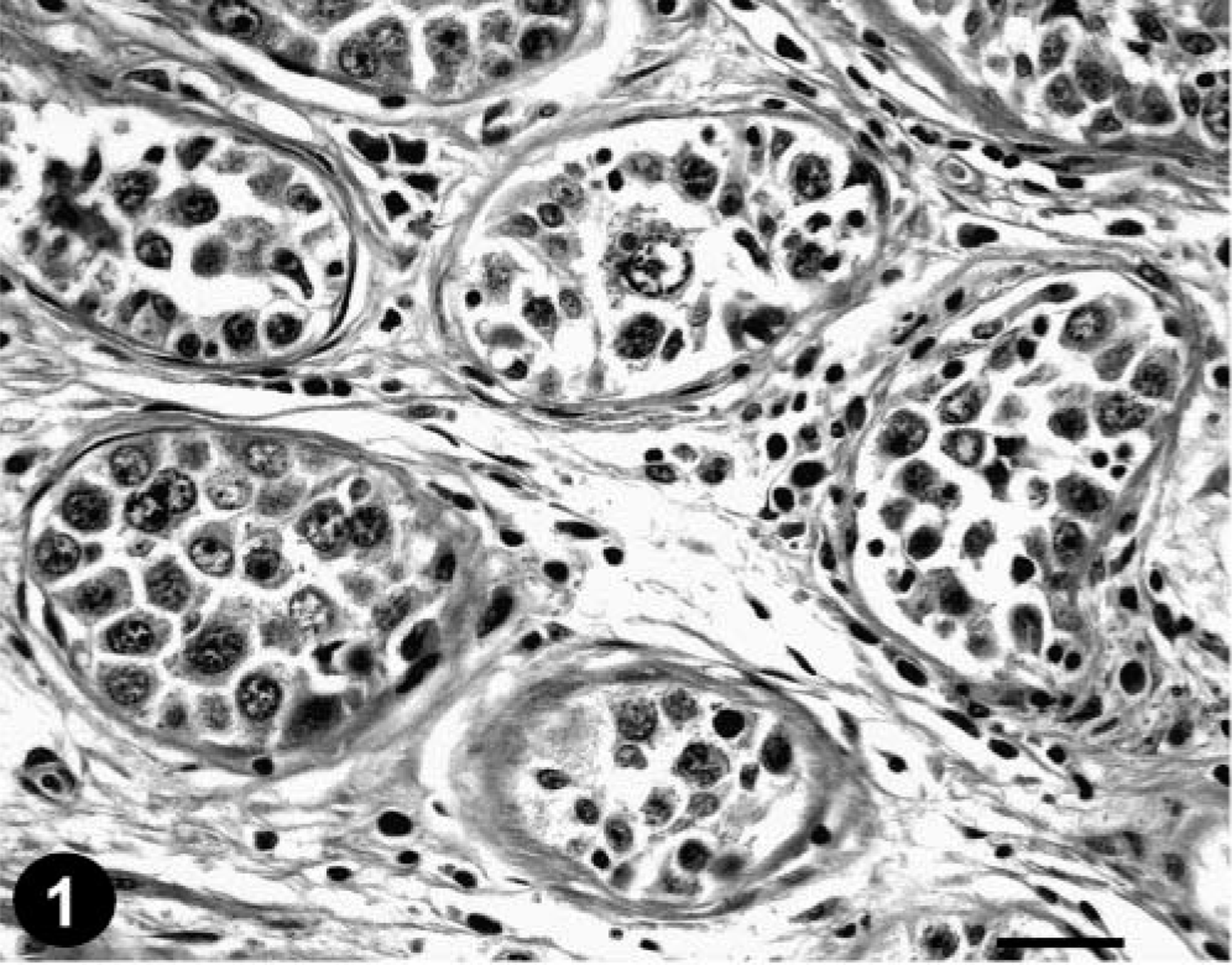
Testis; dog. Intratubular seminoma with signs of invasion according to animal WHO classification. HE. Bar = 20 µm. Inset: At higher magnification shows proliferation of fairly uniform neoplastic cells. Bar = 10 µm.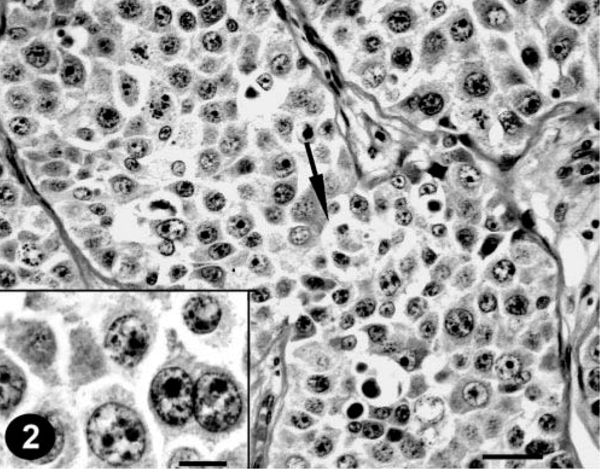
Testis; dog. Diffuse seminoma according to animal WHO classification. HE. Bar = 20 µm. Inset: At higher magnification shows a proliferation of polymorphic neoplastic cells. One cell with spireme pattern of chromatin (arrow). HE. Bar = 10 µm.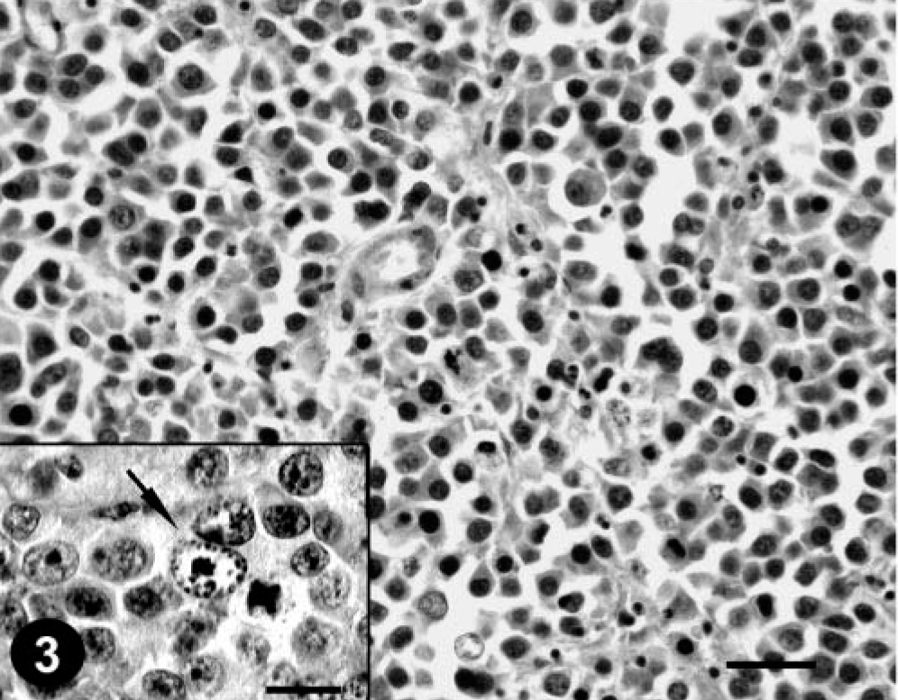
The mean values for the total (t) number of seminomas are given in Table 1, and those for seminomas of different histologic types are given in Table 2. MNA, MNP, and their standard deviations were greater in intratubular with signs of invasion seminomas than in the diffuse and in the intratubular without invasion types, respectively; typical seminomas also showed a greater MNA and MNP compared with the spermatocytic ones. On the contrary, SDNA and SDNP show lower values in spermatocytic seminomas than that reported in typical seminomas. However, the MNA, MNP, and their standard deviations showed significant differences (P < 0.01) only between intratubular with signs of invasion and the other two types (according to the animal WHO) and between spermatocytic and typical seminomas (according to the human WHO), respectively. The mean values of the respective FF and SDF did not reveal statistically significant differences between histopathologic types (P > 0.01).
Mean values of the morphometric nuclear parameters in the total number of seminomas examined.∗.
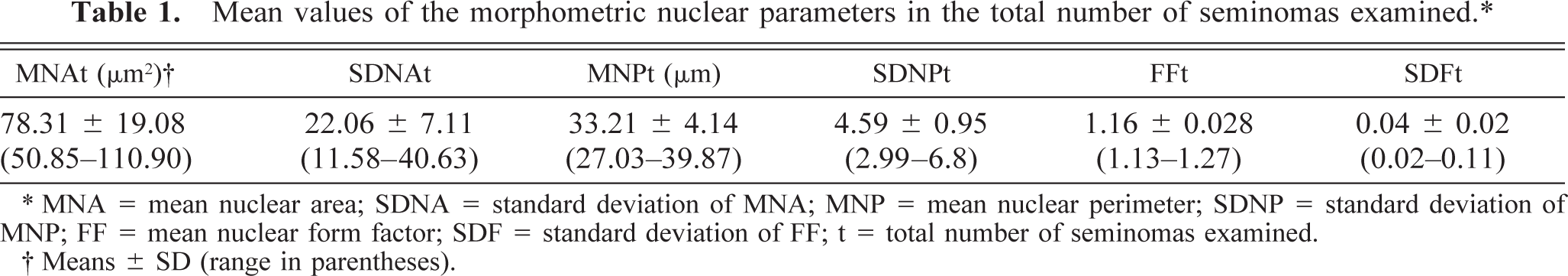
∗ MNA = mean nuclear area; SDNA = standard deviation of MNA; MNP = mean nuclear perimeter; SDNP = standard deviation of MNP; FF = mean nuclear form factor; SDF = standard deviation of FF; t = total number of seminomas examined.
† Means ± SD (range in parentheses).
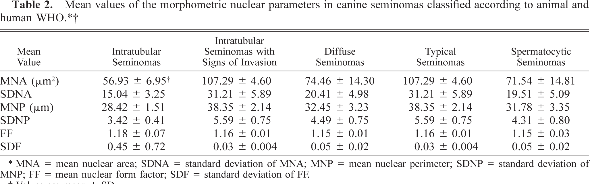
∗ MNA = mean nuclear area; SDNA = standard deviation of MNA; MNP = mean nuclear perimeter; SDNP = standard deviation of MNP; FF = mean nuclear form factor; SDF = standard deviation of FF.
†Values are mean ± SD.
Discussion
Our study demonstrated that the canine seminomas classified according to animal WHO criteria as intratubular with signs of invasion were morphologically similar to human typical seminomas; on the contrary, those classified as intratubular and diffuse were more similar to human spermatocytic seminomas. In this context, morphometry showed that canine seminomas classified as typical (corresponding to the five intratubular with signs of invasion cases) had MNA and MNP significantly and consistently greater than those classified as spermatocytic seminomas (corresponding to the five intratubular cases and 13 diffuse ones). These quantitative findings reinforced our subjective histopathologic evaluation and further suggested that in the dog, two different seminoma subpopulations could exist, which correspond to two morphologic and morphometric different histological types. In this regard, it is possible that, among the canine seminomas of our study, the intratubular ones without signs of invasion and the diffuse ones originated from cells undergoing spermatogenesis (spermatocytes) and that the canine intratubular seminomas with signs of invasion derived from undifferentiated germ cells (spermatogonia). Consequently, according to Benazzi et al., 3 the smaller dimension of the nuclei in canine intratubular without signs of invasion and diffuse seminomas in respect to the larger dimension of the ones observed in canine intratubular seminomas with signs of invasion could reflect differences normally existing between spermatogonia and spermatocytes. These results presumably could demonstrate, as reported previously by Looijenga et al., 7 that most of canine seminomas correspond to human spermatocytic seminomas and could justify the low metastatic potential of canine seminomas, which hence represent a more differentiated type of germ cell neoplasm. The results of the present study also show lower values of SDNA and SDNP in spermatocytic seminomas than that reported in typical seminomas. In this regard, it is well known that the SDNA and SDF could provide useful information about the variability of nuclear size and shape of a given neoplastic population. Nuclear “pleomorphism,” in fact, originates by variation of both nuclear size (polymetrism) and nuclear shape (polymorphism), and assessing nuclear morphometric parameter variability using their SD resulted a very discriminating technique in some neoplastic and nonneoplastic conditions. 1–4 The results obtained in our study regarding the values of SDs also seem to suggest that the well-known nuclear pleomorphism of spermatocytic seminomas could depend on variation in nuclear shape rather than on nuclear size, whereas in typical seminomas, although nuclear form seems to be more regular, a great variability in nuclear size usually occurs.
Because of the small number of cases in which morphometry was evaluated, the data obtained could not be generalized; for example, the observation that the totality of our typical seminomas were intratubular with signs of invasion, with no cases of diffuse type, could be a random event, probably due to the small number of “typical” cases examined.
For this reason, additional studies should be performed to prove the practical value of our results.
Nevertheless, the results obtained reinforce the current opinion that morphometry is a powerful technique able to detect changes not usually visible to the unaided eye, and that morphometric analysis of canine seminomas could be a simple and reproducible method that can identify an aggressive phenotype of canine seminomas (typical seminomas are known to be more aggressive than spermatocytic seminomas in humans) and provide additional diagnostic information that could allow more appropriate therapeutic choices.