
Abstract
The objective of this study was to evaluate by immunohistochemical means the nuclear expression of p27 and p21 proteins in cutaneous mast cell tumors and histiocytomas of dogs. In mast cell tumors, nine of the 13 grade I tumors, 13 of the 19 grade II tumors, and 10 of the 15 grade III tumors showed no detectable or mild p27 immunoreactivity. In contrast, one of the 13 grade I tumors, 12 of the 19 grade II tumors, and 11 of the 15 grade III tumors showed moderate or marked p21 immunoreactivity. Nineteen of the 28 histiocytomas showed no detectable or mild p27 immunoreactivity, and 24 cases showed moderate or marked p21 immunoreactivity. These findings indicate that a loss or absence of p27 expression is an early pathogenic event in mast cell and histiocyte tumorigenesis and that p21 expression may be a marker of mast cell tumor progression and histiocytoma cell proliferation.
Mast cell tumor is the most common skin neoplasm in dogs and is frequently malignant or potentially malignant. Canine cutaneous histiocytoma is a common, benign tumor of young dogs. The tumor is supposed to undergo spontaneous immune–mediated regression, although nuclear pleomorphism and high mitotic index are typical of this tumor. The etiology of these tumors and the molecular and genetic events contributing to tumorigenesis during development and progression of the tumors are largely unknown. Recently, it has been reported that p53 abnormalities 7 and c-Kit overexpression 15 are associated with tumor development or progression in some canine cutaneous mast cell tumors.
Cell proliferation is strictly regulated by cyclin–dependent kinase (CDK) complexes. 6,14,18,19 p27 6,14,18 and p21 19 are members of the CDK inhibitors' Cip/Kip family, which inhibit several CDKs and regulate the cell cycle. Previously, dysregulated p27 expression was demonstrated in a variety of neoplasms. 10 p21 can be induced by p53-dependent 3,4 and -independent pathways, and its expression is related to terminal differentiation. 2,11 Decreased p21 expression caused by a loss of normal p53 function has been reported to predict aggressive behavior in several malignancies. 17 On the other hand, p21 also functions as an assembly factor and an activator of CDK rather than as a kinase inhibitor. 1,6,9 This is believed to explain why overexpression of p21 in carcinomas is associated with tumor progression. 17 In the present study, to better understand mast cell and histiocyte tumorigenesis in the dog, we examined the expression of p27 and p21 proteins in these tumors by immunohistochemistry.
Samples consisted of formalin–fixed, paraffin wax–embedded tissue from 47 mast cell tumors, 28 histiocytomas, and 3 cases of dermatitis. The mast cell tumors were graded according to the histopathologic criteria established by Patnaik et al. 12 as grade I (well-differentiated type), grade II (intermediate type), and grade III (poorly differentiated type). In grade I (13/47 tumors), tumor cells were round and uniform in size and had well-defined cytoplasmic borders. In grade II (19/47 tumors), tumor cells varied in size, were round to ovoid, and had somewhat indistinct cytoplasmic borders. The nuclei were large and slightly vesicular. In grade III (15/47 tumors), tumor cells exhibited a high degree of cellular pleomorphism. The nuclei were large and varied in size and shape. Binucleated cells and mitotic figures were common.
Serial 5-µm sections were mounted on amino–silane–coated slides. The sections were dewaxed and microwave pretreated in 10 mM citrate buffer, pH 6.0, for 6 minutes. All the steps of the reaction were conducted at 25 C. The sections were incubated with normal goat serum for 10 minutes and then with anti-p21 or anti-p27 rabbit polyclonal antibodies (Santa Cruz Biotechnology, Santa Cruz, CA) for 30 minutes. A biotin–streptavidin–immunoperoxidase method was used, with 3-amino-9-ethylcarbazol (AEC) as the substrate chromogen. Sections were counterstained with hematoxylin. Nonimmune rabbit sera were used as negative controls. Normal canine epidermis was used as the positive control. In the keratinocytes of the epidermis, p21 was expressed in the suprabasal compartment. 5 Keratinocytes also showed p27 immunoreactivity. To quantify the reaction, positive cells in 50 fields at 400× magnification were counted; counts were averaged and expressed as percentages. The extent of the nuclear reactivity was classified as absent, mild (1–10% of tumor cells), moderate (10–50%), or marked (50–100%).
Results are summarized in Table 1.
Immunohistochemical reactivity of p27 and p21 in canine cutaneous mast cell tumors and histiocytomas.∗
∗ − = absent; + = mild (1-10%); ++ = moderate (10–50%); + + + = marked (50–100%).
Approximately 60% of reactive mast cells in cases of dermatitis showed p27 immunoreactivity, but the staining was weak (data not shown). In contrast, p21 immunoreactivity was observed only in occasional mast cells in cases of dermatitis (data not shown).
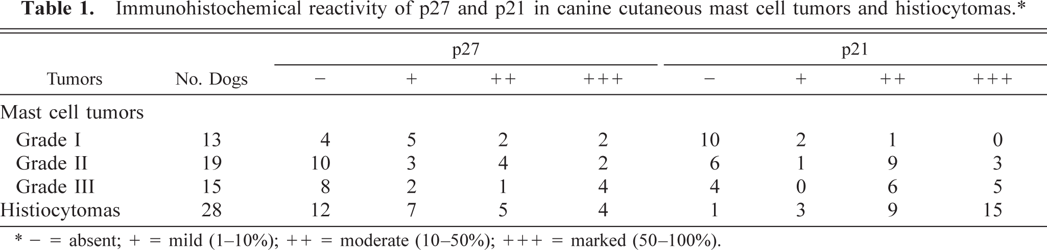
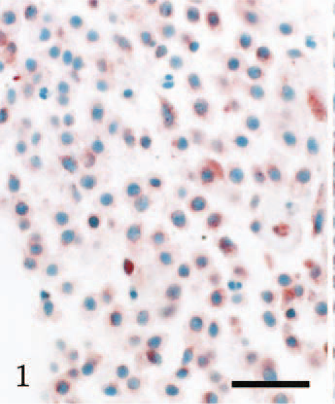
Nine of the 13 grade I mast cell tumors showed no detectable or mild p27 immunoreactivity (Fig. 1), whereas four cases showed moderate or marked immunoreactivity. Thirteen of the 19 grade II mast cell tumors showed no detectable or mild p27 immunoreactivity, whereas six cases showed moderate or marked immunoreactivity. Ten of the 15 grade III mast cell tumors showed no detectable or mild p27 immunoreactivity, whereas five cases showed moderate or marked immunoreactivity. P27 nuclear immunoreactivity was significantly lower in grade I mast cell tumors than in reactive mast cells. Of those cases with no detectable or mild p27 nuclear immunoreactivity, only nine showed cytoplasmic staining (Fig. 1). Twelve of the 13 grade I mast cell tumors showed no detectable or mild immunoreactivity with p21, although one case showed moderate immunoreactivity. Of the 19 grade II mast cell tumors, 12 cases showed moderate or marked p21 immunoreactivity, although seven cases showed no detectable or mild immunoreactivity. Of the 15 grade III mast cell tumors, 11 cases showed moderate or marked immunoreactivity (Fig. 2), although four cases showed no detectable immunoreactivity. The p21 nuclear immunoreactivity increased with histologic grade in mast cell tumors. Of those cases with no detectable or mild p21 nuclear immunoreactivity, 13 showed cytoplasmic staining.
Cutaneous mast cell tumor (grade I); dog. Mild p27 immunoreactivity is present. Biotin–streptavidin–immunoperoxidase method/AEC, hematoxylin counterstain. Bar = 110 µm.
Cutaneous mast cell tumor (grade III); dog. Marked p21 immunoreactivity is present. Biotin–streptavidin–immunoperoxidase method/AEC, hematoxylin counterstain. Bar = 110 µm.
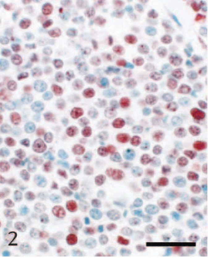
Nineteen of the 28 histiocytomas showed no detectable or mild p27 immunoreactivity, whereas nine cases showed moderate or marked immunoreactivity. Of those cases with no detectable or mild p27 nuclear immunoreactivity, only five showed cytoplasmic staining. In contrast, 24 of the 28 histiocytomas showed moderate or marked p21 immunoreactivity (Fig. 3), whereas four cases showed no detectable or mild immunoreactivity. There was no detectable cytoplasmic staining with p21 in those histiocytomas that showed no detectable or mild p21 nuclear immunoreactivity.
Cutaneous histiocytoma; dog. Marked p21 immunoreactivity is present. Biotin–streptavidin–immunoperoxidase method/AEC, hematoxylin counterstain. Bar = 110 µm.
In the present study, low levels of nuclear p27 expression were present in canine cutaneous mast cell tumors and histiocytomas. Loss or reduction of expression of p27 was observed even in grade I mast cell tumors and appeared to occur independent of tumor grade. In contrast, approximately 60% of reactive mast cells in cases of dermatitis showed p27 immunoreactivity. Nineteen of the 28 histiocytomas had no detectable or mild p27 immunoreactivity. These results suggested that reduced expression of p27 was an early pathogenic event in mast cell and histiocyte tumorigenesis. In epithelial neoplasms, decreased p27 expression is mostly associated with tumor invasiveness and metastasis but not with tumor development. 10 Lack or reduction of p27 expression may be more frequent in mast cell tumor and histiocytoma than in epithelial neoplasms.
Ubiquitination is the principal mechanism regulating p27 protein degradation. It has been reported that loss of expression of p27 in tumors occurs at the posttranslational level, because of increased protein degradation by way of the ubiquitin-proteasome pathway. 10 A study of malignant peripheral nerve sheath tumors and neurofibromas, however, showed that loss of nuclear p27 expression was involved in tumor progression and that strong cytoplasmic p27 staining reflected delayed proteasome degradation and cytoplasmic accumulation of the protein. 8 In the present study, cytoplasmic p27 staining was identified in only some of the cases with no detectable or mild p27 nuclear immunoreactivity. Further studies of cytoplasmic p27 staining in canine tumors are needed.
Nuclear p21 expression increased with histologic grade in mast cell tumors, and occasional reactive mast cells in dermatitis showed p21 immunoreactivity. The level of expression of p21 was higher in histiocytomas than in grade III mast cell tumors. These results suggested that p21 expression might be associated with mast cell tumor progression and histiocytoma cell proliferation. In addition, a high level of p21 expression may be useful as a marker of grading in mast cell tumor. Loss of expression of p21 has been associated with tumor progression in some carcinomas, 17 mantle cell lymphoma, 13 and acute lymphoblastic leukemia. 16 However, in other carcinomas, p21 is frequently expressed, occasionally with the highest expression in more malignant lesions. 17 The relationship between aberrant expression of p21 and tumor development and progression is poorly understood.
In conclusion, loss or absence of p27 expression is an early event in mast cell and histiocyte tumorigenesis, and p21 expression may be a marker of mast cell tumor progression and histiocytoma cell proliferation.