
Abstract
A 10-year-old terrier crossbreed presented with a change in bark intonation of 3–4 month's duration and pronounced panting. Four variably sized masses were observed within the oral cavity. The largest mass was located within the parenchyma at the caudal region of the tongue. Others were located on the left arytenoid, within the soft palate, and in the oropharynx above the soft palate. Histopathologic specimens consisted of large round to polygonal cells occasionally containing multiple nuclei and rare faint cytoplasmic cross striations. Staining was weakly positive with periodic acid-Schiff. Immunocytochemistry was strongly diffusely positive for muscle-specific actin, myoglobin, and desmin and scattered positive for S-100 and vimentin. Phosphotungstic acid-hematoxylin staining enhanced cytoplasmic cross striations. The cytoplasm of all neoplastic cells was filled with mitochondria on electron microscopy. The final diagnosis was multifocal/metastatic rhabdomyosarcoma.
A 10-year-old crossbreed, spayed female terrier presented to Avondale Animal Hospital with a change in bark intonation of 3–4 month's duration and pronounced panting. Four variably sized masses were observed within the oral cavity. The largest mass was located within the muscle parenchyma at the caudal region of the tongue. Others were found on the left arytenoid, within the soft palate, and within the nasopharynx above the soft palate. Fine-needle aspirates from the tongue and arytenoid masses were submitted, stained with Wright's stain, and examined microscopically. The four specimens contained similar polygonal cells consisting of a round, vesicular nucleus with a prominent nucleolus often with anisonucleoliosis. The cytoplasm was abundant, containing numerous minute reddish granules. Moderate to marked anisocytosis and anisokaryosis were present. Multinucleated cells also were observed frequently. Granular cell tumor, oncocytoma, rhabdomyoma, or rhabdomyosarcoma were considered cytologically, with the granular cell tumor most likely due to location; however, because of the unusual multifocal or metastatic nature of the neoplasm, rhabdomyosarcoma also was considered.
The dog was then referred to the Iowa State University veterinary teaching hospital. The dog appeared healthy without dyspnea. Hematologic and serum biochemical parameters were within the established reference values. On urinalysis, a 4+ proteinuria was present, with a urine protein-creatinine ratio of 4.21. Blood pressure was elevated at 208 mm Hg. Increased blood pressure and proteinuria findings were not associated with the oral masses.
Surgical removal of the four masses was attempted. After debulking of the masses, laser treatment was performed to cauterize the surrounding regions that could not be removed. Tissue specimens were fixed in 10% buffered formalin for histopathology and also in glutaraldehyde for electron microscopy (EM). Formalin-fixed tissues were prepared and initially stained with hematoxylin and eosin (HE)(Fig. 1) and periodic acid–Schiff (PAS).
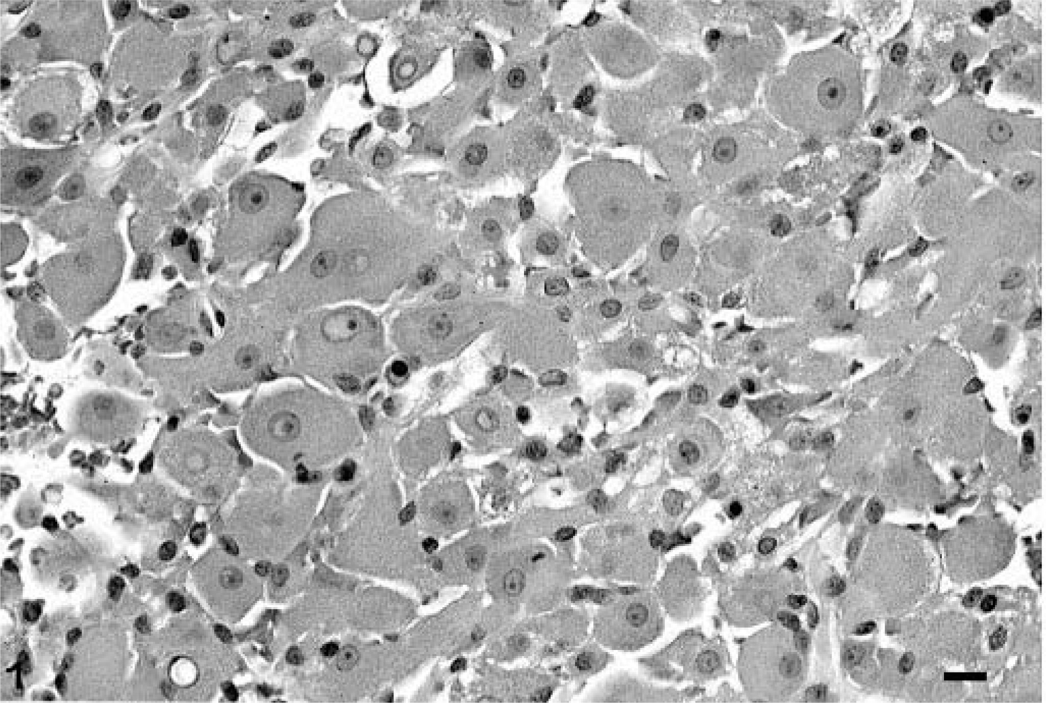
Tongue mass; canine. Pleomorphic and disorganized polyhedral cells present in tongue and oropharyngeal masses. HE-stained tissue sections identified as a rhabdomyosarcoma. Bar = 10 µm.
On HE-stained tissue sections all masses examined within the nasopharynx and the tongue were similar and were composed of large, pleomorphic, round to polygonal cells containing a round nucleus with a prominent nucleolus and extremely abundant eosinophilic granular cytoplasm. Numerous multinucleated and infrequent straplike cells with faint cross striations were present. Other spindle-shaped cells separating clusters of these polygonal cells also were observed. Weak staining was observed with PAS. The mitotic rate was low. The morphologic features of the cells were consistent with rhabdomyoma or rhabdomyosarcoma, which occasionally can be very difficult to differentiate morphologically and behaviorally. 2 Behaviorally, rhabdomyomas differ from rhabdomyosarcomas in that they are usually benign and do not reoccur locally after removal. 2,6
Tissue specimens were stained for S-100, vimentin, desmin, myoglobin, and muscle-specific actin. Also, phosphotungstic acid–hematoxylin (PTAH) staining was done. Vimentin and S-100 staining was scattered positive, whereas strong diffuse staining was observed for muscle-specific actin, myoglobin, and desmin. PTAH staining was diffusely positive, occasionally enhancing irregular disorganized cross striations within neoplastic cells (Fig. 2). EM revealed neoplastic cell cytoplasms filled with mitochondria. The combination of immunohistochemistry, PTAH, and EM findings; the pleomorphic appearance of the cells; multiple locations (especially lingual); tissue invasiveness; and, finally, the reoccurrence within 10 months prompted a final diagnosis of a multifocal rhabdomyosarcoma versus the morphologically similar laryngeal rhabdomyoma.
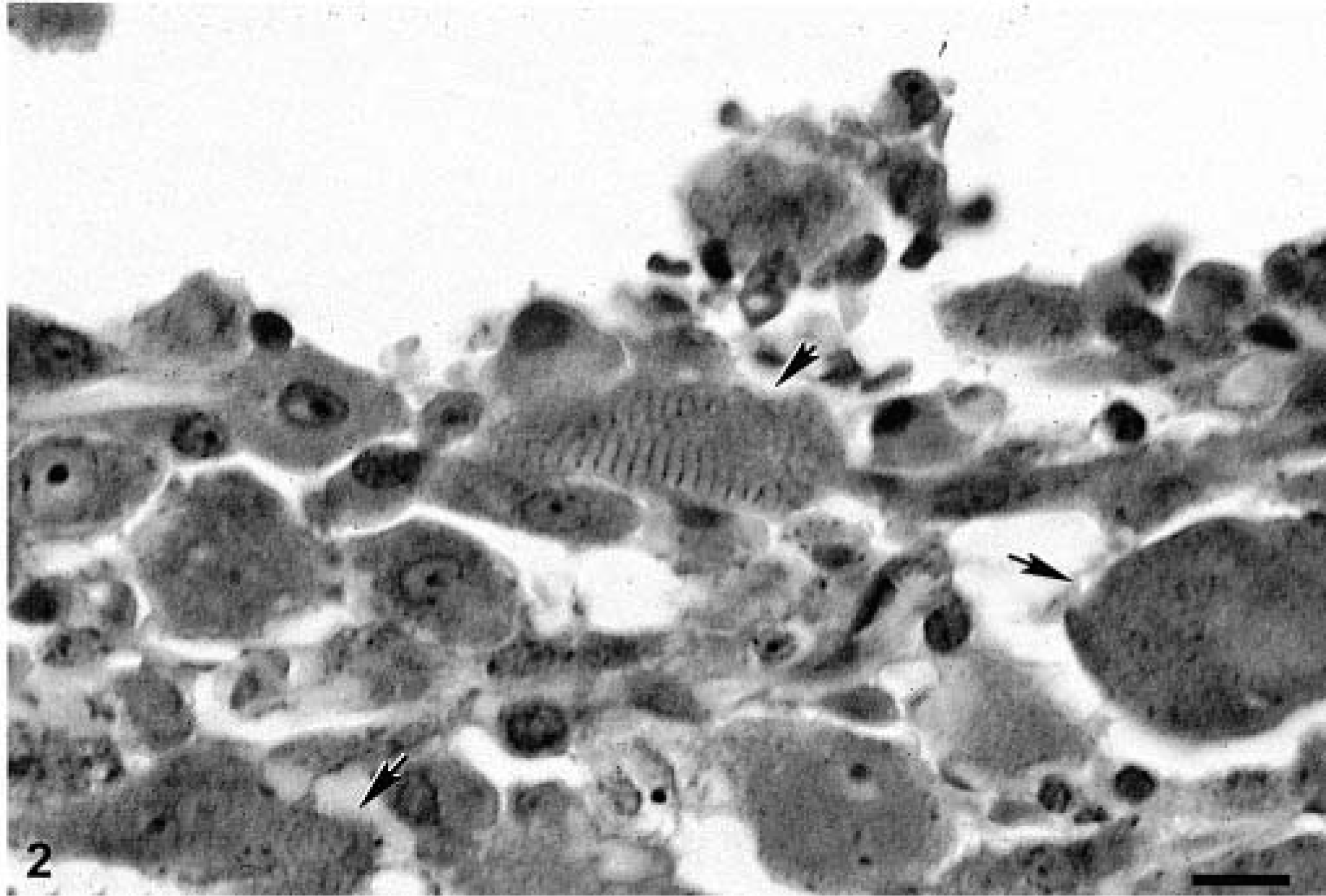
Tongue mass; canine. Enhanced but disorganized cross striations present in tissue sections of tongue mass stained with PTAH. Bar = 10 µm.
Tumors of the tongue are uncommon, comprising 4% of all oral and pharyngeal neoplasms in a review of 469 cases, of which 57 were lingual. 3 Ages ranged from 4 to 15 years, with the median age dependent on the tumor type present. 3 Squamous cell carcinomas, granular cell tumors, mast cell tumors, and malignant melanomas are the most frequently reported tumors within the canine tongue. 1 Rhabdomyosarcoma has been reported once previously within the tongue of a dog. 5 This tumor was initially reported as an oncocytoma but later changed to rhabdomyosarcoma after immunocytochemical staining. 5 In that report surgical removal, radiation, and chemotherapy were unsuccessful, with local reoccurrence. 5
Rhabdomyosarcomas are composed of cells arising from striated muscle, striated muscle progenitor cells, or primitive mesenchymal cells capable of differentiation into striated muscle cells. 2 These tumors are rare and may occur in any part of the body. 2 In the dog they have been reported within striated muscle, pharynx, gingiva, urinary bladder, cardiac muscle, larynx, greater omentum, urethra, skin, and trachea. 5 Depending on the variant type, they are locally invasive neoplasms with potential to metastasize. 2 Dissemination to lungs, lymph nodes, heart, spleen, adrenal glands, kidneys, and skeletal muscles can occur. 2 This report represents a multifocal or locally metastatic rhabdomyosarcoma. Whether these tumors developed individually in multifocal areas or by metastasis is impossible to determine. Metastasis is most likely, but it is not known whether spread occurred through hematogenous or lymphatic routes. The masses were separated without obvious anatomic connection.
A combination of immunocytochemistry, PTAH, and EM is used to differentiate rhabdomyosarcomas and rhabdomyomas from granular cell tumors and oncocytomas. Granular cell tumors in dogs usually stain strongly positive for PAS and occasionally stain positive for vimentin, cytokeratin, and S-100. 2 Desmin and muscle-specific actin are nearly always negative, although originally these cells were thought to originate from myoblasts. 2 On EM, granular cell tumor cytoplasm is laden with lysosomes, whereas rhabdomyomas, rhabdomyosarcomas, and oncocytomas are filled with mitochondria. 4,6 Although on EM, oncocytomas are similar to striated muscle cell tumors, they stain negatively for desmin, myoglobin, and muscle-specific actin, and cross striations are not observed.
This represents only the second reported case of lingual rhabdomyosarcoma in a dog and is unusual in that presentation was multifocal or locally metastatic in nature. In the previous report regrowth of the tumor was relatively rapid after surgical debulking, radiation therapy, and chemotherapy (6 and 8 months). 5
Enalapril had been administered for hypertension, and a recheck of the urine protein-creatinine ratio was within the established reference interval at 0.9. Chemotherapy treatment was offered but refused. After surgery the dog recovered her bark, but the tumors reoccurred in the nasopharyngeal and lingual regions nearly 10 months later, and euthanasia was elected. Only local regrowth of the tumors was observed at necropsy. The reoccurring oral neoplasms also were identified as rhabdomyosarcoma by histopathology, immunocytochemistry (positive for desmin and muscle-specific actin), and other special stains (PAS and PTAH—both positive).