
Abstract
Cutaneous smooth muscle tumors may arise from arrector pili muscles and from smooth muscles of the dermal vasculature. This report describes histologic and immunohistochemical features of eight arrector pili hamartomas in 8 dogs, 15 piloleiomyomas in 10 dogs and 3 cats, 10 piloleiomyosarcomas in 9 dogs and 1 cat, 1 angioleiomyoma in 1 cat, and 9 angioleiomyosarcomas in 6 dogs and 3 cats. Hamartomas and tumors arising from arrector pili muscles preferentially originated from the dorsal trunk. 5/5 (100%) arrector pili hamartomas, 10/12 (83%) piloleiomyomas, 4/5 (80%) piloleiomyosarcomas, 1/1 (100%) angioleiomyoma, and 6/7 (86%) angioleiomyosarcomas were positive for smooth muscle actin. 5/5 (100%) arrector pili hamartomas, 10/12 (83%) piloleiomyomas, 4/5 (80%) piloleiomyosarcomas, 1/1 (100%) angioleiomyoma, and 1/7 (14%) angioleiomyosarcomas were positive for desmin. Two incompletely excised canine angioleiomyosarcomas recurred locally. Metastases were not reported.
Keywords
Dermal smooth muscle tumors may arise from vascular, arrector pili, or deep dermal smooth muscle of genital areas. 5 They are identified as angioleiomyomas and angioleiomyosarcomas, piloleiomyomas and piloleiomyosarcomas, or genital leiomyomas and leiomyosarcomas, respectively. Cutaneous smooth muscle hamartomas and neoplasms are infrequently described in dogs and cats. 4,7,8,12,22,25,26 In domestic animals, they have been reported most often in the dog and ferret, 2,4,8,16,21 but thus far there have been no studies examining age, breed, or sex predilections.
In dogs, leiomyomas of arrector pili muscles were described in 5 4 and 10 8 dogs, respectively. In the latter report, these were described as solitary, generally less than 1-cm tumors that occurred predominantly on the head and neck. Histologically, these were composed of interwoven bundles of well-differentiated smooth muscle cells with elongated blunt-ended nuclei and eosinophilic cytoplasm. Histochemical (Masson trichrome) or immunohistochemical (smooth muscle actin, desmin) staining confirmed smooth muscle origin in these studies.
In cats, there is one documented case of what was termed “multiple piloleiomyomas” but that was actually a discrete, multinodular dermal tumor on the right lateral neck. 7 An unusual finding was the presence of foci of osseous metaplasia. There is also a single case of a feline cutaneous intravascular leiomyosarcoma, which presented as a deep mass involving digits of the left hind foot. 12 Histologically, this consisted of bundles of round to spindle cells that were often growing into vascular lumina. Positive immunohistochemistry for vimentin and smooth muscle actin confirmed the diagnosis. This tumor recurred locally on an adjacent digit after initial excision, but a second excision was apparently curative.
Piloleiomyosarcomas have been described in two separate groups of ferrets. 16,21 These were solitary dermal masses that consisted of a nodular proliferation of interwoven large spindle cells, displaying marked anisokaryosis, a low mitotic rate, and positive staining for vimentin, actin, and desmin. There were no reports of regional or distant metastasis, and in all cases in which clinical follow-up was available, complete surgical excision was curative. Solitary cutaneous tumors of smooth muscle origin are also described in several farm and exotic animal species, including a fibroleiomyoma in a sow, 17 a leiomyosarcoma in a cow, 9 and a leiomyosarcoma in a Peruvian squirrel monkey. 3
The goals of the present study were to define the morphologic and immunohistochemical features of arrector pili muscle hamartomas and dermal smooth muscle neoplasms in dogs and cats as well as to describe the biologic behavior of these tumors and to assess for the presence of age, breed, and sex predilections.
Materials and Methods
A search was performed in the databases of IDEXX Veterinary Services (West Sacramento, CA) for specimens submitted from 1 January 1993 to 1 August 2001. Tissues were submitted in 10% buffered formalin. They were trimmed, embedded in paraffin, sectioned at 5 µm, and stained with hematoxylin and eosin (HE). All cases were reviewed by both authors to confirm the initial diagnosis (Table 1). The lesions were categorized according to the human literature. 5,11,28
Major clinical, histological, and immunohistochemical features of dermal smooth muscle tumors in 33 dogs and 8 cats.
∗ C = dog; F = cat.
† F = female; FS = spayed female; M = male; MC = castrated male.
‡ Diameter (mm).
§ d = the tumor was restricted to the dermis; d/sc = the tumor extended from the dermis into the subcutis.
∥ + = Positive immunolabeling; — = negative immunolabeling.
# ns = Not specified.
¶ nd = Not done.
Paraffin blocks of 5 arrector pili muscle hamartomas, 12 piloleiomyomas, 5 piloleiomyosarcomas, 1 angioleiomyoma, and 7 angioleiomyosarcomas were available for immunohistochemistry. Five-µm-thick sections were cut from routinely processed paraffin blocks, mounted on poly-
Results
Hamartomas of arrector pili muscles
Eight hamartomas of arrector pili muscles were identified in eight dogs (Table 1). There was no breed predilection. Most tumors were present on the dorsal lumbar region (5/8). One was on a forelimb, and no site was available in two dogs.
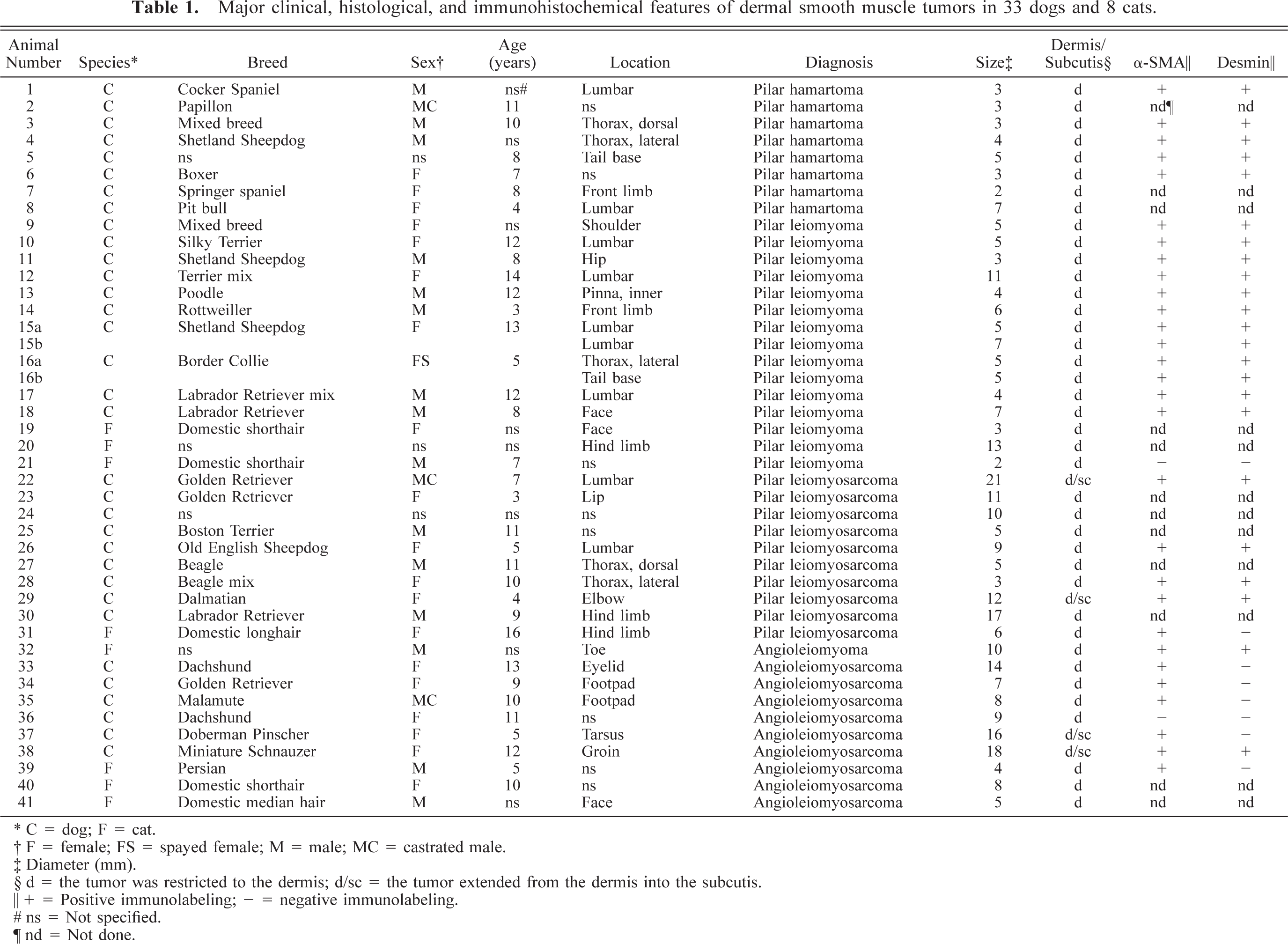
Histologically, these were solitary dermal lesions, which in all dogs were arising directly from arrector pili muscles (these muscles were normally prominent in skin from the dorsal trunk, a common site of occurrence; Fig. 1). Hamartomas were unencapsulated and well demarcated. They were composed of abnormally large or thick but well-differentiated smooth muscle bundles, which encompassed dermal appendages. These were separated by small to moderate amounts of mature collagen. There was no evidence of cytologic atypia or mitotic figures. The overlying epidermis was moderately acanthotic, and a few hyperplastic sebaceous glands were entrapped in all hamartomas. Excision was curative in the two dogs for which clinical follow-up was available (mean follow-up interval 425 days).
Arrector pili muscle hamartoma; dog. Bundles of smooth muscle fibers arise from arrector pili muscles and form a nodular dermal mass made of fascicles separated by dermal collagen. There is moderate acanthosis of the overlying epidermis. HE. Bar = 500 µm.
Piloleiomyomas
Twelve cases were diagnosed in 10 dogs, and three cases were diagnosed in three cats. In dogs, the dorsal trunk was the most common site (6/11), followed by the limbs (2/11) and head (2/11). In cats, the thigh (1/3) and dorsal muzzle (1/3) were affected. Sites were not available in three cases. There was no breed predilection in either species.
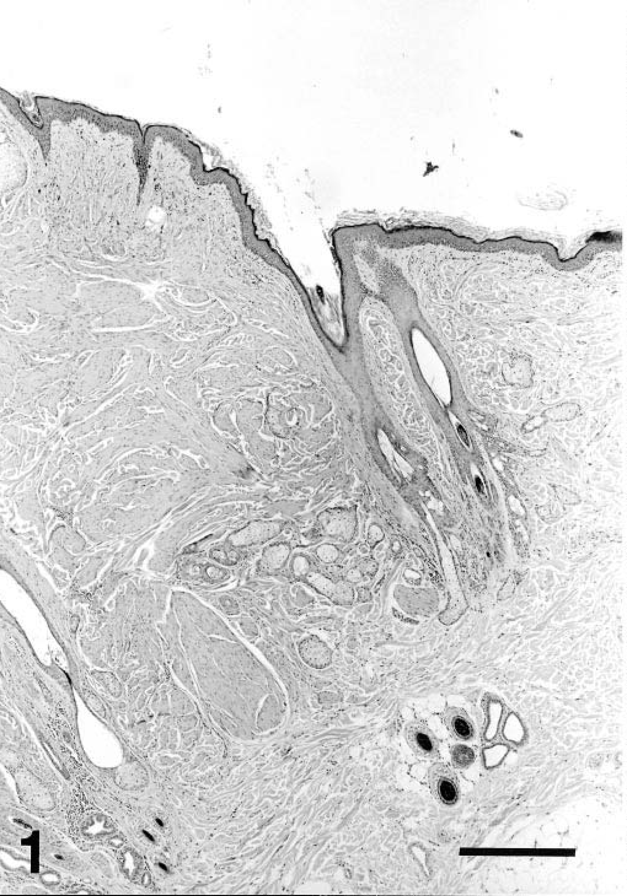
Tumors in both dogs and cats were solitary except for two dogs with two separate masses. These were nodular, well-demarcated, unencapsulated, expansile dermal masses, which showed a connection to arrector pili muscles in most dogs (10/12; Fig. 2) but not in cats. Tumors either entrapped (11/15) or displaced (4/15) dermal appendages and contained scant stroma. They were composed of narrow to broad, interlacing bundles of well-differentiated smooth muscle cells, which were narrow to plump and spindloid, with elongated cigar-shaped nuclei containing sparse, finely granular chromatin. The cytoplasm was homogenous to slightly fibrillar and deeply acidophilic. Mitoses were not observed. Rarely, tumors were inflamed (2/15), ulcerated (1/15), or hemorrhagic (1/15).
Piloleiomyoma; dog. Bundles of neoplastic cells near a hair follicle run parallel to it and merge with the arrector pili muscle. There is no evidence of atypia or of mitotic activity within neoplastic cells, which form closely packed bundles. HE. Bar = 100 µm.
Excision was curative in the five animals for which clinical follow-up was available (four dogs, one cat; mean follow-up interval 288 days).
Piloleiomyosarcomas
Nine cases were diagnosed in dogs, and one case was diagnosed in a cat. In dogs, the dorsal trunk was the most common site (4/9), with two cases involving limbs and one on the muzzle. No site was available for two dogs. In the cat, the tumor occurred on the thigh. There was no breed predilection.
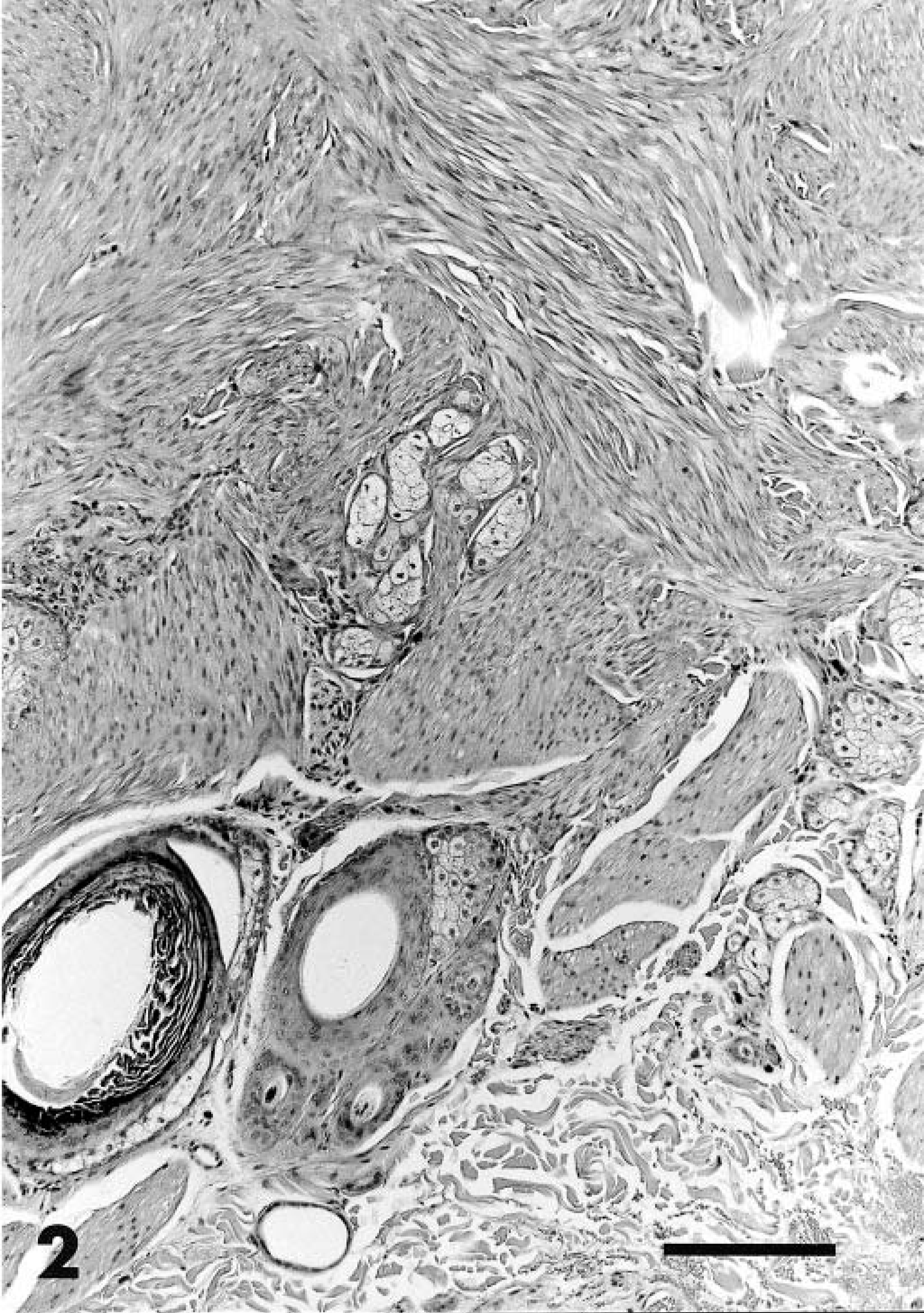
Tumors varied from well circumscribed (2/9 canine and 1/1 feline) to poorly circumscribed and invasive (7/9 canine). Neoplastic cells showed a connection to arrector pili muscles in one cat and four dogs. Tumors either entrapped (6/9 canine, 1/1 feline) or displaced (1/9 canine) dermal appendages. They had a variable histologic appearance. Three canine tumors were composed of well-differentiated smooth muscle but, unlike benign lesions, were invasive and displayed a mitotic rate of >2 per 10 high power fields (HPFs). Five canine tumors included poorly differentiated areas. These regions were hypercellular and contained streaming and interlacing bundles of narrow spindle cells with pale eosinophilic to amphophilic cytoplasm, which was often less abundant than that present in piloleiomyomas. Neoplastic cells occasionally showed nuclear palisading and packeting in the least-differentiated regions. In all cases, these cells displayed mild anisokaryosis (Fig. 3). Nuclei were elongated and cigar shaped. The feline tumor did not contain areas of well-differentiated muscle. Instead, it had streaming and interlacing bundles of slender spindle cells. Some tumors had areas of central coagulation necrosis (2/9 canine, 1/1 feline), lymphoplasmacytic inflammation (2/9 canine; 1/1 feline), mild mastocytic infiltration (2/9 canine), hemorrhage (1/9 canine), ulceration (2/9 canine, 1/1 feline), pyogranulomatous inflammation centered on ruptured hair follicles (1/9 canine), or mild myxomatous stromal degeneration (1/9 canine). The feline tumor contained lymphoplasmacytic and neutrophilic inflammation secondary to ulceration. Excision was curative in the three cases for which clinical follow-up was available (all canine; mean follow-up interval 304 days).
Piloleiomyosarcoma; dog. The tumor is composed of large, intersecting fascicles. Neoplastic cells have abundant cytoplasm, and nuclei display mild anisokaryosis and have prominent nucleoli. HE. Bar = 160 µm.
Angioleiomyomas
An angioleiomyoma was diagnosed on a front digit of a domestic shorthaired cat. This tumor was nodular, well delineated, unencapsulated, expansile, and moderately cellular. It was made of closely packed interlacing bundles of neoplastic smooth muscle fibers originating from the wall of blood vessels (Fig. 4). These blood vessels included variable proportions of capillaries, veins, arteries, and thin, cleftlike vascular spaces haphazardly distributed throughout the tumor. Neoplastic bundles gradually blended into the wall of blood vessels. Neoplastic cells were plump to fusiform, with indistinct cell borders and a moderate to large amount of fibrillar acidophilic cytoplasm. The nucleus was elongated to cigar shaped with blunt ends. Anisokaryosis and anisocytosis were mild. Mitoses were not found. Endothelial cells lining vascular spaces did not show evidence of atypia or of mitotic activity. Excision was curative (522 days postexcision).
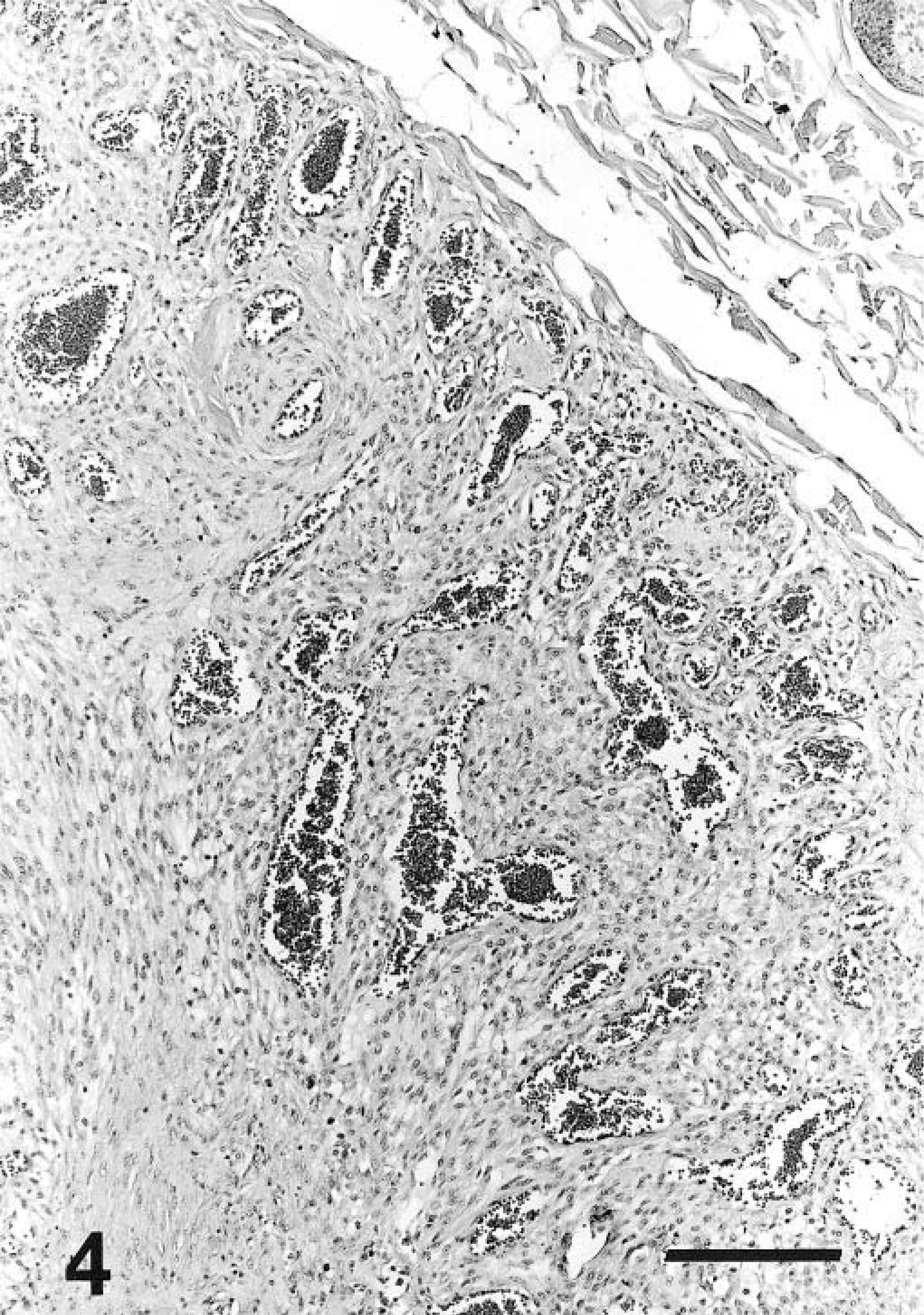
Angioleiomyoma; cat. An expansile tumor is made of bundles of plump neoplastic cells among which numerous veinlike vascular spaces are scattered. HE. Bar = 100 µm.
Angioleiomyosarcomas
Nine angioleiomyosarcomas were diagnosed in six dogs and three cats. There was no breed predilection. In cats, one tumor was located on the face and one on a limb. In dogs, they were predominantly located at the extremity of the limbs (two tumors from foot pads and one tumor from the tarsus) but were also found elsewhere on the body (a tumor from an eyelid and a tumor from the groin area). The site of the tumor was not available for one cat and one dog.
Angioleiomyosarcomas were differentiated from angioleiomyomas by a higher mitotic rate (0–46 mitoses per 10 HPFs; mean = 7), atypia, and a higher cellularity (Fig. 5). They had an expansile (n = 4) or invasive (n = 5) growth pattern. Neoplastic cells in angioleiomyosarcomas were also smaller and had an increased nuclear : cytoplasmic ratio compared with that of angioleiomyomas. Neoplastic cells blended into or seemed to originate from the media of blood vessels in all tumors. Endothelial cells lining the vascular spaces did not show atypia.
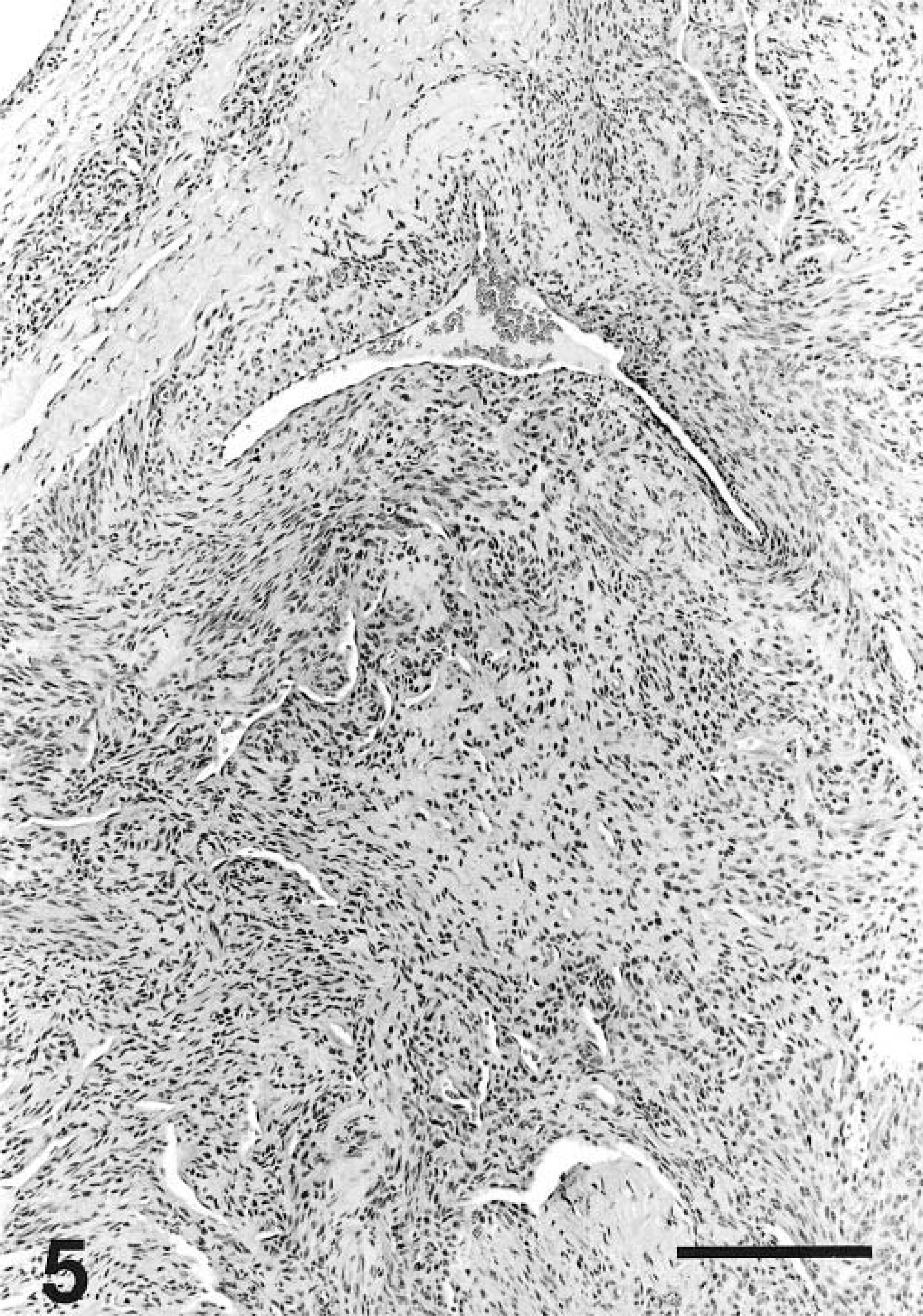
Angioleiomyosarcoma; dog. Densely cellular, haphazardly arranged bundles of neoplastic spindle cells are associated with slitlike vascular spaces. HE. Bar = 200 µm.
Follow-up information was available for one cat (840 days) and three dogs (mean follow-up of 244 days). One incompletely excised angioleiomyosarcoma from near a foot pad in one dog recurred and resulted in the euthanasia of this patient 383 days after the primary excision because of pain upon walking. A second poorly differentiated canine angioleiomyosarcoma from the groin recurred approximately 2 cm from the original surgery site 487 days after excision. There was no indication of local recurrence or metastasis for the three other angioleiomyosarcomas with follow-up.
Immunohistochemistry
All hamartomas (n = 5) and the angioleiomyoma were strongly positive for both desmin and α-SMA (Fig. 6). A subset of piloleiomyomas (n = 10), piloleiomyosarcomas (n = 4), and angioleiomyosarcomas (n = 1) were positive with both markers. One piloleiomyosarcoma and five angioleiomyosarcomas were positive for α-SMA but negative for desmin. The remaining tumors (two piloleiomyomas and an angioleiomyosarcoma) were negative for both markers.
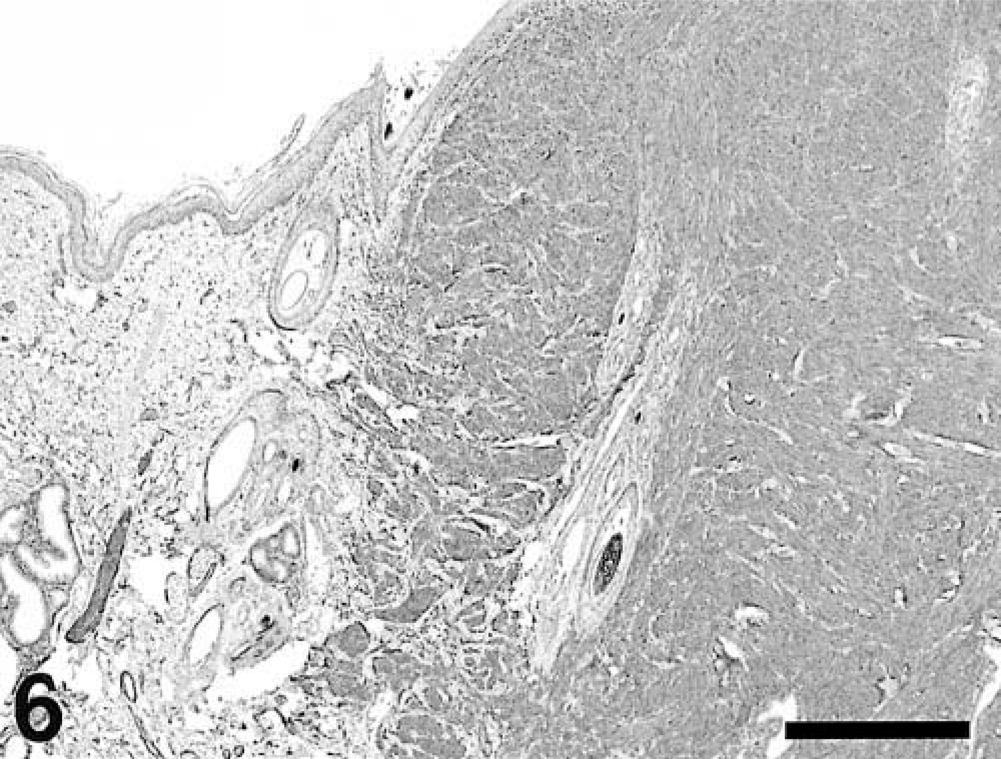
Piloleiomyoma; dog. Neoplastic cells and nearby arrector pili muscles strongly express α-smooth muscle actin. α-Smooth muscle actin, avidin–biotin–peroxidase complex method. Bar = 400 µm.
Discussion
This is the first comprehensive study of cutaneous smooth muscle tumors in dogs and cats. It provides the first description of arrector pili muscle hamartomas and dermal vascular smooth muscle tumors in animal species and shows that dermal leiomyosarcomas are low-grade malignancies with low potential for local recurrence.
The diagnosis of smooth muscle hamartomas and benign and well-differentiated tumors is straightforward: cells in these lesions characteristically have large amounts of acidophilic cytoplasm and a cigar-shaped nucleus. These features are unique to smooth muscle neoplasms. Other important features include connection of the tumor with arrector pili muscles or with the wall of blood vessels and juxtaposition of longitudinally and transversally cut fascicles. Hamartomas are distinguished from piloleiomyomas by the presence of broad, well-defined muscle bundles that maintain a normal anatomic relationship with hair follicles. In contrast, muscle bundles in piloleiomyomas are often narrower and more tightly interlacing, and these tumors may compress adjacent tissue and encompass or displace follicles, with loss of a distinct perifollicular orientation of muscle cells. However, this difference may be arbitrary and lesions that are presently identified as pilar hamartomas may actually represent early stages of piloleiomyomas.
Differential diagnoses for moderately well-differentiated and poorly differentiated malignant dermal smooth muscle neoplasms include fibrosarcoma, malignant peripheral nerve sheath tumor (MPNST), and hemangiopericytomas. Neoplastic cells in leiomyosarcomas more commonly form broad, discrete fascicles oriented at right angles to one another than neoplastic cells in fibrosarcomas and MPNSTs. Also, compared with the cells of piloleiomyosarcomas, cells of fibrosarcomas are more conical, whereas cells of MPNSTs are more wavy and asymmetric. However, poorly differentiated leiomyosarcoma cannot be differentiated from other spindle cell sarcomas on histologic grounds alone: in this series, the diagnosis of poorly differentiated leiomyosarcoma rested on the presence of focal areas of distinct smooth muscle differentiation of neoplastic cells.
The tumors in this series showed consistent immunohistochemical staining for smooth muscle actin and more variable expression of desmin. α-SMA is a cytosolic intermediate filament that is involved in the mechanism of contraction and that is specific to smooth muscle cells. Desmin is a cytoskeletal intermediate filament that is expressed in skeletal, cardiac, and smooth muscle. Both are used in human and veterinary pathology as cell markers for smooth muscle tumors. 4,11,13 However, several references have suggested that α-SMA may be a more reliable marker of smooth muscle tumors than desmin in both human beings and dogs. 4,13 One study examining canine smooth muscle tumors reported positive desmin staining in only 5 of 11 sarcomas. 1 It is notable that only one of seven angioleiomyosarcomas in this study was positive for desmin, versus four of five piloleiomyosarcomas. Similarly, human vascular smooth muscle tumors often fail to express desmin. 13
Neoplastic cells of leiomyosarcomas in this study showed positive staining for α-SMA and desmin in 83% and 42% of cases, respectively. Similarly, in humans, 68–100% and 43–100% of dermal leiomyosarcomas have been reported to label for α-SMA and desmin, respectively. 10,18,20,24,29 The absence of labeling for these two markers does not definitively rule out a diagnosis of leiomyosarcoma. Also, other canine spindle cell tumors, such as hemangiopericytomas 14,19 and a variety of tumors in humans (including primitive neuroectodermal tumors, neuroblastomas, giant cell tumors of tendon sheaths, mesotheliomas, tumors of myofibroblast origin, and osteosarcomas), have been reported to express α-SMA and/or desmin. 5 Thus, a diagnosis of leiomyosarcoma cannot rely exclusively on positive labeling for these markers.
h-Caldesmon and calponin are cytoskeleton-associated actin-binding proteins that may be more specific myogenic markers than either desmin or α-SMA. In human beings, both benign and malignant smooth muscle tumors have been shown to express these proteins. 15,27 These markers are not currently in widespread use in veterinary diagnostic pathology.
In this study, two angioleiomyosarcomas recurred locally, but no metastases were recorded. In human beings, local recurrence and distant metastases for cutaneous leiomyosarcomas have been reported in 0–44% and 0–50% of cases, respectively. 6,10,20,23 The prognosis in human cases differs significantly between cutaneous (i.e., dermal) and subcutaneous tumors, with the latter having much higher potential for both local recurrence and metastasis. 11,13,20 All tumors in this study were arising within the dermis, with secondary involvement of the subcutis in 4 of 41 cases (two piloleiomyosarcomas and two angioleiomyosarcomas). In the two cases in which local recurrence was reported, neither had subcutaneous involvement. These numbers are likely to be too small to draw any definitive conclusions about the prognostic significance of subcutaneous involvement in our study.
Necrosis and mitotic rate are the other histologic criteria that are often used as potential predictors of tumor malignancy. In human cutaneous leiomyosarcomas, neither of these seemed to correlate with the development of local recurrence. 6,11,23 Similarly, no correlation was found in this study, although again, the overall number of recurrent cases may not be large enough to be representative.
Tumors described in the human literature often are larger than those of dogs and cats from this series, which may account for the difference in clinical behavior. However, the paucity of subsequent clinical information and autopsy information in this study may have resulted in an underestimation of the actual rate of metastasis of dermal leiomyosarcomas in dogs and cats.
In summary, cutaneous smooth muscle tumors of vascular and arrector pili origin in dogs and cats are typically solitary lesions that infrequently invade into the subcutis. Immunohistochemistry for α-SMA, and to a lesser extent desmin, may aid in their diagnosis, particularly in poorly differentiated tumors. In most cases, surgical excision is curative; incompletely excised tumors had a low rate of local recurrence, and no metastases were reported.
Footnotes
Acknowledgements
We express our sincere thanks to Mr. R. Havens, Ms. M. Litton, Ms. S. Puerner, and Ms. A. Whittington for outstanding technical assistance. We are very grateful to Drs. Atwater, Burtch, Cocanoor, Dayton, Dietrich, Dykehouse, Fenster, Gervais, Good, Ingram, Johnson, McGuire, Metzler, Nanbu, Nugent, Ohler, Samaroo, Schroeder, Schwartz, Smith, Steinkruger, Sullivan, Sutter, Ueno, Waterhouse, and Werkes for follow-up information. We offer special thanks to Dr. Steve Smith for his review of the manuscript.