
Abstract
Most feline cutaneous mast cell tumors (CMCT) are behaviorally benign; however, there is a subset of these tumors with marked pleomorphism (previously termed poorly differentiated) that have been reported to be more aggressive. In this study, pleomorphic CMCT from 15 cats were identified from surgical biopsy submissions, and follow-up clinical data were obtained for 14 of these cats. Pleomorphic CMCT were discrete dermal nodules composed of sheets of pleomorphic round cells. Tumors from all 15 cats contained markedly cytomegalic and karyomegalic cells; 9/15 tumors (60%) contained multinucleated tumor giant cells. Typical mast cell granules were easily identified in sections stained with hematoxylin and eosin and with metachromatic stains and based on ultrastructural evaluation in cytomegalic as well as smaller tumor cells, indicating that the tumors were not poorly differentiated. The mitotic rate was very low (<1 mitosis per 10 high-power fields [hpf]) in 14 of 15 tumors (93%). Affected cats were 6–19 years old (mean age = 11.5 years), and there was no breed or sex predilection. Two cats had local recurrence. The only cat that had a pleomorphic CMCT with a high mitotic rate (1–2 mitoses/hpf) subsequently developed numerous other dermal neoplasms and was euthanatized. In this study, the large majority of feline pleomorphic CMCT were behaviorally benign. Mitotic rate is likely an important prognostic indicator of CMCT behavior.
Cutaneous mast cell tumors (CMCT), also referred to as mastocytomas, are common in the cat, accounting for 21% of feline skin neoplasms in a recent survey.13 Feline CMCT have been generally classified into two broad histologic types: the more common mast cell type and the uncommon histiocytic type, which occurs primarily in young Siamese cats.7,8,19 Some authors have further subclassified mast cell type CMCT as either well differentiated or poorly differentiated.6,7,19 Histopathologic grading schemes similar to those used for grading canine mast cell tumors have not proven prognostically useful for feline CMCT.2,14 Most investigators have concluded that the majority of feline CMCT are behaviorally benign.2,4,6,14,15,19 However, in one study seven of 65 cats (11%) with mast cell type tumors had marked anisocytosis, nuclear pleomorphism, mononuclear and multinucleated tumor giant cells, and high mitotic activity that recurred or spread within 3 months.18 The purpose of this study was to describe the histologic features and determine the biologic behavior of feline pleomorphic CMCT.
Materials and Methods
Animals and case inclusion criteria
From 1997 to 2001, 14 cases were selected for inclusion in this study from surgical biopsies examined by one pathologist (F. Y. Schulman) as a consultant for Marshfield Veterinary Diagnostics Laboratory, (Marshfield, WI). One case from 1995 was retrieved from the archives at the Armed Forces Institute of Pathology (Washington, DC). The prevalence of this tumor cannot be determined because the number of feline skin tumors examined over the collection period is not known. For the purpose of this study, feline pleomorphic CMCT was defined as a dermal nodule composed of atypical mast cells with cytoplasmic granules identifiable on sections stained with hematoxylin and eosin (HE) and metachromatic stains and exhibiting both cytomegaly (cells at least three times the diameter of neighboring cells) and karyomegaly (cell nuclei at least three times larger than neighboring cell nuclei) with nuclear pleomorphism. Signalments were taken from biopsy submission forms or were obtained via telephone conversations with the submitting clinicians. Additional follow-up information obtained from telephone interviews with the clinicians included present health status of the cat, interval between initial observation and surgical excision, ancillary treatment, and incidence of local recurrence, metastasis, and additional cutaneous neoplasms.
Histology and electron microscopy
Formalin-fixed, paraffin-embedded tissue samples were sectioned at 4–6 μm and stained with HE, Giemsa, Luna mast, and toluidine blue stains for light microscopic evaluation.
For ultrastructural studies, tumor tissue from cat No. 14 was deparaffinized, hydrated, postfixed in 1% osmium tetroxide, dehydrated, cleared, and embedded in epoxy resin. One-micrometer sections were cut and stained with toluidine blue for preliminary light microscopic examination. Thin sections (80–90 nm) were cut, stained with uranyl acetate and lead citrate, and examined with a Zeiss EM10 transmission electron microscope.
Results
Of the 15 cats included in the study, eight were male and seven were female. There were 10 domestic short- or longhair cats, two Siamese, and one Maine Coon cat; breed information was unavailable for two cats. The most common occurrence sites were abdomen (five), shoulder (three), back (two), flank (two), and head (two). Ages at time of surgical excision ranged from 6 to 19 years; the average age was 11.5 years. Twelve cats had a single CMCT, and three had two CMCT at initial presentation. Of these latter three cats, two had a second tumor that was a typical nonpleomorphic CMCT; the other cat had a second pleomorphic CMCT. Case summaries are presented in Table 1.
Case summaries for 15 cats with pleomorphic cutaneous mast cell tumors (CMCT).
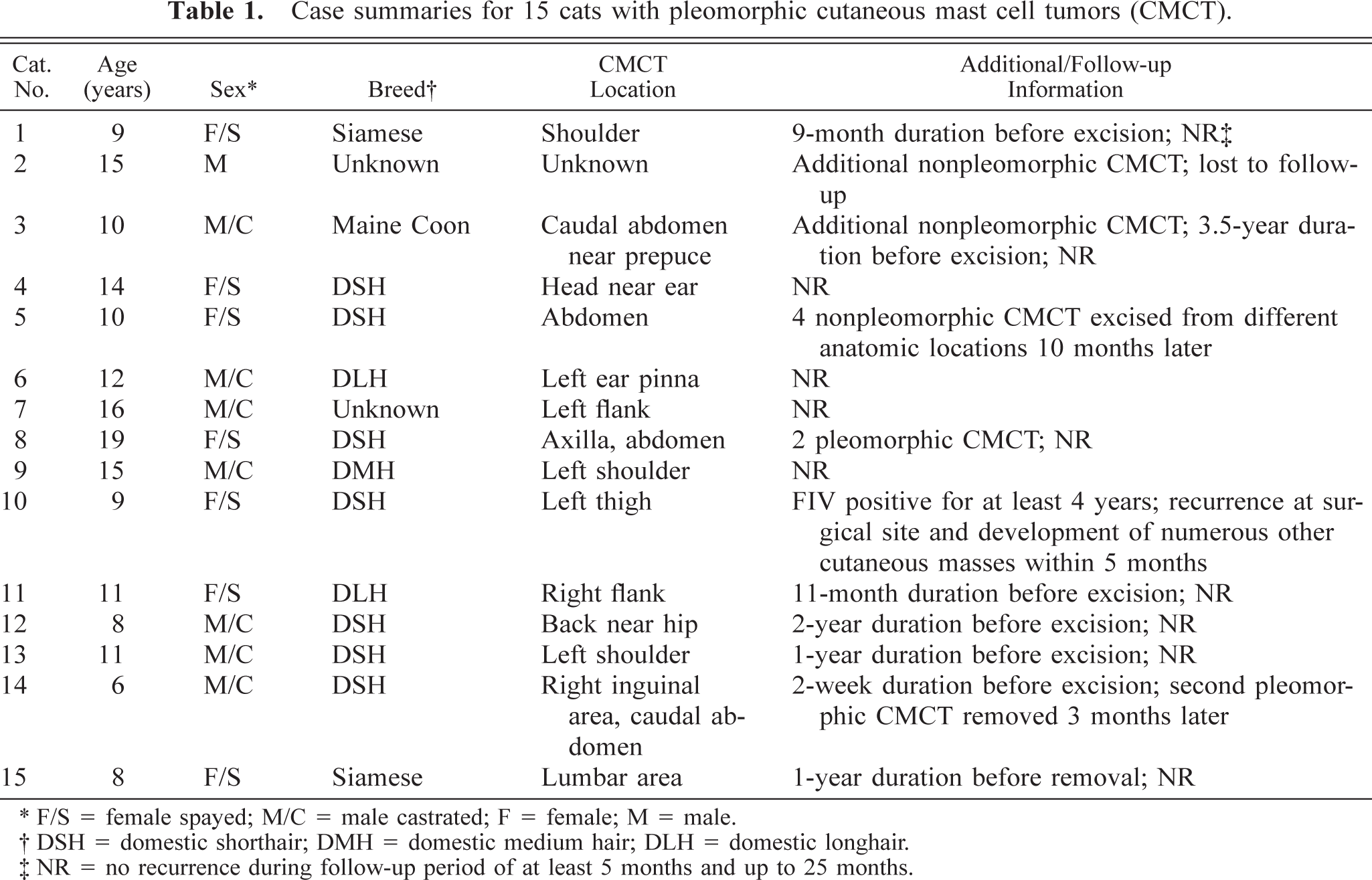
F/S = female spayed; M/C = male castrated; F = female; M = male.
DSH = domestic shorthair; DMH = domestic medium hair; DLH = domestic longhair.
NR = no recurrence during follow-up period of at least 5 months and up to 25 months.
The histologic appearance was similar in most tumors. The tumors were nonencapsulated, generally well-circumscribed, round nodules extending to the deep dermis and elevating the overlying epidermis (Fig. 1). Sheets of neoplastic round cells entrapped or effaced adnexal structures and were multifocally separated by thin bands of collagenous stroma or myxomatous matrix. Tumors from five cats were less well circumscribed, with neoplastic cells extending into the subcutis. Neoplastic cells had distinct cell borders and widely variable amounts of pale to brightly eosinophilic cytoplasm. All tumors had neoplastic cells containing numerous fine eosinophilic to basophilic cytoplasmic granules consistent with mast cell granules.
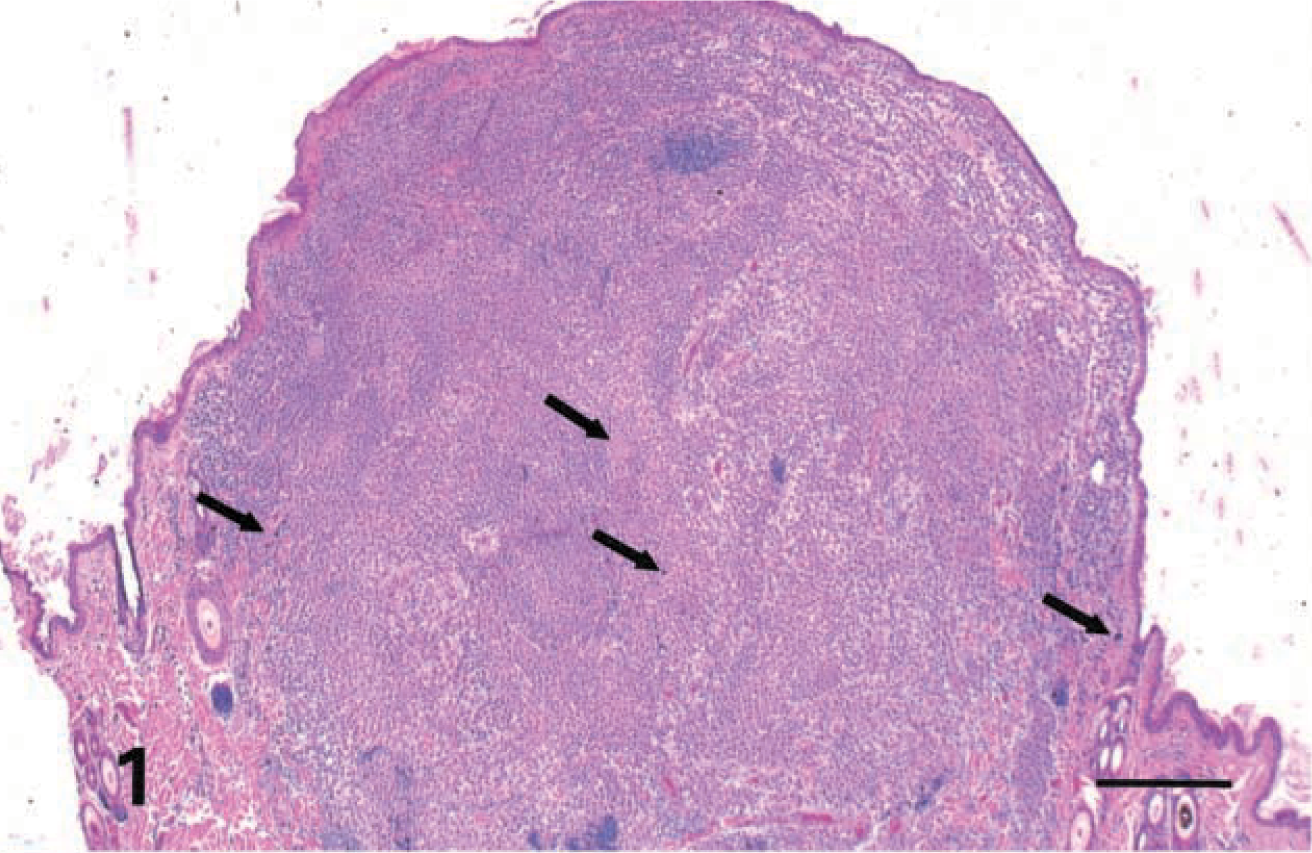
Haired skin; cat No. 4. Well-circumscribed dermal nodule elevates the overlying epidermis. Note cytomegalic and karyomegalic neoplastic cells visible at low magnification (arrows). HE. Bar = 400 μm.
Nuclei of smaller neoplastic cells were generally round and centrally to paracentrally located within the cytoplasm, with moderately stippled chromatin and one or two distinct basophilic nucleoli. The mitotic rate was very low (<1 mitosis/10 high-power fields [hpf]) in tumors from 14 cats. The CMCT from cat No. 10 had a mitotic rate averaging 1–2 mitoses/hpf.
All tumors contained uninucleated giant cells with diameters that were commonly 3–5 times larger than neighboring neoplastic cells, and occasionally 10–20 times larger, with moderate to marked karyomegaly and nuclear pleomorphism (Fig. 2). These cells occasionally had eccentrically located nuclei and a perinuclear pale eosinophilic cytoplasmic vacuole or hof. Tumor giant cell nuclei were round, elongate, or irregularly shaped, with finely stippled to coarsely clumped chromatin; many nuclei contained one to three prominent magenta nucleoli.
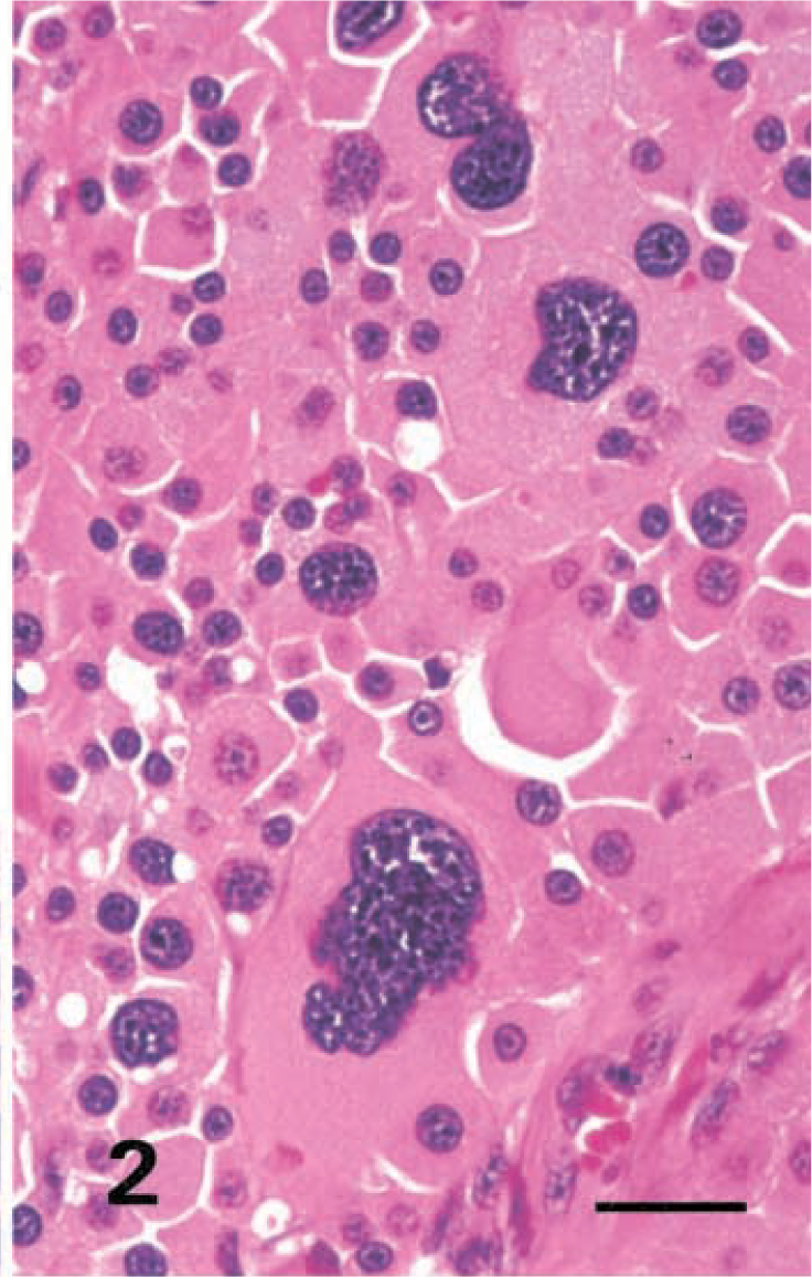
Haired skin; cat No. 14. Uninucleated giant cells are 10 times larger than neighboring tumor cells. These giant cells exhibit karyomegaly and marked nuclear pleomorphism. HE. Bar = 25 μm.
Additionally, nine tumors contained multinucleated giant cells that were usually round, had distinct cell borders, and contained up to 30 nuclei (Fig. 3). Cytoplasmic and nuclear features of multinucleated cells were usually similar to those of the smaller uninucleate neoplastic cells. There were occasional binucleate and trinucleate tumor giant cells with pleomorphic nuclei.
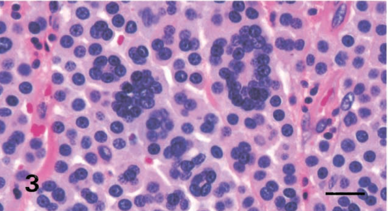
Haired skin; cat No. 6. Multinucleated giant cells with up to 30 nuclei. HE. Bar = 25 μm.
Metachromatic granules within the cytoplasm of small neoplastic cells were readily demonstrated with Luna mast, Giemsa, and toluidine blue stains in all tumors (Fig. 4). Most cytomegalic and karyomegalic tumor cells also contained numerous metachromatic granules within their cytoplasm. There was endocytosis of erythrocytes or neutrophils by some neoplastic cells in tumors from cats Nos. 1 and 14. These cells contained intact erythrocytes, discrete clear vacuoles with cellular debris, or degenerate neutrophils within their cytoplasm (Fig. 5). Twelve of 15 tumors (80%) contained perivascular aggregates of lymphocytes. There were a few infiltrating eosinophils in five neoplasms (33%) and numerous eosinophils in one neoplasm (7%). The tumor from cat No. 10 had numerous infiltrating neutrophils. Tumor excision appeared complete for 14 of the 15 tumors. Tumor cells extended to the deep margin of the section of the tumor from cat No. 13.
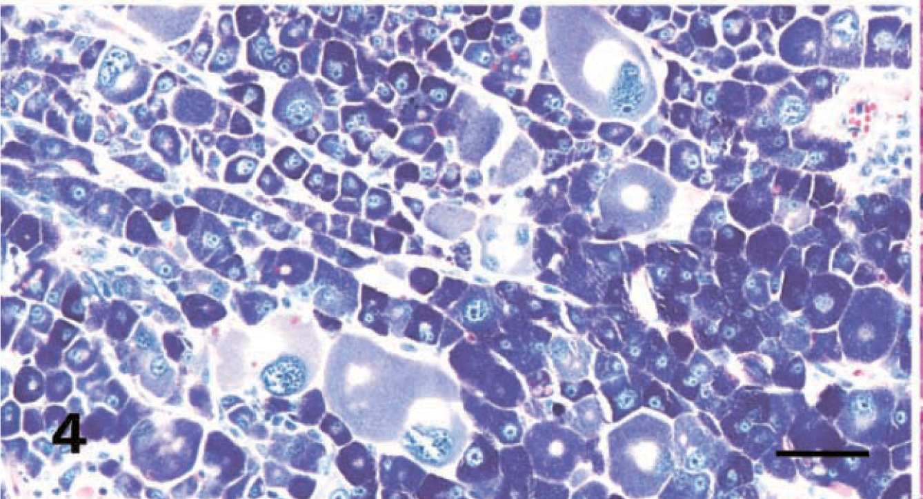
Haired skin; cat No. 14. The cytoplasm of both tumor giant cells and smaller neoplastic cells contain numerous metachromatic granules. Giemsa. Bar = 50 μm.
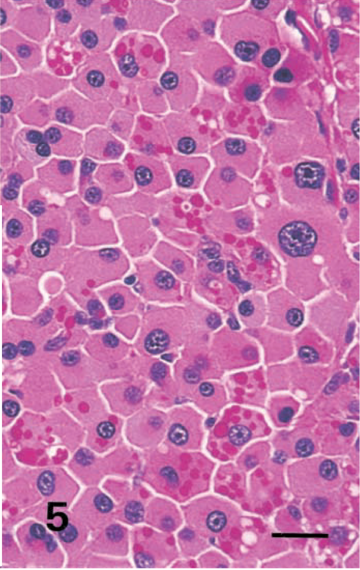
Haired skin; cat No. 14. Endocytosis of erythrocytes by neoplastic mast cells. HE. Bar = 25 μm.
Ultrastructurally, neoplastic mast cells contained numerous round to irregular distinct cytoplasmic granules. These granules were moderately electron dense and were either homogenous or contained amorphous, very electron-dense areas of various sizes within the granule matrix (Fig. 6).
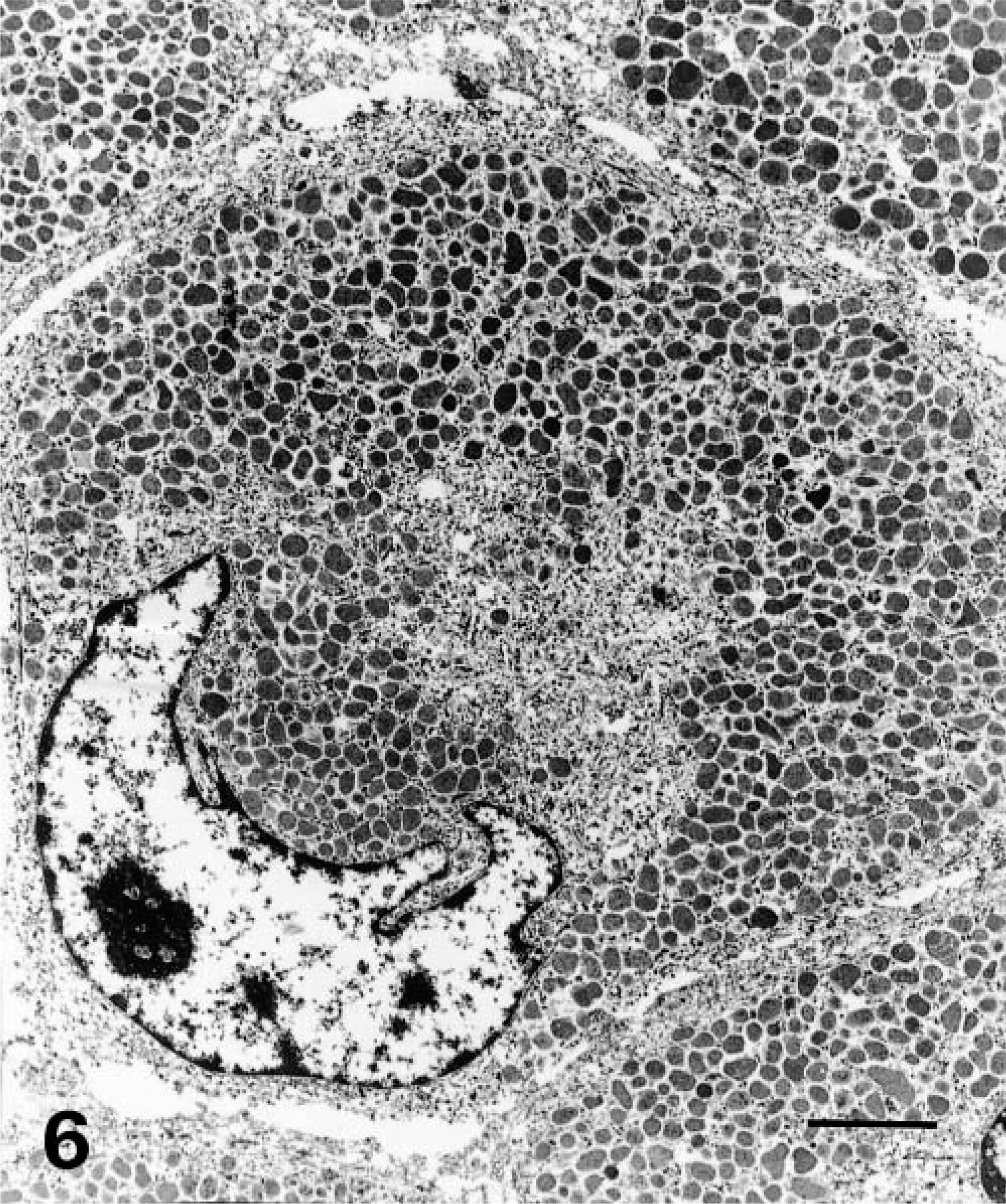
Transmission electron micrograph. Haired skin; cat No. 14. Bizarre pleomorphic nucleus with a prominent nucleolus within a cytomegalic neoplastic mast cell. The cytoplasm contains numerous moderately electron-dense granules. A central zone devoid of granules corresponds to the perivascular hof noted at light microscopic evaluation. Bar = 4 μm.
Follow-up information was obtained for 14 cats and is summarized in Table 1. The follow-up period ranged from 5 to 25 months. Eleven of 14 cats (79%) had no evidence of recurrence, additional cutaneous tumors, or metastasis in the follow-up period. Cat No. 5 developed four additional typical nonpleomorphic CMCT in other anatomic sites after the initial surgery. Cat No. 14 had a second pleomorphic CMCT arise close to the initial surgery site 3 months after excision of the first tumor but no additional occurrences. Cat No. 10 had apparent local recurrence and subsequent development of 30–40 additional cutaneous masses within 5 months of initial excision. The additional masses were firm, occasionally ulcerated cutaneous nodules up to 0.5 cm in diameter. Fine-needle aspirates yielded pleomorphic round cells. This cat had a positive enzyme-linked immunosorbent assay result for feline immunodeficiency virus (FIV) 4 years earlier. There was mild weight loss after development of the tumors but no palpable lymph node enlargement. The cat was euthanatized because of the numerous skin tumors and positive FIV status. No necropsy was performed. CMCT resulted in death or euthanasia of only one cat (No. 10).
Three cats had ancillary medical treatment in addition to surgical tumor removal. Corticosteroids were administered to cat No. 1 for 9 months prior to surgical tumor removal, but little change in tumor size was noted. Corticosteroids were administered to cat No. 10 following the recurrence and spread of the CMCT, but no clinical improvement was noted. Cat No. 14 was placed on a tapering dose of oral prednisone following removal of the second pleomorphic CMCT.
Discussion
Although in recent studies most feline CMCT have been considered benign,2,4,6,14,15,19 a subset of tumors termed poorly differentiated have exhibited aggressive biologic behavior.18,19 Those tumors were described as meeting criteria of malignancy, including anisokaryosis, anisocytosis, nuclear pleomorphism, high mitotic rate, and uninucleated and multinucleated giant cells. In our study, only the tumor from cat No. 10 exhibited all these characteristics, including high mitotic rate, and this cat experienced tumor recurrence and spread. The outcome in this cat supports the conclusions of previous researchers that tumors meeting cytomorphologic criteria of malignancy with high mitotic activity are behaviorally aggressive. However, the remaining 14 tumors included in our study had similar histologic features except for mitotic rate, which was very low, yet the 13 of these tumors for which follow-up information was available were behaviorally benign. These findings suggests cellular pleomorphism alone is not indicative of aggressive behavior of feline CMCT.
There was an extended time interval (9–42 months) from initial observation until surgical excision in six of the seven tumors for which the time interval was known, yet none of these cats had recurrence or spread in the follow-up period. There was apparent incomplete excision of the tumor of cat No. 13 but no local recurrence. These findings further support the benign nature of most pleomorphic CMCT.
Mast cell granules were readily identified in the cytoplasm of cytomegalic and karyomegalic pleomorphic neoplastic cells and in smaller neoplastic cells in sections stained with HE and metachromatic stains, indicating cellular differentiation. The single representative tumor selected for ultrastructural evaluation had cytoplasmic granules that were typical for differentiated mast cells. Therefore, because these tumors are not poorly differentiated and the term poorly differentiated implies malignancy, we recommend that neoplasms of this type instead be termed pleomorphic.
Previous researchers have reported feline CMCT recurrence rates of 13–56%2,8,14,18 and an occurrence rate for multiple CMCT at initial diagnosis of 13–43%.2,14 The rate of CMCT recurrence and the rate of multiple CMCT at initial presentation in the present study were both 21% (3/14), falling within the ranges of rates previously reported.
In the present study, there was no sex predilection for CMCT. Although some authors have reported a predilection in male cats,2,10 others have reported no sex predilection.5,8,13,18 The abdomen and shoulder were the most common sites of pleomorphic CMCT in the present study, followed by the back, flank, and head. In contrast, a predilection for the head and neck was reported by some investigators2,3,5 but was not found by others.8,14,18 No clear breed predilection is evident in our study. The distribution of breeds in this study is likely a reflection of the demographics of the pet population sampled. The average age of affected cats in this study was 11.5 years (range, 6–19 years), consistent with the findings of previous studies that CMCT occur more commonly in older cats.2,5,13,14,18
The single cat (No. 10) with an aggressive pleomorphic CMCT had tested positive for FIV. A definitive relationship between FIV and CMCT development has not been demonstrated. However, in one study a relationship between FIV and development of multiple CMCT was observed,5 similar to the situation for cat No. 10 in the present study. Other neoplastic diseases have been reported in FIV-infected cats, including lymphoma, fibrosarcoma, myeloproliferative disease, and systemic mastocytosis, but the pathogenesis of FIV-associated tumorigenesis is poorly understood.1,9,17
Neoplastic mast cell phagocytosis of erythrocytes in cats and humans has been previously reported.11,12,16,19 Neoplastic cells from two tumors in this study contained erythrocytes, clear cytoplasmic vacuoles with particulate debris, neutrophils, or nuclear debris within their cytoplasm. Erythrophagocytosis and endocytosis of other inflammatory cells are seemingly uncommon histopathologic features of feline CMCT.
In this study, the large majority of feline CMCT with cytomorphologic features such as anisocytosis, anisokaryosis, cytomegaly, nuclear atypia, and multiple nuclei had benign biologic behavior. High mitotic rate was the only characteristic that was an indicator of aggressive behavior.
Footnotes
Acknowledgements
We thank R.-A. V. Ferris, MFS, A. Morataya, and D. Miles for photomicroscopy, and J. Jenkins for electron microscopy. T. O. Johnson and L. D. Yantis are majors in the US Army. The opinions or assertions contained herein are the private views of the authors and are not to be construed as official or as reflecting the views of the Department of the Army or the Department of Defense.