
Abstract
Specific oncogenes that contribute to the pathogenesis of canine osteosarcoma (OS) have not been identified. In the process of characterizing four OS cell lines, we have found one cell line, CO8, that overexpresses the sis oncogene, which encodes the platelet-derived growth factor (PDGF)-α. The expression of an important downstream transcriptional target of the PDGF signaling pathway, c-myc, is also elevated fourfold. Conditioned medium from CO8 alone specifically induces tyrosine phosphorylation and therefore the activation of the PDGF-α and PDGF-β receptors on murine 3T3 cells. All of the canine OS lines tested contain PDGF receptors and therefore are capable of responding to PDGF. Given the importance of PDGF in promoting cell proliferation, migration, and cell survival, the activation of the sis oncogene and the resultant growth factor autocrine loop potentially contribute to the pathogenesis of a subset of canine osteosarcomas.
The production of growth factors and the establishment of autocrine loops by tumor cells play an important role in the pathogenesis of many human tumors, including osteosarcoma (OS).1,5 Platelet-derived growth factor (PDGF) is a homo- or heterodimeric protein consisting of A or B chains that acts as a potent mitogen, chemoattractant, and survival factor for many cell types.6 Homodimers of PDGF B chains (PDGF-β) bind and activate PDGF-α and PDGF-β receptors. The PDGF-β gene (c-sis) was originally identified as the cellular homolog of the v-sis oncogene and is expressed along with its receptor in several human tumor cell lines.3,6 Overexpression of c-sis or v-sis in mouse 3T3 cells results in a transformed phenotype.2,6
We previously have described the isolation and characterization of several canine OS cell lines (CO).8 To measure sis expression, equal quantities (2 µg) of poly A+ mRNA were analyzed by northern blotting and hybridization with a human sis probe. The sis transcripts are present in all of the OS cell lines (Fig. 1). However, only CO8 expresses high levels of sis mRNA. Low-level expression of sis mRNA frequently is detected in mesenchymal tumors and may be a general consequence of transformation.6 The high level of sis expression in CO8 is likely a consequence of an activating mutation such as an amplification or translocation involving the sis gene, or a mutation within the sis promoter.9
We were unable to detect sis protein in CO8 cells by using several commercially available antibodies directed against human PDGF-β. As an indirect measure of sis protein activity, we measured the expression of an important downstream transcriptional target of the PDGF signaling pathway, c-myc, by northern blotting (Fig. 1). Levels of c-myc mRNA are elevated fourfold in CO8 relative to the other OS cell lines, whereas cdk4 mRNA levels remain relatively constant. Taken together, these results indicate that CO8 expresses high levels of sis mRNA and suggest that sis overexpression activates PDGF signaling pathways and myc transcription.
A characteristic of cells transformed by sis is the secretion of active PDGF-β into the culture medium.4 We assayed conditioned medium (CM) from CO8 cells for a PDGF–β–like activity by using mouse 3T3 fibroblasts as an indicator cell line. In these cells, serum starvation results in the downregulation of PDGF receptor activity, as indicated by the reduction in the amount of phosphotyrosine-containing receptors. After the addition of fetal bovine serum, which contains high concentrations of PDGF-β, an increase occurs in phosphotyrosine-containing PDGF-α and PDGF-β receptors (Fig. 2). CO8 CM also contains a factor that activates both PDGF receptors. This effect is specific to PDGF receptors because the epidermal growth factor (EGF) receptor is not activated by CO8 CM. Activation of PDGF receptors is not detected when using CM from CO2, another OS cell line. These results indicate that CO8 synthesizes and secretes a factor that specifically activates PDGF receptors.
We next used western blotting to determine whether the OS cell lines express PDGF receptors. Control canine fibroblasts express high levels of the PDGF-β receptor, but not the PDGF-α receptor (Fig. 3). All of the OS cell lines express the PDGF-α receptor. CO7 also expresses the PDGF-β receptor. Therefore, all of the OS cell lines are able to respond to PDGF-β.
Autocrine loops involving several growth factors have been identified in human OS cell lines and tumors.1,5 In this report, we present evidence that PDGF-β and the PDGF-α receptor are coexpressed in canine OS cell lines. Furthermore, one OS cell line expresses high levels of sis mRNA, coincident with increased expression of the myc oncogene, a downstream transcriptional target of the PDGF signaling pathway. Based upon our findings, sis oncogene activation is unlikely to occur frequently in canine OSs. However, given the importance of PDGF in promoting cell proliferation, migration, and cell survival, sis activation potentially contributes to the pathogenesis of a subset of canine OSs.
The specific mechanism by which PDGF-β contributes to OS progression is, as yet, unclear. Of interest is that all of the OS cell lines except CO8 contain mutations in the tumor suppressor gene, PTEN.7 It is possible that overexpression of sis antagonizes PTEN action, and provides an alternative, nonmutational mechanism to inactivate this important tumor suppressor.
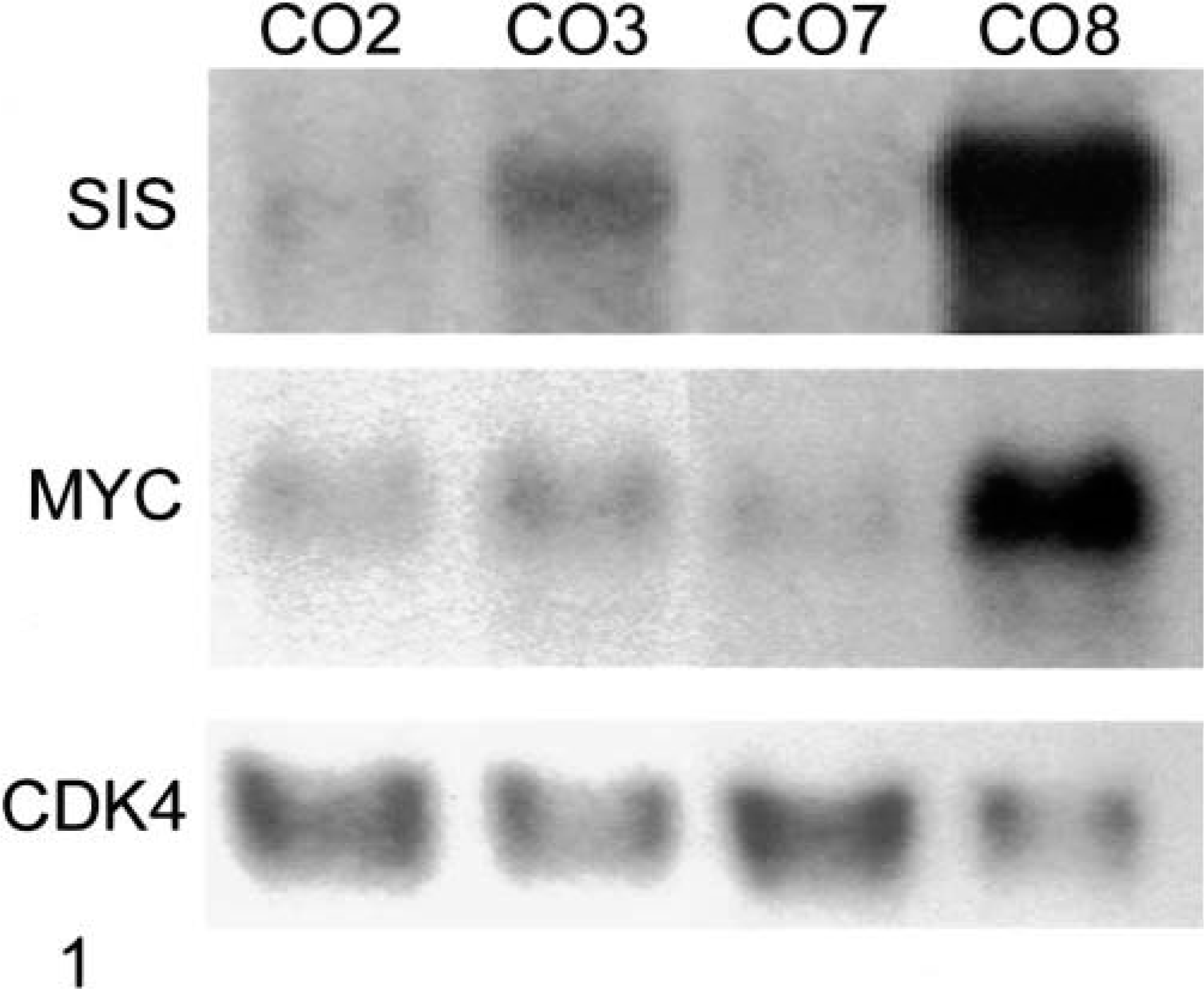
Overexpression of sis and myc mRNA in CO8. Two micrograms of poly A+ (sis probe) and 20 µg of total RNA (myc and cdk4 probes) from each cell line were analyzed by northern blotting.
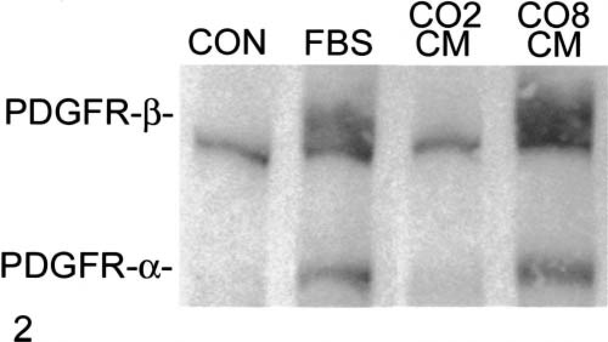
Conditioned medium (CM) from CO8 induces the tyrosine phosphorylation of PDGF-α and PDGF-β receptors in 3T3 cells. Equal numbers of CO2 and CO8 cells were washed twice with serum-free Dulbecco modified Eagle medium (SF-DMEM) and incubated with SF-DMEM. After 36 hours of incubation, the CM was centrifuged and filtered to remove intact cells and cellular debris. Serum-starved murine 3T3 cells were incubated with SF-DMEM, FBS, and CM, obtained from equal numbers of CO2 and CO8 cells for 20 minutes. Protein extracts were prepared and analyzed by western blotting (20 µg/lane) with antibodies (sc7020, Santa Cruz Biotechnology Inc., Santa Cruz, CA) that recognize phosphotyrosine containing proteins. The positions of PDGF-α and PDGF-β receptors are indicated. The EGF receptor migrates just below the PDGF-β receptor.
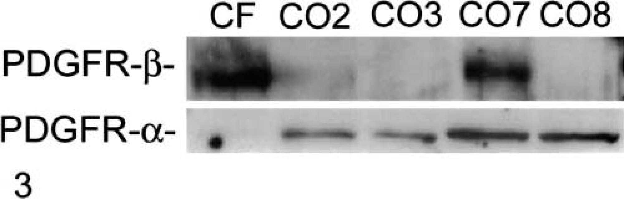
Platelet-derived growth factor receptors are expressed in canine OS cell lines. Protein extracts were prepared from exponentially growing cells and analyzed by western blotting (20 µg/lane) with antibodies to PDGF-α (sc338) and PDGF-β (sc432) (Santa Cruz Biotechnology Inc., Santa Cruz, CA) receptors.
Footnotes
Acknowledgements
This study was supported by a grant from the Morris Animal Foundation (97CA-21).