
Abstract
Liver cytology was evaluated in 28 healthy dogs 1–14 years of age with normal liver structure and function. Smears were stained with May-Grünwald-Giemsa. Hepatocytes had distinct cell borders, and cells did not overlap. Cells with two nuclei and cells with intranuclear crystalloid structures were observed regularly. Cytoplasm contained small numbers of vacuoles characteristic of glycogen and lipid and small amounts of pigment consistent with ceroid or bile. Nuclei were uniform. Small numbers of biliary epithelial cells were seen in most samples. Lymphocytes and neutrophils occurred in small numbers, with lipocytes, mast cells, fibrocytes, mesothelial cells, eosinophils, and Kupffer macrophages seen less frequently. Mean parenchymal cell sizes were significantly greater in older dogs, but no age-related differences were observed in nuclear size. Older dogs also had a significantly increased number of nuclei per cell. There were more neutrophils in young and old dogs than in middle-aged dogs.
The clinician faces some difficulties when examining a patient with hepatic disease. Although the presence of liver disease may be detected with physical or biochemical examinations or with different imaging methods, the precise diagnosis of the specific disease requires histopathology in most cases. 3 For this purpose, surgical or percutaneous liver biopsies are obtained with large-bore needles. However, these methods have some disadvantages. Apart from reported mortality rates of 0.9–3.5% for blind percutaneous liver biopsies in the dog associated with the use of large-bore needles, the major disadvantage is the relatively long period necessary for the preparation of paraffin-embedded tissues for histologic examination. 6 As an alternative, the cytologic examination of hepatic cells obtained by fine-needle aspiration biopsy is being used with increasing frequency in companion animal medicine. 1,2,8,14–15 Advantages of this technique are minimal invasiveness, ease of the technique, no need for anesthesia or sedation, and the fact that results can be known within 30 minutes.
Although an experienced cytologist may achieve good diagnostic accuracy, well-described objective cytologic criteria for normal livers and different liver diseases need to be established. Without such criteria, it is virtually impossible to standardize the cytologic diagnoses made by different investigators. The first step in the cytologic evaluation of a liver smear is to determine whether it is normal or abnormal. In healthy humans, there may be changes that could be easily interpreted as abnormal but are actually part of normal variation. 5,10 For the cytologic examination of the liver of dogs, no reference values have been established. Diagnostic criteria for liver cytology have not been developed but rather have been adopted from experience, human references, and extrapolated histologic criteria.
The purpose of this study was to analyze cytologic characteristics of the liver in healthy dogs of different ages to get reference values for cytologic parameters and to detect the influence of age on these parameters.
Liver tissues from 28 healthy dogs, both Beagles and mixed-breed dogs, were examined. Ages of dogs were from 1–14 years. The possible influence of age was evaluated by comparing young (1–2 years, n = 8, group I), middle-aged (5–6 years, n = 10, group II), and old (10–14 years, n = 10, group III) dogs. The dogs were judged to be in good health by the absence of clinical signs or abnormalities at physical examination. The dogs included had normal liver histology and blood values for liver-related parameters that were within reference ranges. Blood parameters examined alkaline phosphatase and alanine aminotransferase activities, the total fasting bile acid concentration in plasma, the prothrombin time, the activated partial thromboplastin time, and the fibrinogen concentration. Liver histology of large-bore biopsies was evaluated by one experienced liver histopathologist (TvdI) and was normal in all dogs included.
Liver tissue was obtained by a blind aspiration technique 9 using a Menghini cannula with an inner diameter of 1.2 mm. The dogs were placed in right lateral recumbency, skin was clipped, and disinfected, and the abdominal wall and skin were locally anesthetized with 3% lidocaine. Through a small incision made with a surgical blade through the abdominal wall 1–2 cm caudal to the xyphoid, the Menghini needle attached to a saline-filled 5 ml syringe was passed into the abdomen until the left lateral liver lobe was reached. Liver tissue was then aspirated with slight vacuum. Part of each piece of tissue uniform in size for all dogs was taken for a cytologic impression smear and the other part was fixed in 10% neutral buffered formalin. The fixed tissue was routinely embedded in paraffin, and 4-µm-thick sections were stained with hematoxylin and eosin, Van Gieson's stain, and the reticulin stain according to Gordon and Sweet. The cytologic smears were air-dried and stained with May–Grünwald–Giemsa stain.
During the examinations, the smears were coded to prevent the observer from knowing the age of the dog. On initial evaluation of a smear, several representative parts of the slides were selected for further examination. The slides were analyzed for general cell criteria, nuclear criteria, cytoplasmic criteria, and the incidence of cell populations other than hepatocytes (Table 1). The number of intracellular vacuoles and amount of bile pigment were evaluated and scored 0–3 (0 = no vacuoles; 1 = small amounts of vacuoles in a minority of cells; 2 = variable amounts of vacuoles in a majority of cells; 3 = large amounts of vacuoles in almost every hepatocyte). For calculating incidence rates, the number of different cell types per 250 hepatocytes was used. Cell sizes and nucleus sizes were measured with an ocular micrometer.
Description of cytologic criteria for evaluating liver preparations in healthy dogs.
Statistical analysis was performed with the SPSS statistical packages. 12 For cytologic parameters, the mean and 95% confidence intervals or range were reported. The influence of age was analyzed by comparing the three age groups with respect to the cytologic parameters with a one-way analysis of variance for interval data, the Kruskal–Wallis test for ordinal data, and the chi-square test for bivariate data. A P-value of <0.05 was considered significant.
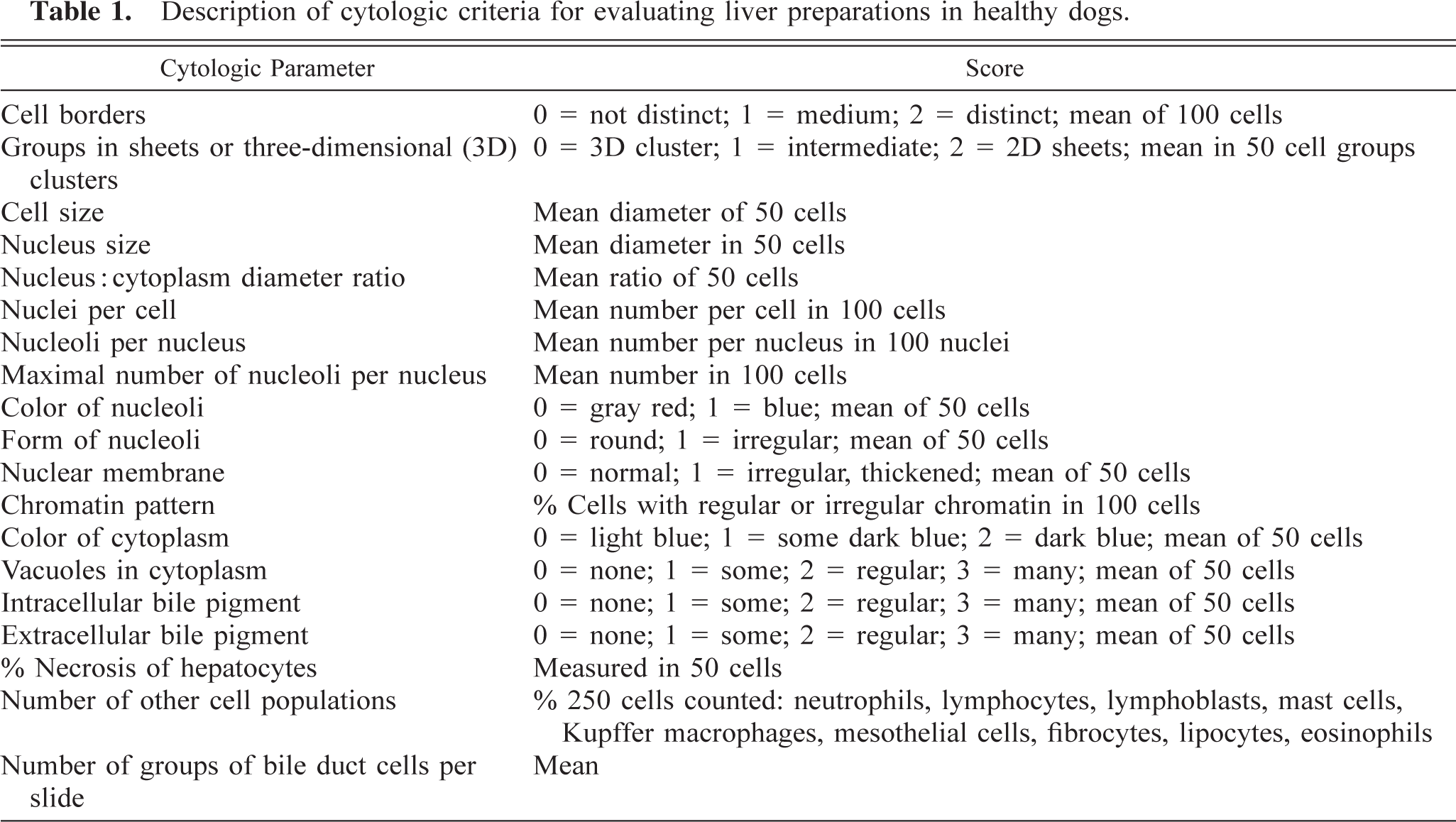
The results of individual cytological parameters are given in Table 2. Hepatocytes in healthy dogs had mostly distinct cell borders and were organized in two-dimensional sheets (Fig. 1). The cytoplasm was light blue with few small gray-green granules in a minority of the cells, consistent with ceroid or bile pigment. Furthermore a small number of cells contained small, well-delineated vacuoles consistent with lipid or an indistinct vacuolar cytoplasmic change consistent with glycogen. Extracellular bile pigment or necrosis were not seen. The mean nucleus:cytoplasm ratio was 1:2.9. There were one or two nuclei per cell and one to three nucleoli per nucleus. Nucleoli were round; 70% had a gray-red color, and 30% had a blue-red color. The nuclear chromatin was mostly regular; it was slightly irregular in fewer than 10% of the cells. Chromatin condensation was not observed. The nuclear membrane was not thickened in any of the cells. Occasionally, an intranuclear rectangular crystal was seen but was not associated with age.
Results of cytologic criteria applied to group of young dogs (1–2 years; group I), middle-aged dogs (5–6 years; group II), and old dogs (10–14 years; group III).
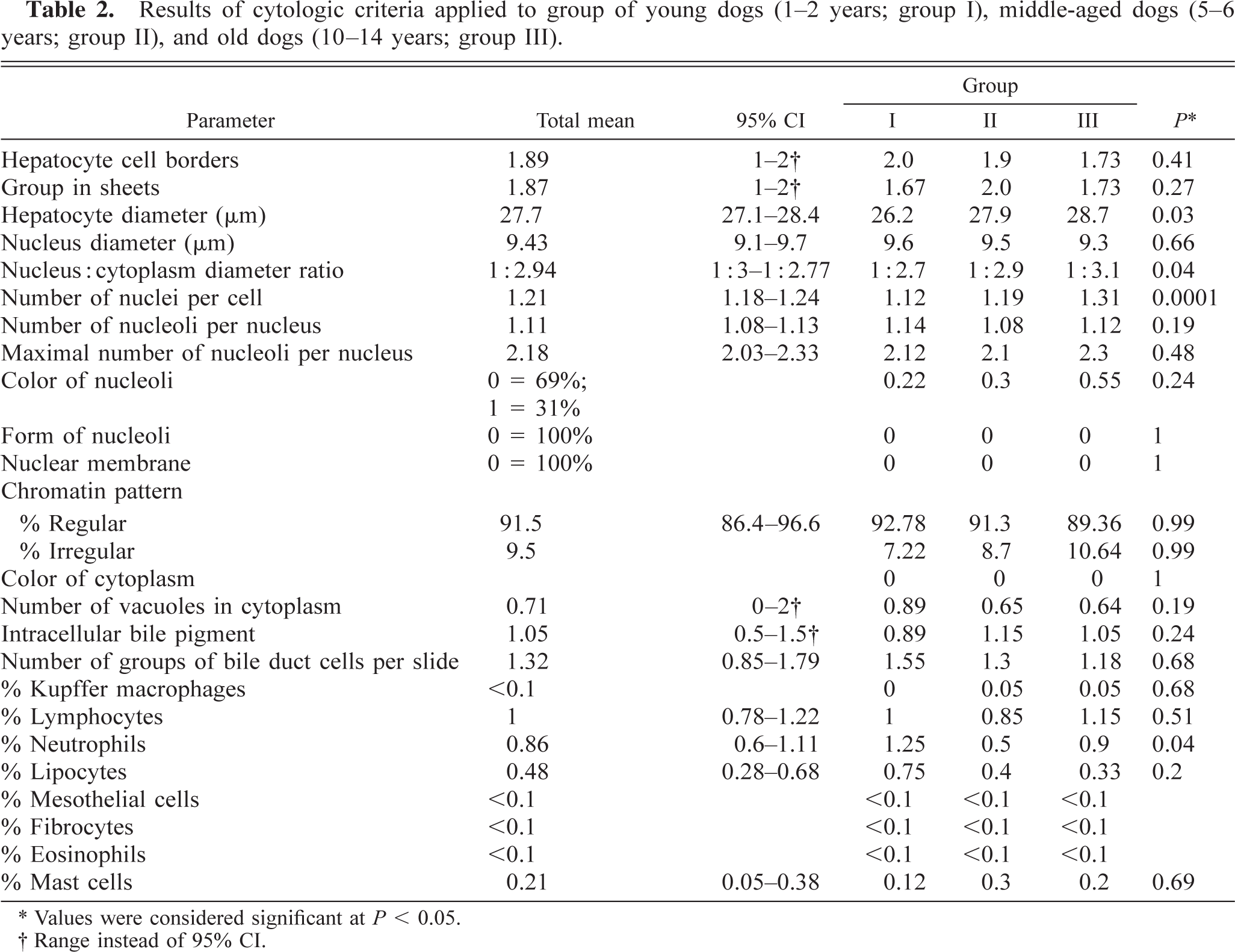
∗ Values were considered significant at P < 0.05.
† Range instead of 95% CI.
Normal hepatocytes; dog. Two-dimensional sheets of cells. May–Grünwald–Giemsa. 1,000×. Bar = 9 µm.
The most frequently encountered nonhepatocytes were lymphocytes, neutrophils, and lipocytes. Mast cells were less frequent. Kupffer macrophages, fibrocytes, mesothelial cells, and eosinophils were observed very rarely. Lymphoblasts did not appear in any of the samples. Small numbers of groups of cuboid biliary epithelial cells were detected regularly.
Significant age-associated differences were observed for cell size, number of nuclei per cell, frequency of neutrophils, and nucleus:cytoplasm ratio. Cell size was greatest in group III and lowest in group I. All differences between groups I, II, and III were significant. Because the nucleus size did not change, the nucleus:cytoplasm ratio decreased with age. There were also higher numbers of nuclei per cell with increasing age. The number of neutrophils per slide was highest in young dogs (group I), less high in old dogs (group III), and lowest in the middle-aged dogs (group II). For the other parameters, no age relationship could be demonstrated.
The presence of liver disease can be detected with physical examination and laboratory screening tests, and the distribution pattern of liver disease can be visualized with imaging methods such as ultrasonography. In the present study, ultrasonography was not used, but histopathologic examination of liver tissue was performed to confirm that the patients had no evidence of liver disease. Although fine-needle aspirates of the liver are becoming more popular, objective cytologic criteria for identifying normal liver aspirates in the dog have so far not been established. The present study provided cytologic reference values for healthy dogs and indicated the association of age with some parameters.
Hepatocytes were mostly organized in two-dimensional sheets with distinct cell borders. This is also an important cytologic characteristic of normal hepatocytes in humans, whereas overlapping of cells with less well-defined cell borders indicates the presence of a hepatoma. 4
Small amounts of intracellular pigment, fat, and glycogen could often be detected in hepatocytes of healthy dogs. These substances represent physiologic metabolic activity in hepatocytes and were also detected in cytologic samples of normal human liver aspirates. 5 Because only May–Grünwald–Giemsa and no other special stains were used, the distinction of granules such as bile or ceroid can be difficult based on cytologic examination. Neither extracellular accumulation of bile nor pigmented Kupffer cells could be detected in normal liver smears. As in humans, this finding may be restricted to dogs with liver disorders including cholestasis. 10
Nuclear structures were mostly uniform in healthy dogs, with only slight variation in nucleus size. The nucleoli were pale in the majority of cases, and the chromatin was regular or slightly irregular without condensation, slits, vacuoles, or thickened nuclear membranes. This description is in agreement with that of normal human liver cytology and is considered a criterion for distinguishing normal liver cells from degenerative liver changes or neoplastic diseases. 4,5,7 Intranuclear rhomboid crystalloid structures were often seen with no relationship to age of dog. This phenomenon was not observed in healthy humans, and the specific cause is unknown.
As in humans, cell populations other than hepatocytes could be detected in cytological liver smears from healthy dogs. Mostly neutrophils and lymphocytes in approximately equal numbers were detected, whereas other cells were rarely observed. In contrast to human studies, Kupffer macrophages were seldomly detected. 10
In healthy dogs, only a few biliary epithelial cell clusters were detected. This low number of clusters was also observed in cytologic samples of healthy humans and was recommended as a criterion to distinguish normal livers from those with bile duct proliferation, as observed in liver cirrhosis or cholestasis. 10 This diagnostic criterion must be used carefully because the number of biliary epithelial cell clusters on cytologic slides is affected by the thickness and size of cytologic samples.
For the cytologic parameters cell size, nucleus:cytoplasm ratio, frequency of neutrophils, and number of nuclei per cell, a significant difference between age groups was found in healthy dogs. The larger cell size and higher number of nuclei per cell in older dogs might be associated with more frequent occurrence of diffuse and nodular hyperplasia of hepatocytes. In humans, these regenerative aspects are also known to increase with age. 11,13 The decreased nucleus:cytoplasm ratio is caused by an increase of the cytoplasm as nuclear size remains constant. A higher frequency of neutrophils predominantly in young and to a lesser extent in old dogs (age groups I and III) compared with group II was obvious. Neutrophils might increase in the liver in response to nonspecific reactive hepatitis. Young and very old dogs may be more frequently affected by subclinical parasitic or other infections.
Reference values for cytologic parameters of liver aspirates were evaluated in healthy dogs. For the parameters cell size, number of nuclei per cell, nucleus:cytoplasm ratio, and percentage of neutrophils, an influence of age must be taken into account. Cytologic characteristics of hepatocytes and the presence of other cell populations or extracellular bile accumulation might be of diagnostic importance in cytologic preparations in dogs as in human medicine.