
Abstract
Seventy-three mammary tumors and three mammary tissue specimens were examined to elucidate the expression of bone morphogenetic protein (BMP)-6 in the myoepithelial cells of canine mammary gland tumors. Morphologically, the myoepithelial cells were classified into four types: resting and proliferating cells inside the basement membrane, and spindle- and star-shaped cells proliferating in the outer area of the basement membrane. The characteristics of these myoepithelial cells were confirmed by immunohistochemistry using antibodies raised against keratin, cytokeratin 19, alpha-smooth muscle actin, and vimentin. In simple adenoma, a small number of resting myoepithelial cells was immunopositive for BMP-6. In complex adenomas and benign mixed tumors, all types of myoepithelial cells, depending in some cases on their specific location within the tumor, were immunopositive for BMP-6, but almost all of the tubular epithelial cells were immunonegative. Foci consisting of a proliferation of BMP-6–positive star- and spindle-shaped cells had mucinous stroma with marked hyaline and chondroid changes. In contrast, the foci with BMP-6–negative spindle- and star-shaped cells tended to have mucinous stroma without chondroid change. Several types of mesenchymal cells including chondrocytes, osteoblasts, and fibroblastlike cells in the mixed tumors, showed an intense immunopositive reaction for the BMP-6 antibody, and were located close to the ectopic cartilage and bone matrix. No significant immunoreactivity for BMP-6 was observed in most of the malignant mammary tumors; only one malignant mixed tumor was examined. All of these findings indicate that BMP-6 expression in myoepithelial cells may increase in complex adenomas and benign mixed tumors in canine mammary glands, and that BMP-6 expression is most intense in the vicinity of chondroid matrix in these tumors.
Mammary gland tumors are the most commonly occurring neoplasm in female dogs. 1,17 In canine mammary mixed tumors, dominant ectopic cartilage and bone formation accompanied by abundant proliferation of myoepithelial cells is observed frequently. 15–17,21 The prominent proliferation of myoepithelial cells is considered to be one of the unique features of canine mammary tumors, although similar lesions are also occasionally described in mammary tumors of other animals. 17 In human breast tumors, myoepithelial proliferations are rare, and benign tumors resembling canine mammary mixed tumors are known as pleomorphic adenomas. In canine complex adenomas and mixed tumors, four morphologically different types of myoepithelial cells are observed: resting and proliferative suprabasal myoepithelial cells, and spindle- and star-shaped interstitial cells. 5,22 The myoepithelial differentiation may culminate in the formation of various mesenchymal tissues, including cartilage and bone in canine mammary mixed tumor.
Recent studies of bone morphogenetic proteins (BMPs), initially isolated from bovine bone extracts, 3 have revealed that BMPs play an important role in bone formation, including the process of ectopic ossification. 9,11,19,24 These proteins are known to be part of the superfamily of transforming growth factor (TGF)-β–related growth factors, and induce endochondral bone formation when implanted ectopically into experimental animals. 3,24 Heterotopic endochondral ossification within mixed tumors of the foot pad in C3 (1)/Tag transgenic mice is reported to be associated with elevated TGF-β-1 and BMP-2 expression, and the bone is thought to be formed by ossification of cartilage resulting from metaplasia of myoepithelial cells. 14 The mRNA and its product Vgr-1, which is the murine homologue of BMP-6, are normally located mainly in the central nervous system, epidermal suprabasal layer, and hypertrophic cartilage of the growth plate. 8,13 BMP-6 is also known to be involved in the development of the embryonic urinary system 2 and the differentiation of keratinocytes. 6 A previous report 24 indicated that BMP-6 acts in an autocrine manner to promote withdrawal from the cell cycle or cell differentiation. Immunohistochemical studies of BMP expression in mixed tumor of the skin and salivary pleomorphic adenomas 10,12,25 have shown that myoepithelial cells are intensely positive for BMP, suggesting that BMP expression on these cells contributes to the formation of chondroid tissues in these tumors.
The purpose of the present study was to elucidate the expression of BMP-6 in canine mammary tumors, especially mixed tumors, and to evaluate the possible role of BMP-6 in mesenchymal tissue formation, that is, cartilage and bone formation.
Materials and Methods
Tissue Samples
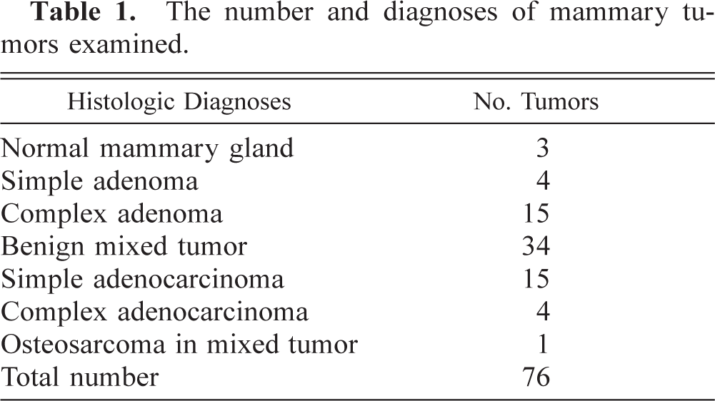
Surgical specimens from 73 canine mammary tumors and from 3 normal mammary glands from necropsied dogs were collected for BMP-6 immunostaining. All of the tissue specimens were fixed in methanol Carnoy's solution for 12–24 hours. The samples then were immersed in methanol for 6 hours, toluene for 4 hours, and paraffin for 2 hours. Paraffin sections (4 µm thick) were prepared and stained with hematoxylin and eosin. Histopathologic diagnoses were made using the tumor classification of the World Health Organization. 15 The histologic diagnoses of all tumors used in this study are given in Table 1. Of the 76 samples, sections from 21 tumors and 3 samples of normal mammary tissues were also examined immunohistochemically to confirm the characteristics of the myoepithelial cells.
The number and diagnoses of mammary tumors examined.
Antibodies
Immunohistochemistry for BMP-6 was performed using an avidin–biotin–peroxidase complex (ABC) kit (PK4000, Vectastain, Burlingame, CA). Goat serum against human BMP-6 (1:20, Santa Cruz, CA) was used as a primary antibody. Biotinylated rabbit serum against goat immunoglobulin (1:20, DAKO Japan, Kyoto, Japan) was used as secondary antibody. Immunohistochemistry was also performed using an envision polymer (DAKO Japan) method with mouse monoclonal antibodies against human alpha-smooth-muscle actin (SMA, 1:50, DAKO Japan), human vimentin (1:100, DAKO Japan), and cytokeratin 19 (1:10, Boehringer Mannheim, Boehringer, Germany), and rabbit serum against keratin, for wide-spectrum screening (DAKO Japan).
Immunohistochemistry
Deparaffinized sections were incubated with 0.5% hydrogen peroxide in methanol for 10 minutes to inhibit endogenous peroxidase activity. For BMP-6 immunostaining, sections were incubated with bovine serum albumin (BSA) and the primary antibody for 30 minutes at 37 C. The sections were also incubated with secondary antibody for 30 minutes at 37 C. Then sections were reacted with ABC reagents for 30 minutes at 37 C. For SMA, cytokeratin 19, and vimentin immunostaining, sections were incubated with BSA for 30 minutes at 37 C and the primary antibodies for 30 minutes at 37 C, and then followed by reaction with envision polymer reagents for 30 minutes at 37 C. Finally, the sections were exposed to the chromogen, 3,3-diaminobenzidine-4 HCl, and counterstained with Mayer's hematoxylin. The results of the immunohistochemistry were expressed semiquantitatively as follows: − = negative; ± = 0–5% positive cells; + = 5–10% positive cells; 2 + = 10–50% positive cells; 3 + = 50–90% positive cells; and 4 = >90% positive cells as previous reports. 22
Results
The immunoreactivity of mammary gland cells for keratins, SMA, and vimentin is indicated in Table 2. In normal mammary glands, resting myoepithelial cells in the basement membrane were intensely positive (3+ to 4+) for keratin and cytokeratin 19, and moderately (2+) positive for SMA and vimentin. In both complex adenoma and benign mixed tumors, resting and proliferating myoepithelial cells within the basement membrane showed similar immunoreactivity to the resting myoepithelial cells of normal mammary tissues. The spindle- and star-shaped cells proliferating in the interstitial area with various amounts of mucinous stroma were intensely (4+) positive for vimentin, whereas immunoreactivity for keratin, cytokeratin 10, and SMA was markedly weak (+) as compared with that of resting myoepithelial cells (Table 2). In addition, chondrocytes and osteoblasts around the cartilage or bone in benign mixed tumors were positive only for vimentin.
Immunoreactivity of myoepithelial cells for keratin, actin, and vimentin in mammary tissues and tumors.
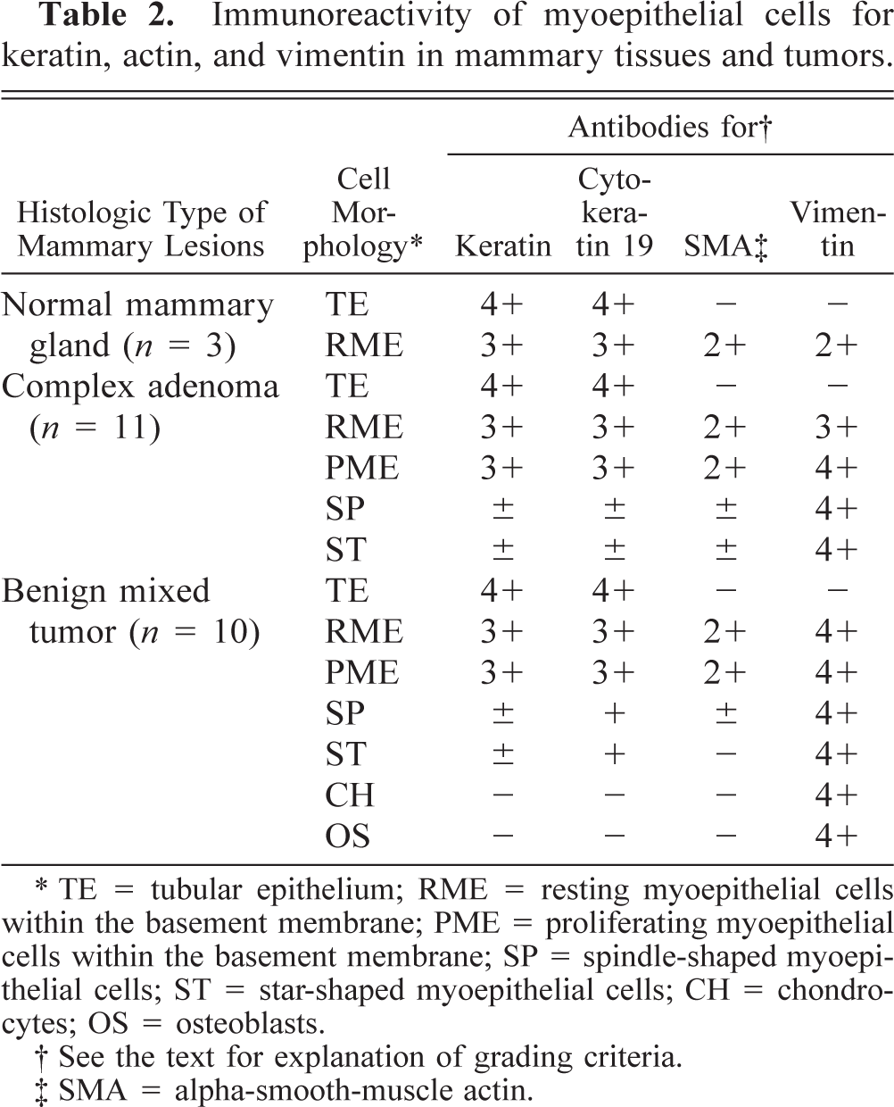
∗ TE = tubular epithelium; RME = resting myoepithelial cells within the basement membrane; PME = proliferating myoepithelial cells within the basement membrane; SP = spindle-shaped myoepithelial cells; ST = star-shaped myoepithelial cells; CH = chondrocytes; OS = osteoblasts.
† See the text for explanation of grading criteria.
‡ SMA = alpha-smooth-muscle actin.
The results of the immunohistochemistry for BMP-6 of mammary cells in normal mammary tissues and tumors are summarized in Table 3. The details in each sample are as follows.
Immunoreactivity of mammary tumors for BMP-6.
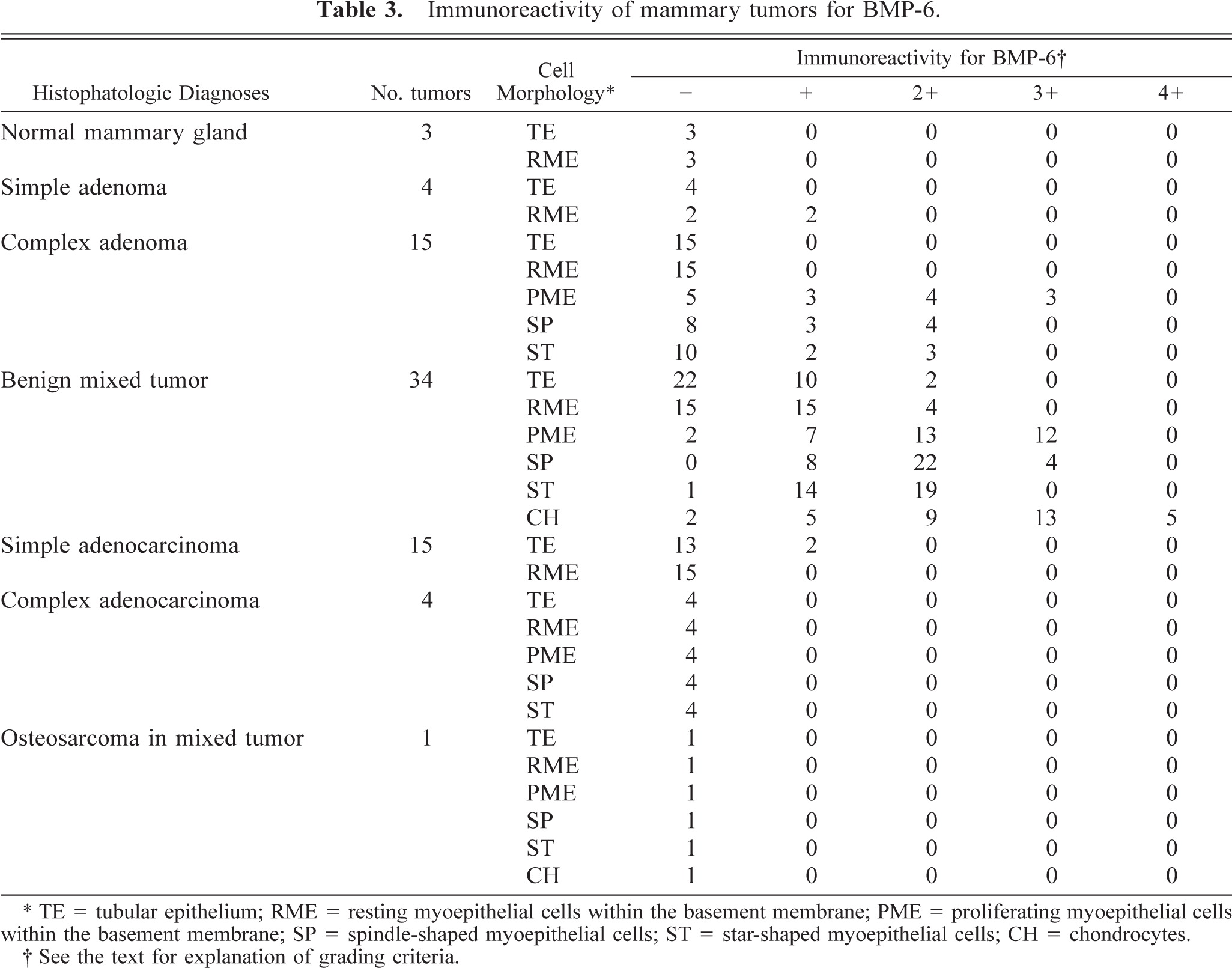
∗ TE = tubular epithelium; RME = resting myoepithelial cells within the basement membrane; PME = proliferating myoepithelial cells within the basement membrane; SP = spindle-shaped myoepithelial cells; ST = star-shaped myoepithelial cells; CH = chondrocytes.
† See the text for explanation of grading criteria.
Normal Mammary Tissues
In the normal mammary tissue, tubular epithelial cells were negative for BMP-6. The resting myoepithelial cells within the basement membrane were also BMP-6 immunonegative (Table 3).
Simple adenoma
These tumors were composed of a pure proliferation of well-differentiated tubular epithelial cells. Immunohistochemically, the resting myoepithelial cells exhibited weak immunopositive reaction (+) for BMP-6 in two out of four cases (Fig. 1), whereas the proliferating tubular epithelial cells were completely negative for BMP-6.
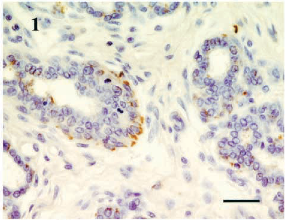
Mammary gland; simple adenoma; dog. Immunoreactivity of resting myoepithelial cells for BMP-6. Immunostaining for BMP-6, numerical grade (+), Bar = 100 µm.
Complex adenoma
These tumors consisted of a mixed proliferation of tubular epithelial cells and myoepithelial cells with a large amount of mucinous stroma. The immunoreactivity of myoepithelial cells for BMP-6 varied between tumors. The most intense immunoreactivity for BMP-6 was confirmed in the proliferating myoepithelial cells within the basement membrane (Fig. 2). Of 15 complex adenomas, intense positive reaction (3+) for BMP-6 of the myoepithelial cells was found in 3 tumors, and weak to moderate immunoreactivity (+ to 2+) was confirmed in 7 tumors (Table 3). In the remaining five tumors, no significant positive reaction for BMP-6 was confirmed. Moreover, spindle- and star-shaped interstitial myoepithelial cells showed weak to moderate immunoreactivity (+ to 2+) for BMP-6 in seven and five tumors, respectively (Fig 3a, b), whereas the reactivity was relatively weaker than that of proliferating myoepithelial cells within the basement membrane. The mucinous stroma containing BMP-6–positive star-shaped myoepithelial cells tended to show marked hyalinous or chondroid changes. No BMP-6 immunoreactivity was found in tubular epithelial cells and resting myoepithelial cells in complex adenomas examined.
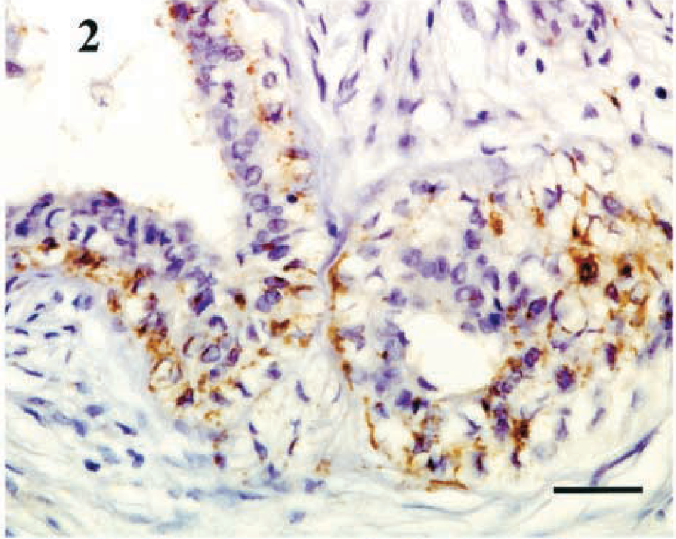
Mammary gland; complex adenoma; dog. Intense immunoreactivity of proliferating myoepithelial cells within the basement membrane for BMP-6. Immunostaining for BMP-6, numerical grade (3+). Bar = 100 µm.
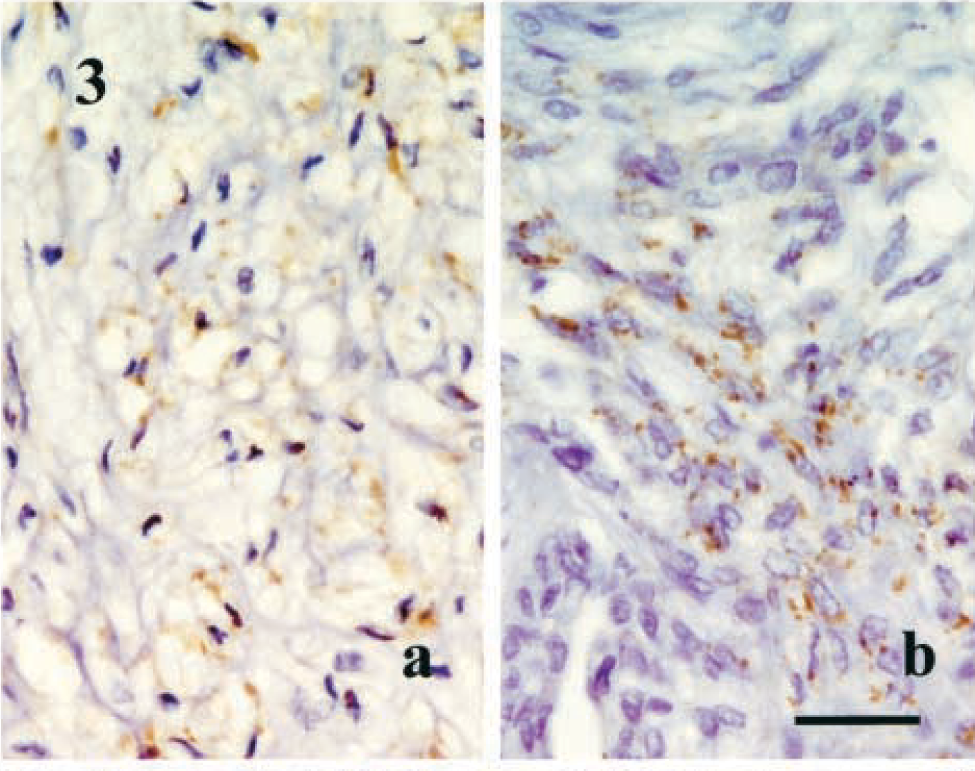
Mammary gland; complex adenoma; dog. Fig. 3a Interstitial star-shaped myoepithelial cells with mucinous stroma showing positive immunoreactivity for BMP-6. Fig. 3b Spindle-shaped myoepithelial cells showing positive immunoreactivity for BMP-6. Immunostaining for BMP-6, numerical grade (2+). Bar = 100 µm.
Benign Mixed Tumor
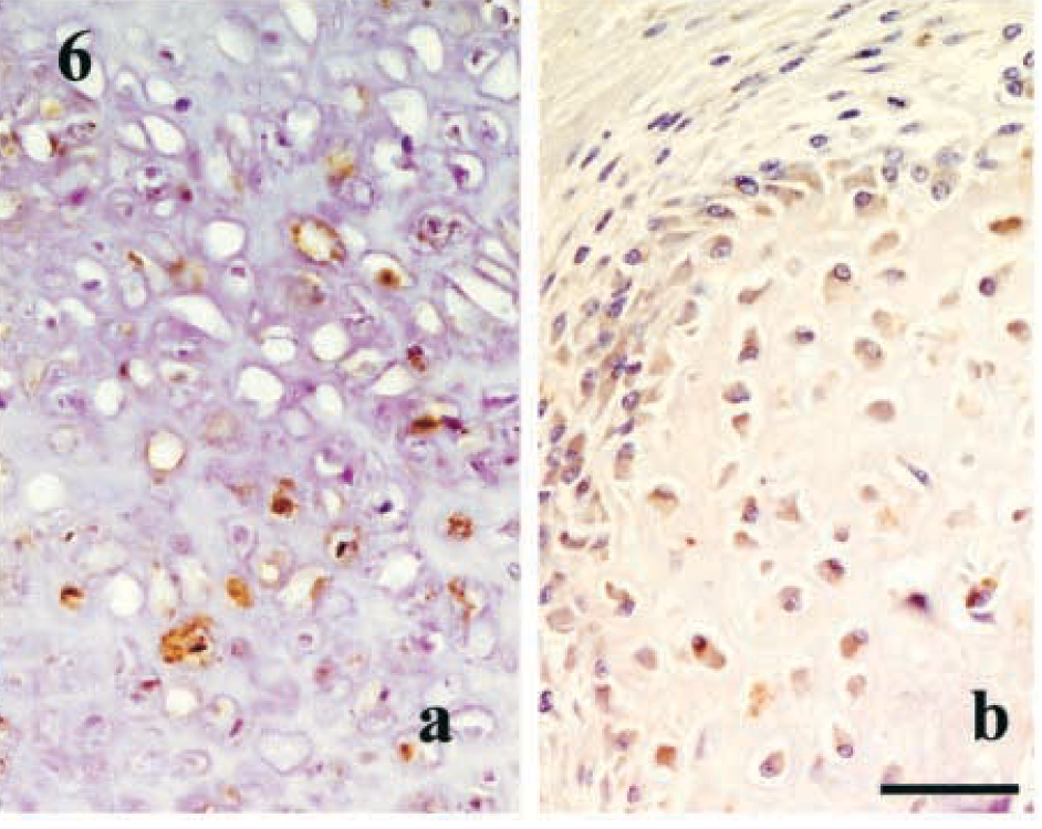
These tumors had a complex morphology that was characterized by mixed proliferation of tubular epithelial cells, myoepithelial cells with abundant mucinous stroma, and mesenchymal components that might include cartilage, bone, bone marrow, and fatty tissues. BMP-6–positive cells were most frequently found in this tumor (Table 3). In almost all the tumors examined, proliferating myoepithelial cells within the basement membrane, spindle- and star-shaped interstitial myoepithelial cells (Fig. 4), and chondrocytes in the ectopic cartilages showed mild to intense immunoreactivity for BMP-6. In addition, a small number of tubular epithelial cells and resting myoepithelial cells within the basement membrane showed weak immunoreactivity (+) for BMP-6 in 12 and 19 tumors, respectively (Table 3). In benign mixed tumors, the relationships between chondroid or bone tissues and BMP-6–positive myoepithelial cells also were examined. In almost all tumors, star-shaped myoepithelial cells showing intense immunoreactivity (2+ to 3+) for BMP-6 were located within mucinous stroma, in association with marked hyalinous and chondroid change (Fig. 5). On the other hand, the stroma containing BMP-6–negative star-shaped myoepithelial cells tended to exhibit either no, or very mild hyalinous or chondroid changes. However, in some benign mixed tumors, strong hyalinous and chondroid changes were observed around BMP-6–negative star-shaped myoepithelial cells. In such areas, a small number of tubular epithelial cells and resting myoepithelial cells expressing BMP-6 (+ to 2+) were observed in close association with the stroma that had chondroid changes. Large spindle-shaped cells with abundant collagen, considered to be stromal fibroblasts, were also distributed around mucinous stroma and cartilage. These cells were sometimes positive for BMP-6. In some mature cartilage, chondrocytes exhibited marked immunoreactivity (+ to 4+) for BMP-6 (Fig 6a) Osteoblasts around bone tissues were also weakly to moderately positive (+ to 3+) for BMP-6 (Fig 6b).

Mammary gland; benign mixed tumor; dog. BMP-6–positive proliferating myoepithelial cells within the basement membrane, and some interstitial star- and spindle-shaped myoepithelial cells showing positive reaction for BMP-6. Immunostaining for BMP-6, numerical grade (3+). Bar = 150 µm.
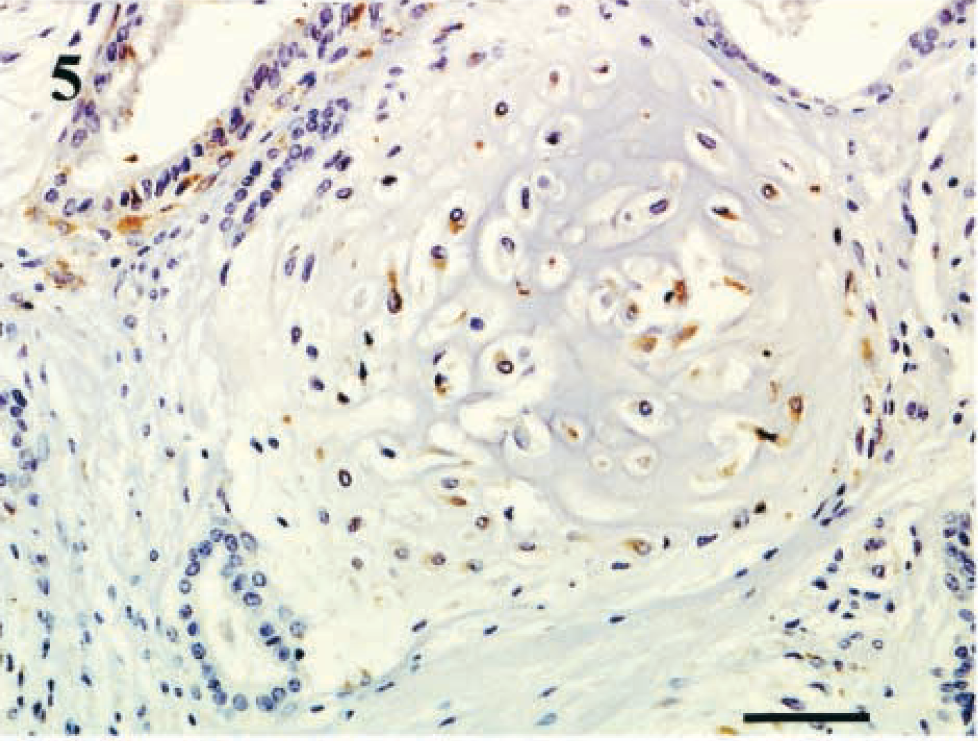
Mammary gland; benign mixed tumor; dog. The mucinous stroma with chondroid change containing BMP-6–positive star-shaped myoepithelial cells. Immunostaining for BMP-6, numerical grade (2+). Bar = 150 µm.
Malignant Mammary Tumors
Fifteen simple adenocarcinomas, four complex adenocarcinomas, and one sarcoma in mixed tumor were collected and examined for BMP-6 expression. Among these 20 tumors, a small number of tubular epithelial cells in only two simple adenocarcinomas showed weak immunoreactivity (+) for BMP-6. In the remaining 18 tumors, no significant immunoreactivity for BMP-6 was detected (Table 3).
Discussion
The histogenesis of mesenchymal elements, such as bone and cartilage, in canine mixed tumors remains unclear, whereas three possible origins of mesenchymal tissues have been proposed: metaplasia from epithelial cells, interstitial connective tissues, and myoepithelial cells. 15 Moulton 17 described that the bone in mixed tumors arises by endochondral ossification of the cartilage formed by myoepithelial cells and stated that it is doubtful that bone arises by intramembranous ossification of stromal connective tissues. The present study confirmed BMP-6 expression in myoepithelial cells in canine mammary tumors, especially in complex adenomas and benign mixed tumors. The results were almost in conformity with those of skin mixed tumors and salivary pleomorphic adenomas in humans. 10,12,25 In this study, myoepithelial cells were morphologically classified into four types: resting and proliferative myoepithelial cells within the basement membrane, and spindle- and star-shaped interstitial myoepithelial cells. Immunohistochemistry for keratin, cytokeratin 19, SMA, and vimentin revealed that myoepithelial cells within the basement membrane maintain their normal immunohistochemical characteristics, 5,18,22 whereas spindle- and star-shaped myoepithelial cells proliferating in the interstitial area tend to show arrested expression of keratin and SMA. Proliferating interstitial myoepithelial cells may eventually become fibroblast-like cells, showing only vimentin immunoreactivity. In mixed tumors, advanced mesenchymal tissue formation including cartilage and bone is thought to occur by further metaplastic change of proliferating myoepithelial cells, which become fibroblast-like cells. Mammary mesenchymal cells such as interstitial myofibroblasts or fibroblasts possibly also contribute to mesenchymal tissue formation, together with metaplastic myoepithelial cells.
Interestingly, all types of myoepithelial cells in canine mammary adenomas and benign mixed tumors showed immunoreactivity for BMP-6. In particular, proliferating myoepithelial cells within the basement membrane and interstitial spindle- and star-shaped cells were immunoreactive for BMP-6, together with chondrocytes and osteoblasts. Among these myoepithelial cells, proliferating cells within the basement membrane exhibited the most intense immunoreactivity for BMP-6. When myoepithelial cells started to proliferate in the interstitial areas, the immunoreactivity for BMP-6 declined. This finding might indicate that when myoepithelial cells start to proliferate within the basement membrane, the cells immediately express BMP-6 at a high level. With the advancement of the proliferation of myoepithelial cells in the interstitial area, some spindle- and star-shaped cells arrest BMP-6 expression, together with the expression of keratin and SMA filaments. In addition, BMP-6–positive interstitial myoepithelial cells seem to produce a large amount of mucinous stroma with chondroid change, unlike BMP-6–negative cells. Mucinous stroma showing chondroid changes was observed in both benign mixed tumors and complex adenomas, but a greater number of BMP-6–positive myoepithelial cells and chondroid tissues were found in the mixed tumors. In this regard, BMP-6 expression on myoepithelial cells seems to be closely associated with the appearance of chondroid tissues in canine mammary tumors. In addition, in some benign mixed tumors, fibroblast-like large spindle cells around cartilage and bone showed immunoreactivity for BMP-6. Fibroblast-like mesenchymal cells and chondrocytes have been known to express high level of BMPs during experimentally induced osteogenesis. 19 With respect to ectopic bone and cartilage formation in canine mammary tumors, the BMP-6–positive mesenchymal cells might be also involved, although the origin of these mesenchymal cells in canine mixed tumors remains to be established.
In a previous report on BMP-6 expression in embryogenesis, Wall et al. 23 described that BMP-6 appeared in the cutaneous suprabasal cells, and was absent from the basal layer and hair follicles. They suggested that BMP-6 acts in an autocrine manner to promote the cell cycle or differentiation of cutaneous suprabasal cells, and that BMP-6 also acts in a paracrine fashion to regulate the proliferation and differentiation of basal cells. Other reports concerning human esophageal squamous cell carcinoma with extensive chondroid differentiation showed that BMP-positive carcinoma cells affect other tumors cells via a paracrine action and might lead to the chondroid metaplasia of epithelial cells. 4,20 Analyses of these previous data indicate that the BMP-6 expressed on myoepithelial cells may promote not only metaplastic changes including cartilage and bone formation, but also differentiation of tubular epithelial, myoepithelial, and mesenchymal cells in canine mammary tumors in both an autocrine and paracrine manner. Several reports 14,23 have revealed that BMPS, which are known to be part of the superfamily of TGF-β–related growth factors, regulate the proliferation and differentiation of many types of cells. Intracellular TGF-β expression is known to be reduced when the epithelial cells begin to proliferate. In our study, no significant BMP-6 expression was detected in malignant tumors, whereas weak immunoreactivity for BMP-6 of tubular epithelium was recognized in two adenocarcinomas. This may indicate that BMP-6 promotes the differentiation of canine mammary cells, resulting in the inhibition of proliferation of these cells. The loss of BMP-6 expression on myoepithelial cells may permit the malignant transformation of neoplastic cells. To confirm this hypothesis, further studies using a larger number of malignant cases are necessary. BMPs are already known to induce endochondral bone formation when implanted ectopically into experimental animals, 3,24 and applications in the clinical setting for several bone disorders including bone fracture and defects are being developed. Because BMP seems to promote the differentiation of mammary cells, together with cartilage and bone formation, this protein may have a clinical application for the treatment of canine mammary tumors.
In conclusion, the present study is the first to demonstrate the expression of BMP-6 on myoepithelial cells in canine mammary tumors. Our findings will be useful for clarifying the genesis of ectopic cartilage and bone formation in canine mammary gland tumors, especially mixed tumors.