
Abstract
A juvenile female loggerhead sea turtle (Caretta caretta) stranded in Gran Canaria was submitted for necropsy. The turtle had exhibited anorexia and lethargy for 2 weeks prior to its death. At necropsy, the thymus was enlarged by two white and firm nodules. White nodules similar to those in thymus were observed in the plastron, thyroid gland, heart, aorta, left lung, spleen, liver, kidneys, stomach, and small intestine. Histopathology revealed a neoplastic proliferation of round cells identified as lymphoid cells. Ultrastructurally, the neoplastic cells were consistent with lymphoblastic cells, and viruses were not detected. The diagnosis was multicentric lymphoblastic lymphoma. This is the first report of a lymphoid neoplasm in a sea turtle.
Descriptions of neoplasia in reptiles are uncommon in comparison to prevalences reported for mammals and birds. 7 Except for fibropapillomatosis, neoplastic disease is very infrequently seen in sea turtles. Within the class Reptilia, lymphoid neoplasias are more frequently found in snakes, 1,8 although there are also descriptions in lizards, 1,11,12,14 crocodilians, 13 and terrestrial chelonians. 3,4,6,10 Here, we describe the macroscopic, cytologic, histopathologic, and ultrastructural findings in a loggerhead sea turtle (Caretta caretta) with multicentric lymphoblastic lymphoma.
A juvenile (straight carapace length = 34 cm; weight = 5 kg) female loggerhead sea turtle stranded in Gran Canaria was submitted to the College of Veterinary Medicine, University of Las Palmas of Gran Canaria, for necropsy. After stranding, the turtle had been housed in an aquarium at the Center of Rehabilitation of Wild Fauna, Gran Canaria, for 2 weeks. On physical examination, the turtle had been depressed, anorexic, and lethargic. The mucous membranes of the oral cavity appeared paler than normal. Blood was not collected for hematology, and it is not known whether the turtle was leukemic. Despite force feeding and intramuscular administration of the antimicrobial enrofloxacin, the turtle died.
At necropsy, the turtle was cachectic. The ventral aspect of the plastron was infiltrated with white and firm irregular masses, ranging in size from 2 to 4 cm. The thymus glands were bilateral and were located in the cervical region in the angle formed by the subclavian and common carotid arteries. The thymus of each side was white and firm and consisted of several lobes. Each lobe contained multiple nodules that were of similar size, irregular, and homogeneous in color and texture on cut section. The total weight of the thymus was 25 g, and it appeared to be larger than those of previous turtles we had necropsied.
White nodules similar to those described in thymus were observed in the thyroid gland. Several irregularly shaped, sharply demarcated white foci up to 2 cm in diameter were present in the ventral pectoral muscles. Similar masses were observed in the coelomic wall. The coelomic cavity contained approximately 200 ml of thin, yellow, transparent fluid.
The liver was friable, with rounded edges and numerous firm white nodules ranging in diameter from 2 mm to 1 cm throughout the parenchyma. Three similar masses were observed in the ventricle, left atrium, and aortic arch. The spleen was enlarged, and cut sections revealed numerous small white nodules. The left lung had three white, firm nodules, 1 cm in diameter, showing elevation on the pleural surface. The kidneys contained numerous poorly defined white foci of small size (3–5 mm in diameter) that in cut sections effaced the architecture. Nodules of various sizes were also observed in the serosa and mucosa of the stomach and small intestine. The mucosa of the stomach and small intestine contained a few depressed ulcers with reddened rims that were coated by fibrinonecrotic exudate. No parasites were observed in blood vessels or digestive tract.
Direct smears from a fine-needle aspirate of the nodular masses of the thymus and plastron were moderately cellular with a population of mildly pleomorphic immature lymphoid cells. These cells were generally large and somewhat variable in size. The cytoplasm was basophilic, moderate in amount, and occasionally vacuolated. These cells had centrally located, mildly hyperchromatic nuclei. The nucleoli were prominent, and there were few mitotic figures. In addition, smears from the plastron contained few erythrocytes and an occasional heterophil.
Representative tissues from all organs were fixed in neutral buffered 10% formalin, embedded in paraffin, sectioned at 5 µm for light microscopy, and stained with hematoxylin and eosin (HE), periodic acid–Schiff (PAS), and Giemsa stains. Selected samples from thymus and epicardium were fixed in 2.5% buffered glutaraldehyde, routinely processed for transmission electron microscopy, and embedded in epoxy resin. Ultrathin sections were mounted on grids, stained with uranyl acetate and lead citrate, and examined with a Zeiss 910 transmission electron microscope.
Histologic examination revealed numerous parenchymal organs and tissues containing nodular, unencapsulated masses or monotonous sheets composed of hyperchromatic neoplastic lymphoid cells (Figs. 1, 2). Larger nodules were centrally necrotic. These cells had round or irregularly shaped hyperchromatic nuclei with coarse chromatin clumping and moderate to abundant basophilic cytoplasm. Cytoplasmic granules were not detected in these cells in sections stained with PAS, Giemsa, or HE. Mitotic figures were not numerous, and individual cell necrosis was common.
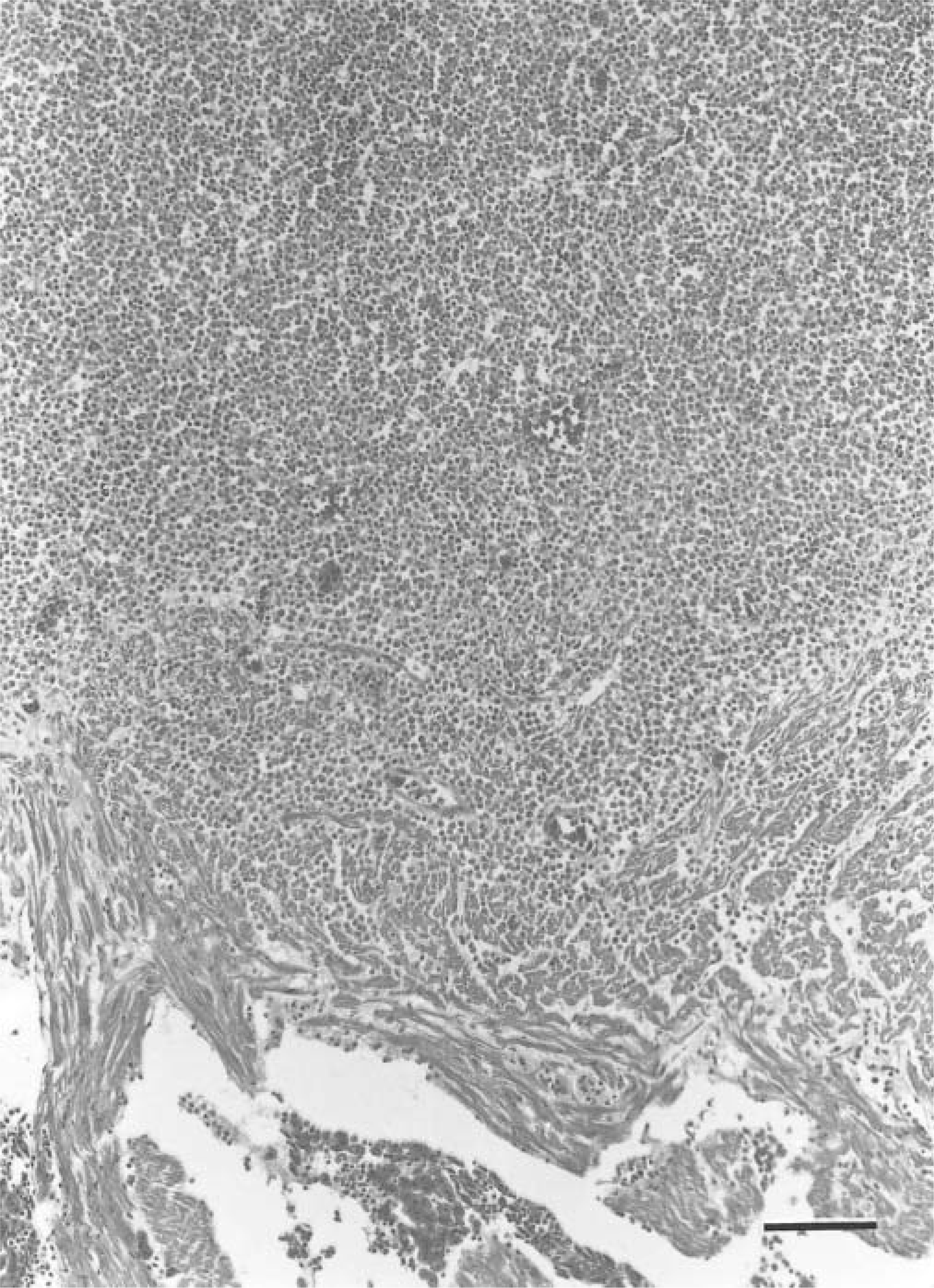
Heart; loggerhead sea turtle. The epicardium contains a nodule composed of neoplastic lymphoblasts. The myocardium was also infiltrated by these cells. HE. Bar = 190 µm.
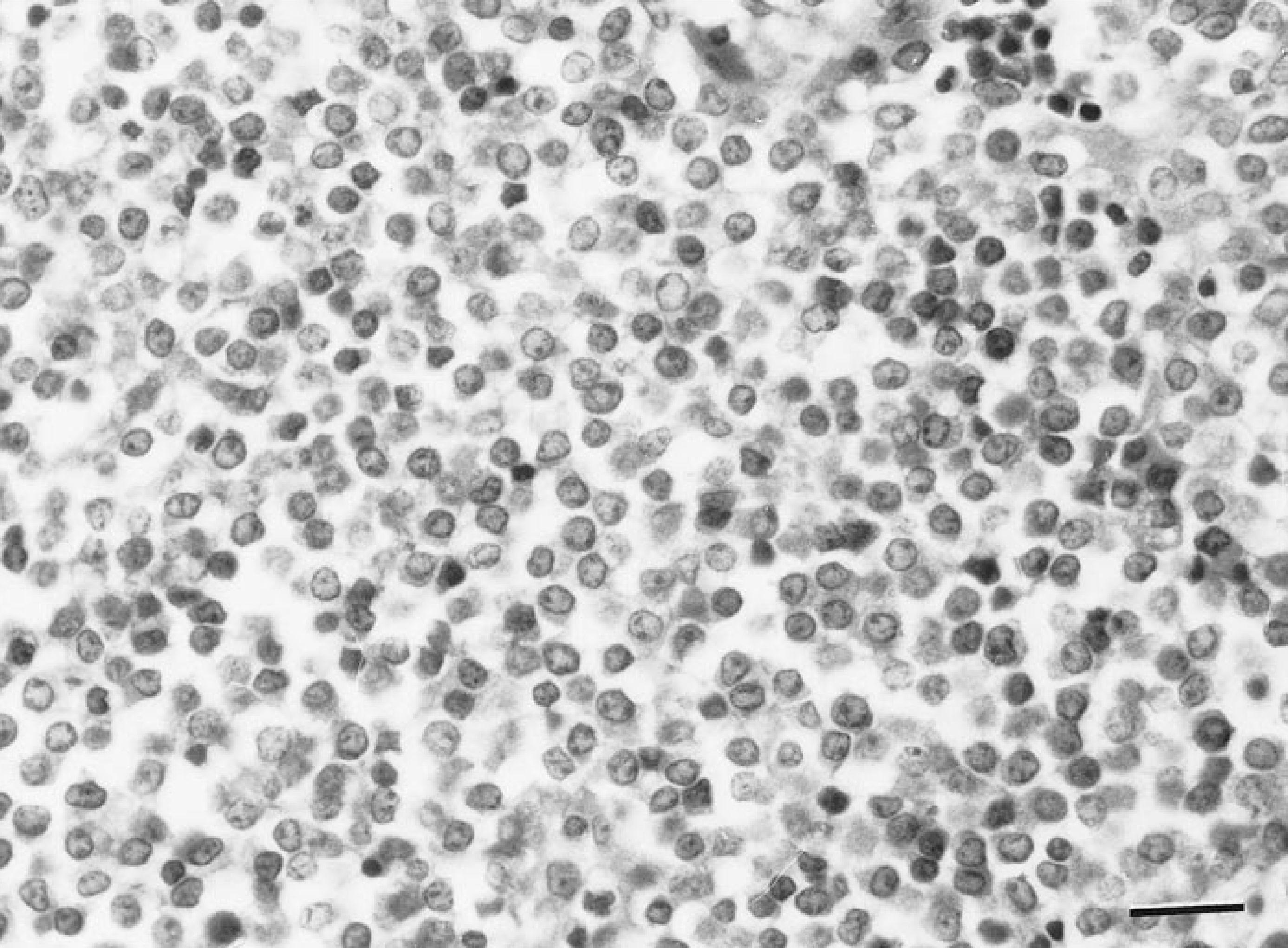
Heart; loggerhead sea turtle. The nodule in Fig. 1 is composed of hyperchromatic neoplastic lymphoid cells. HE. Bar = 17 µm.
The masses observed in the plastron consisted of sheets of neoplastic lymphoid cells infiltrating the bone of the epiplastron, resulting in diffuse osteolysis. The bone marrow of the epiplastron was densely infiltrated by neoplastic lymphoblasts with smaller numbers of granulocytic hematopoietic cells admixed. The nodules detected in the ventral pectoral muscles and myocardium were composed of neoplastic lymphoid cells with entrapped necrotic myofibers. Immature lymphocytes were also randomly distributed in the adventitia of the aortic arch, with small foci infiltrating the tunica media. Masses of similar cells were found in the pulmonary interstitium.
In the liver, neoplastic cells were present in large numbers in the sinusoids and within the space of Disse. In the stomach and small intestine, there was multifocal transmural infiltration by neoplastic lymphoblasts and mucosal ulcers coated by fibrinonecrotic debris. A transmural infiltration by neoplastic lymphoblasts was also observed in the urinary bladder. Because of infiltrates of lymphoid cells, there was a loss of the normal architecture of many affected organs, including thymus, thyroid gland, parathyroids, spleen, and kidneys. No neoplastic lymphoid cells were observed in sections of brain, spinal cord, skin, trachea, gall bladder, eye, salt glands, pancreas, or large intestine.
Transmission electron microscopy revealed a moderately polymorphous population of round and oval cells with central rounded or oval nuclei, peripherally clumped heterochromatin, and two or more nucleoli. Most cells had a moderate to abundant amount of cytoplasm and moderate numbers of organelles, including mitochondria, lysosomes, endoplasmic reticulum, and dilated vacuoles (Fig. 3). Viruses were not detected.
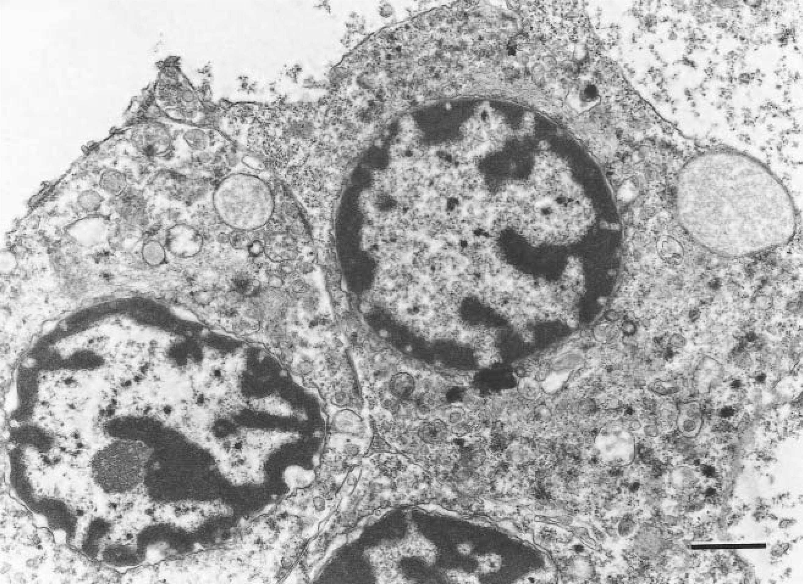
Heart; loggerhead sea turtle. Lymphoid cells of the nodule have central rounded or oval nuclei, peripherally clumped heterochromatin, moderate to abundant amount of cytoplasm, and moderate numbers of organelles. Uranyl acetate counterstain. Bar = 1.25 µm.
The morphology and ultrastructural features of neoplastic cells and the pattern of tumor proliferation support a diagnosis of lymphoblastic lymphoma in this sea turtle. The absence of cytoplasmic granules helped to rule out a myelogenous neoplasia, and the rounded nuclei, peripherally clumped heterochromatin, moderate to abundant amount of cytoplasm, and moderate numbers of cytoplasmic organelles were consistent with lymphoid neoplasms described in other species. 9,11
Neoplasia has been reported in 2–10% of reptile necropsies, and almost 25% of those were lymphoid neoplasms. 1 In reptiles, lymphoid neoplasms most commonly have a multicentric distribution. 2,6,9,11,12,14 Lymphoid neoplasms are more frequent in snakes and lizards than in other reptiles. Confirmed multicentric lymphoblastic lymphoma has been reported in an anaconda (Eunectes murinus), 2 a California king snake (Lampropeltis getulus californiae), 9 an eastern king snake (Lampropeltis getulus getulus), 8 a green iguana (Iguana iguana), 11 a savannah monitor lizard (Varanus exanthematicus), 12 and an East Indian water lizard (Hydrosaurus amboinensis). 14
Multicentric lymphoblastic lymphoma reported in a male Greek land tortoise (Testudo hermanni) involved the liver, heart, kidneys, spleen, pancreas, and intestinal serosa. 6 Gray-white nodules in the liver and kidney contained large lymphoid cells with vesiculated nuclei, but ultrastructural study was not attempted. A malignant lymphoma was diagnosed in a Burmese star tortoise (Geochelone platynota), but there are no detailed published pathologic description of that case. 3 Lymphoreticular neoplasia was detected in a Florida soft-shelled turtle (Trionyx ferox), 4 and alimentary tract lymphomas were described in several Galapagos tortoises (Geochelone elephantopus). 10 Although the tissue of origin of reptilian lymphomas is uncertain, the main lymphoid organs in reptiles are the spleen and thymus. 11
Except for fibropapillomatosis, neoplasms are very uncommon in sea turtles. Green turtle fibropapillomatosis (GTFP) is characterized by multiple cutaneous papillomas, fibromas, and fibropapillomas and occasional visceral fibromas. 5 No involvement of lymphoid organs has been described in sea turtles with GTFP. The multicentric lymphoblastic lymphoma described in this loggerhead sea turtle is the first lymphoid neoplasm to be reported in a sea turtle.
Although virus-associated lymphoma has been reported in other reptiles, 7 no viral etiology could be identified by electron microscopic examination in this case. However, this negative finding does not exclude a viral etiology. Culture of neoplastic cells was not attempted, and polymerase chain reaction evaluation of tumors for the presence of conserved retroviral sequences was beyond the ability of our laboratory. To investigate a possible etiology, further studies will be conducted to determine the presence of environmental contaminants, pesticides, and heavy metals in the tissues of this turtle.
Footnotes
Acknowledgements
The survey of the pathology and causes of mortality among sea turtles stranded in the Canary Islands was supported by the project I+D REN2000-1753. We are grateful to members of Viceconsejería de Medio Ambiente, Cabildo Insular de Gran Canaria, for providing us with the turtles. We thank Francisco Freire of the Electron Microscopy Service, University of Las Palmas de Gran Canaria, for preparing specimens for electron microscopy and for preparation of the electron micrographs. We thank Pedro Castro for technical assistance.