
Abstract
Leatherback sea turtles are globally distributed and endangered throughout their range. There are limited data available on disease in this species. Initial observations of solitary large intestinal diverticulitis in multiple leatherbacks led to a multi-institutional review of cases. Of 31 subadult and adult turtles for which complete records were available, all had a single exudate-filled diverticulum, as large as 9.0 cm in diameter, arising from the large intestine immediately distal to the ileocecal junction. All lesions were chronic and characterized by ongoing inflammation, numerous intralesional bacteria, marked attenuation of the muscularis, ulceration, and secondary mucosal changes. In three cases, Morganella morganii was isolated from lesions. Diverticulitis was unrelated to the cause of death in all cases, although risk of perforation and other complications are possible.
The leatherback sea turtle (Dermochelys coriacea) is the largest living chelonian and is endangered throughout its range. 1 The species is highly migratory and globally distributed, extending as far north as Norway and south to New Zealand. 1 Many aspects of leatherback life history remain unknown. Larger immature and adult turtles occur in the open ocean, as well within coastal and shelf waters, where they primarily feed on gelatinous zooplankton, such as jellyfish (hydrozoans and scyphozoans), salps (thaliacians), and other pelagic invertebrates. 1 Major threats to species survival include harvest of eggs and nesting females and incidental capture in fishing gear. 1,7
There is very little information available on necropsy findings in leatherbacks. Opportunities for postmortem examination are relatively infrequent due to the low probability of carcasses washing ashore prior to decomposition. Collaboration among researchers is essential for assembling information and providing meaningful context to observations. In this communication, we describe solitary large intestinal diverticulum formation with diverticulitis in large immature and adult leatherbacks as a frequent necropsy finding and consider cause of death and other relevant case information.
During collaborative discussion of necropsy findings in leatherbacks, it became apparent that many examined animals found in different areas of North America had a solitary, exudate-filled diverticulum emanating from the region of the ileocecal junction. As in other sea turtle species, the cecum of leatherbacks is indistinct, and the ileocecal valve is followed by an increase in luminal diameter at the beginning of the large intestine. 10 Records from several institutions were reviewed for gross and histologic findings related to this lesion, as well as location and circumstances of animal collection, cause of death, sex, and curved carapace length (CCL). Size class categories were applied as previously defined. 1 Inclusion of cases required specific comment in necropsy records that the entire digestive tract, including the ileocecocolic region, was evaluated. Histologic sections were stained with hematoxylin and eosin using routine methods. Additional sections from representative cases were stained using Brown and Brenn, Fite’s acid fast, and Gomori methenamine silver methods.
Forty-four necropsy records were reviewed for leatherbacks necropsied from 2003 to 2013 (34 subadult or adults, 10 juveniles). Three cases, all subadult or adults, were excluded due to incomplete information. Of 31 subadult and adult leatherbacks (CCL >100 cm) with consistent evaluation of the gastrointestinal tract, all had a solitary diverticulum originating from the large intestine at the ileocecal junction. This lesion was not found in any of the 10 juvenile leatherbacks examined, including 5 found stranded on beaches in Florida and 5 incidentally caught by Pacific fishing vessels (CCL ranging from = 17.9–88.5 cm). Case information for individuals with diverticulitis is provided in Supplemental Table S1. Most were caught or were found on the Atlantic coast of North America. Three were recovered on the beach or from waters off California, and 1 (case No. 1) was caught in the Gulf of Mexico.
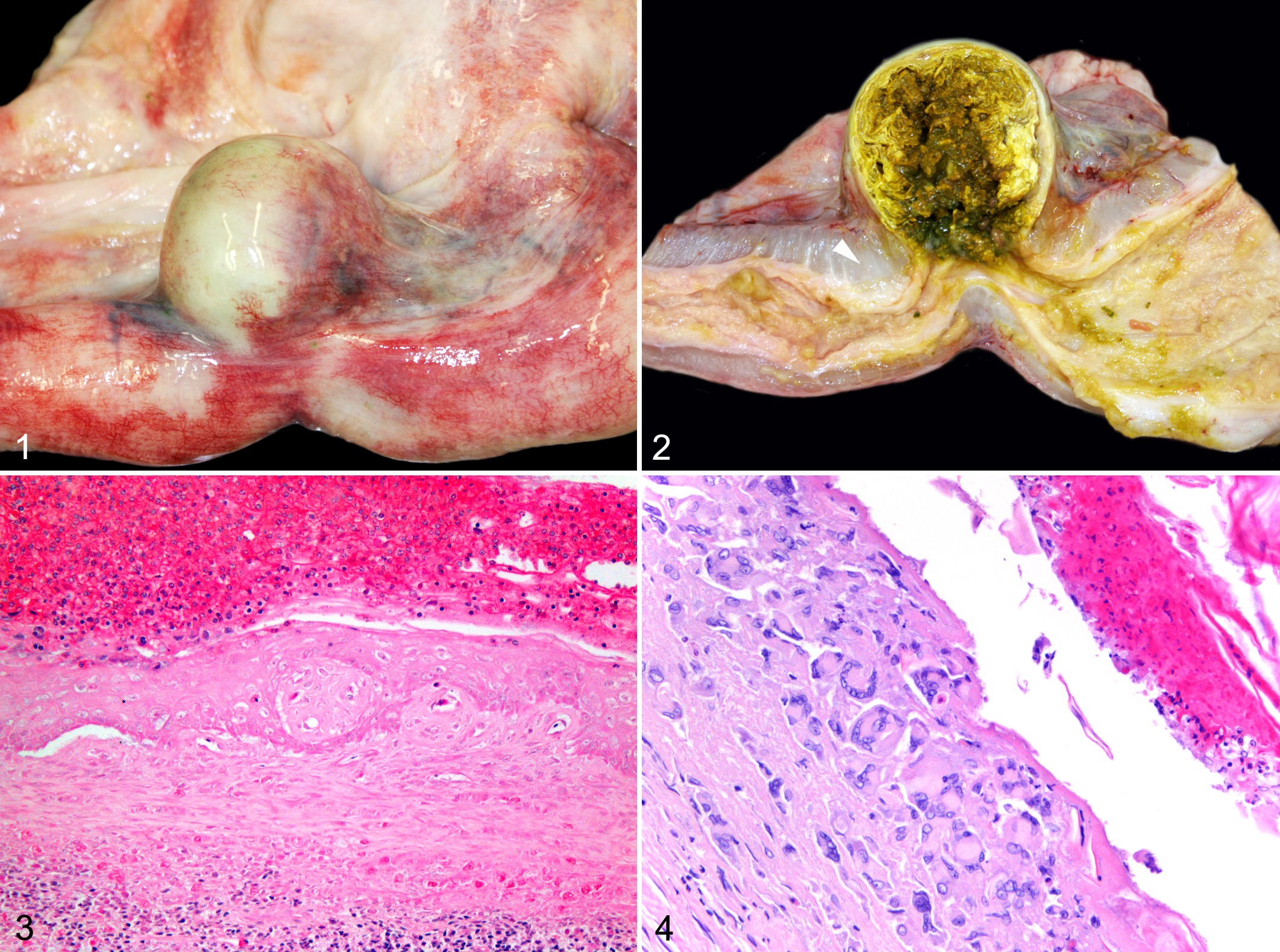
The gross appearance of the diverticulum was similar in all cases and consisted of a solitary mass immediately distal to the ileocecal valve (Fig. 1). The location relative to the mesentery was specifically noted in 11 cases. Ten lesions were adjacent to the mesentery, and 1 was described as antimesenteric. The lesions ranged in diameter from 2.0 to 9.0 cm. There was no correlation between size of diverticula and CCL. Various degrees of congestion of serosal vessels and mesenteric edema were present. On section, the diverticula were distended by firm, lamellated, caseous exudate, and the wall was attenuated to 1.0 to 2.0 mm (Fig. 2). No intralesional foreign material was found. The adjacent bowel lumen was narrowed (Fig. 2). The mucosa surrounding the opening to the diverticulum was locally ulcerated and covered by exudate. There was no evidence of perforation in any case.
Leatherback sea turtles; large intestinal diverticula.
Examination of 21 cases included histology of the diverticula. The caseous material within the center of the lesions was composed of layers of degenerate heterophils and numerous bacteria. Rare trematode ova were interspersed within the exudate in 2 turtles and were considered incidental findings. In most cases, there was multifocal or diffuse ulceration of the mucosa, and the muscularis was markedly attenuated or unapparent. Secondary mucosal changes included squamous metaplasia (Fig. 3) and formation of mucus-filled cysts. In some lesions, the ulcerated mucosa was completely replaced by multinucleated giant cells aggregated around the luminal exudate (Fig. 4). Inflammation within the wall of the diverticula ranged from moderate to intense; was composed of heterophils, lymphocytes, macrophages, and eosinophils; and was associated with fibrosis. The infiltrate extended to the subserosa, although there was no evidence of perforation. Mesothelial hyperplasia overlying the diverticulum was observed in 3 cases.
Bacterial morphology within lesions was highly variable and included coccobacilli, bacilli, and filamentous organisms. Gram-negative bacilli were predominant. Neither fungi nor acid-fast organisms were observed. Due to the postmortem interval and expectation of rapid contamination, aerobic bacterial cultures were obtained from only 3 cases, all of which yielded pure isolates of Morganella morganii. Heavy growth was observed in case Nos. 7 and 21 and light growth in case No. 22.
In most cases, death was attributed to human interaction, and animals were in robust nutritional condition. Nine turtles were found entangled in heavy line and/or fishing gear or had distinct ligature wounds indicative of entanglement. Six turtles died from acute traumatic injuries, including 4 with equidistant parallel chop wounds typical of injuries caused by watercraft propellers and 2 cases with blunt head trauma that also was attributable to watercraft. One leatherback drowned in a trawler net (nonfisheries vessel), and another died from gastric obstruction caused by an 84 × 35–cm piece of plastic sheeting. Three additional cases were suspected to be related to human interaction, including 1 turtle with chronic ligature wounds (case No. 10), another that died unexpectedly following capture during a research study (case No. 29), and a third turtle with a nonfatal incised wound on 1 flipper (case No. 5). Forced submergence is a consideration for an additional leatherback (case No. 22) that was nutritionally robust and had evidence of seawater aspiration without any apparent underlying condition.
Seven leatherbacks had chronic or debilitating disease; however, none was determined to be related to the diverticulitis. Findings in 5 of these cases included poor nutritional condition in combination with significant inflammatory lesions involving 1 or more organ systems, and 2 turtles were underweight without a clear cause. Notably, both of the latter animals had pieces of soft plastic cranial to the pylorus that could have resulted in at least partial obstruction. No circumstantial information or necropsy findings were found to suggest the cause of stranding in the remaining 3 cases.
The consistent finding of solitary diverticulitis caudal to the ileocecal junction in all examined subadult and adult leatherbacks from 2 ocean basins is remarkable. Selection of records was biased for detection of diverticulitis only to the extent that inclusion required confidence that the small intestine and colon were completely examined. All necropsy records available for review from the collaborating institutions were considered, and only a few cases were excluded. In addition to the cases presented in the current series, 1 author (P.-Y.D.) opportunistically observed the lesion in a nesting female leatherback killed near the coast of Cameroon, Africa, in November 2012, and another (M.C.J.) has encountered the lesion in previous field necropsies on the Atlantic coast of Canada. Solitary diverticulitis was photographed but not diagnosed in a single leatherback from the coast of Brazil included in a description of comparative digestive anatomy of sea turtles, 6 and a “hard, clay-like ball” at the junction of the small and large intestine was described in a large male found stranded in Gwynedd, Wales (United Kingdom) in 1988. 2 To our knowledge, similar prevalence of diverticular disease has not been documented in any other animal taxa, nor have we encountered similar lesions at this location in other sea turtle species. There was no evidence that diverticulitis was associated with death or clinical disease in any of the cases. Many were apparently healthy individuals that died from acute traumatic events. There was no apparent relationship between size of the diverticulum or degree of ulceration and other findings, such as nutritional condition or cause of death. Nonetheless, it stands to reason that the diverticula are not completely benign given severe attenuation of the bowel wall, intradiverticular accumulation of exudate and bacteria, and risk of complication, such as perforation or obstruction. Inference of disease and mortality from strandings of predominantly oceanic species is inherently limited due to a low probability of detection and recovery of dead and moribund animals.
The diverticula were diagnosed as proximal large intestine in origin due to difficulty in clearly distinguishing the cecum and colon and the likely involvement of both structures in some cases, especially in larger lesions. Attenuation and loss of the muscularis are features of acquired diverticula. 5 Differences in anatomy and physiology limit comparison with diverticular disease in other animals, including humans. In general, acquired diverticula are thought to be associated with a variety of factors that affect the integrity of the bowel wall and intraluminal pressure, including anatomical features, muscular hypertrophy, age, genetics, diet, and microfauna. 3–5 The consistent anatomical localization of this lesion in leatherbacks suggests that diverticular development is, at least in part, due to regional anatomy, although the mechanism is unclear. The indistinct cecum in this species, as well as the mesenteric orientation of many lesions (whereas the cecum develops embryologically from the antimesenteric border of the large intestine), does not suggest an obvious risk of an anatomical trapping effect, such as may occur in taxa with a prominent blind-ended cecum. Most of the lesions in which location was specifically noted were primarily mesenteric, and thus intrinsic weakness of the muscularis associated with vascular penetration may play a role, as proposed in human colonic diverticula. 9 Dysfunction of the ileocecal valve does not appear to be a likely cause as lesions were large intestinal rather than enteric. 6 Other general anatomical features of this site in leatherbacks include an increase in luminal diameter at the origin of the large intestine and gut-associated lymphoid tissue. The absence of diverticula in juveniles suggests that age or size is a factor in development. Last, adaptation to consume a high-volume, dilute diet is a notable, unique characteristic of leatherbacks and could influence pathologic response to abnormal motility, obstruction, or other disorders. Although other sea turtle species also consume gelatinous zooplankton, diets generally include various other marine organisms, such as vegetation, macroalgae, and invertebrates, depending on species.
In humans, diverticulitis results in clinical symptoms, occurs in a minority of patients with diverticular disease, and has an uncertain pathogenesis. 9 It is hypothesized that fecal impaction within diverticula results in secondary inflammation, obstruction, and mucus accumulation, leading to ulceration and perforation. Although a similar process is plausible in leatherbacks, no turtles had acute lesions with which to infer progression. All were characterized by abundant intradiverticular exudate and chronic, ongoing inflammation. The larger lesions in some turtles suggest that diverticula may enlarge as luminal material accumulates, which could lead to complication.
The postmortem interval and open exposure to the colon lumen resulted in limited opportunity for a conclusive microbiological study and will pose a similar problem in future cases. Isolation of pure cultures of M. morganii, a commensal within the gastrointestinal tract and opportunistic pathogen, 8 from 3 cases was interesting and suggests that this organism thrives within the diverticular exudate and may play a role in ongoing inflammation. Mixed bacterial morphology was evident by histology, and thus culture results may reflect fastidious growth requirements of other organisms present within lesions. Although improved characterization of gastrointestinal microfauna of leatherbacks and within diverticula could be interesting, comparative findings in other taxa suggest that other primary factors are involved in development of the lesion.
Solitary large intestinal diverticulitis occurring caudal to the ileocecal valve is a frequent finding in subadult and adult leatherback turtles examined in North America. Whatever the cause(s), innate or exogenous, the species appears to be broadly susceptible to development of this lesion. Although diverticulitis was unrelated to cause of death in the cases presented in this report, there is potential for complications and related illness. The gastrointestinal tract, including the ileocecocolic region, should be closely evaluated during necropsy and thoroughly documented. Additional cases, especially those with more acute lesions, may provide further insight into pathophysiology and significance.
Footnotes
Acknowledgements
We thank participants in the Sea Turtle Stranding and Salvage Network (United States) and the Canadian Sea Turtle Network (Kathleen Martin), officers of the Department of Fisheries and Oceans Canada, Population Ecology Division (Conservation and Protection Division officers), staff and volunteers of New England Aquarium, the Marine Mammal Center, Moss Landing Marine Laboratories Marine Operations, Jennifer Keene, Robert Prescott, Kara Dodge, Sea Rogers Williams, Bridget Dunnigan, Don Lewis, Michael Moore, Darlene Ketten, Betty Lintell, Kate Sampson, Scott Benson, Craig Harms, Sue Barco, Matthew Godfrey, International Fund for Animal Welfare, Massachusetts Department of Marine Fisheries, Massachusetts Audubon Society, Provincetown Center for Coastal Studies, Paul Doshkov and staff of the Cape Hatteras National Seashore, California Department of Fish and Wildlife, and Salvatore Frasca Jr and the Connecticut Veterinary Medical Diagnostic Laboratory for providing histopathologic evaluation of case material submitted from New England Aquarium. In addition, we thank Thierry Work and George Balazs for necropsy information on juvenile turtles.
Declaration of Conflicting Interests
The author(s) declared no potential conflicts of interest with respect to the research, authorship, and/or publication of this article.
Funding
The author(s) disclosed receipt of the following financial support for the research, authorship and/or publication of this article: Partial funding was provided by grant NA10NMF4720028 from the United States Department of Commerce, National Oceanic and Atmospheric Administration, National Marine Fisheries Service.