
Abstract
Teratomas are histologically complex neoplasms that are composed of structures derived from multiple germ cell layers (ectoderm, mesoderm, and endoderm). These neoplasms are uncommon in domestic animals and are usually found in the gonads. This paper describes teratomas of the adrenal gland in four domestic ferrets (Mustela putorius furo). Three of four of the neoplasms contained tissues from ectodermal, mesodermal, and endodermal germ cell layers; two of four contained rudimentary teeth. In one case, malignant epithelial cells had metastasized to local lymph nodes. Teratomas, although uncommon, should be included in the differential diagnosis for adrenal neoplasms in domestic ferrets.
Teratomas are histologically complex neoplasms that are composed of structures derived from multiple germ cell layers (ectoderm, mesoderm, and endoderm). These neoplasms are uncommon in domestic animals and are usually found in the gonads. This paper describes teratomas of the adrenal gland in four domestic ferrets (Mustela putorius furo).
Ferret No. 1 was a 3-year-old male with a history of anorexia and progressive weakness. Physical examination revealed a palpable abdominal mass; radiographic examination revealed a large ventral thoracic mass and pleural effusion. The animal was euthanatized based on these findings. Necropsy revealed generalized lymphadenomegaly, splenomegaly, and pleural and peritoneal effusion. An incidental finding was unilateral enlargement of the right adrenal gland, which measured 2 × 2.5 × 1.3 cm. Grossly, the adrenal gland had a yellow granular surface. On cut section, there was an oval, cystic mass surrounded by a thin rim of bone and compressed adrenocortical tissue.
Microscopic evaluation of tissues revealed infiltration of numerous lymph nodes by blast lymphocytes that effaced normal architecture and infiltrated liver, lung, urinary bladder, and colon. The neoplasm in the right adrenal consisted of multiple well-differentiated tissues including bone, cartilage, and squamous epithelium containing sebaceous and apocrine sweat glands, hair, and a rudimentary tooth. Compressed adrenocortical tissue surrounded the neoplasm. Based on these findings, a diagnosis of lymphoblastic lymphosarcoma and mature adrenal teratoma was made.
Ferret No. 2 was an adult domestic female of unknown age, which exhibited a 2-week persistent fever of unknown origin. Complete blood count revealed a leukocytosis of 18,500 WBC/mm3 (normal 4,900–13,800/mm3). 10 Abdominal radiography revealed bilateral radiopacity in the adrenal glands. Based on these findings, abdominal exploration was performed. Both adrenal glands measured over 2.5 cm in diameter, and multiple small white foci in the liver were seen. Based on the high index of suspicion of bilateral adrenocortical carcinoma with hepatic metastasis in this patient, the animal was euthanatized intraoperatively. Multiple tissues were taken at necropsy and placed in formalin.
Microscopic examination of tissues revealed bilateral adrenal neoplasms. The neoplasms contained well-differentiated tissues derived from all three germ cell layers. Both neoplasms contained bone, cartilage, respiratory epithelium, and elements of haired skin, including squamous epithelium. The right adrenal gland contained a large cyst-like structure replacing almost 70% of the gland, which contained abundant mineralized debris, keratin, and degenerate neutrophils. The left adrenal neoplasm contained similar small cysts as well as well-differentiated brain tissue, containing neurons, and gut and respiratory epithelium. The white foci in the liver were identified as foci of hepatocellular lipid accumulation, a common incidental finding in ferrets.
Ferret No. 3 was a castrated, color-diluted (silver-mitt) male approximately 1 year of age. The ferret presented with a complaint of anorexia and a distended abdomen. Abdominocentesis yielded a large amount of a dark brown fluid, and abdominal radiographs showed a large fluid-filled cystic mass in the caudal abdomen. Based on these findings, the animal was euthanatized and necropsied. On necropsy, abnormal findings were restricted to the abdomen. A large cystic mass appeared to originate from the dorsal aspect of the abdominal cavity, which when opened, contained abundant brown fluid and several fragments of hair and bone. The lining of the mass contained tufts of hair. A mesenteric lymph node was enlarged. Sections of the mass, the enlarged lymph node, and the liver and kidneys were submitted in 10% neutral buffered formalin to the Armed Forces Institute of Pathology for microscopic examination.
Examination of the submitted tissue revealed a neoplasm contained within an overlying layer of morphologically normal adrenal cortex. The neoplasm contained ectodermal, mesodermal, and endodermal elements, including haired skin and peripheral nerve (ectoderm), bone, cartilage, and adipose tissue (mesoderm), and respiratory glandular epithelium (endoderm). In some areas of the neoplasm, pleomorphic epithelial cells formed solid nests and cords, and in some areas, formed large cystic glands lined by arborizing papillary projections. These cells had a large nuclear:cytoplasmic ratio with a vesicular chromatin pattern, 1–3 large magenta nucleoli, and an average of one mitotic figure per three 40× fields. This population of cells occasionally lined the cyst wall up to three cell layers thick. Similar cells were present within subcapsular sinuses and perinodal lymphatics of the submitted mesenteric node. The pleomorphic cells were immunopositive for cytokeratin. Based on the immunohistochemical results, cellular atypia, and the presence of cells within an adjacent node, a diagnosis of malignant teratoma with tubulopapillary adenocarcinoma was given in this case.
Ferret No. 4 was a clinically normal, 4-month-old spayed female used in a topical ectoparasiticide study. One day after dosing, a firm midabdominal mass was palpated during routine anesthesia for blood sampling. Two days later, the ferret was euthanatized and necropsied.
Necropsy revealed a firm mass dorsal to the colon. The left adrenal gland was not identified; all other tissues appeared to be within normal limits. On cut section, several cystic areas exuded a brown viscous fluid, and an area of cartilage within the neoplasm was noted.
Microscopic examination of the submitted tissue revealed a neoplasm composed of ectodermal, mesodermal, and endodermal elements surrounded by compressed adrenocortical tissue. Elements of well-developed bone, bone marrow, cartilage, peripheral nerve, skin with hair and sebaceous glands, gastrointestinal-type mucosa, and even a well-developed tooth were identified within the neoplasm (Figs. 1–4). The cystic areas contained keratin debris admixed with moderate numbers of neutrophils.
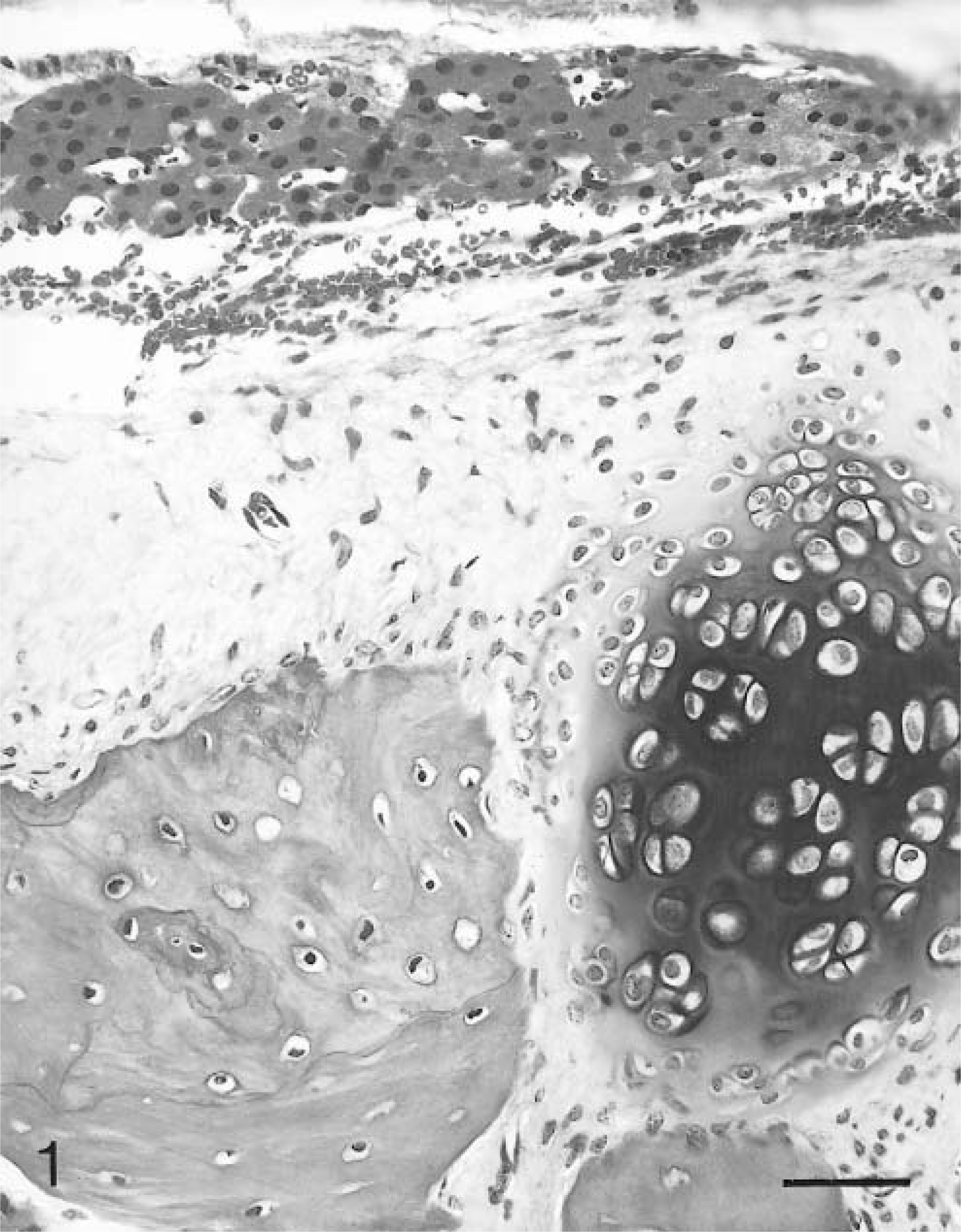
Adrenal teratoma from ferret No. 4. Mature mesenchymal elements (bone, left; cartilage, right) subjacent to a compressed rim of morphologically normal adrenocortical cells (A). HE. Bar = 37.5 µm.
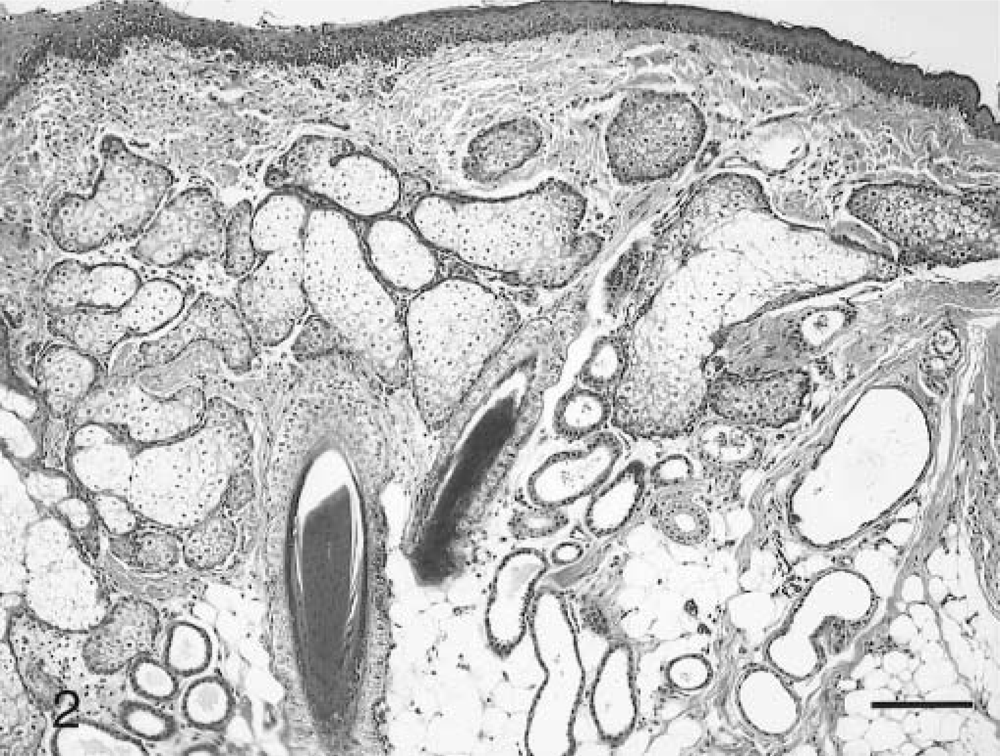
Adrenal teratoma from ferret No. 4. Mature ectodermal element of well-differentiated haired skin with overlying stratified squamous epithelium, hair follicles, sebaceous glands, and dilated apocrine glands. HE. Bar = 75 µm.
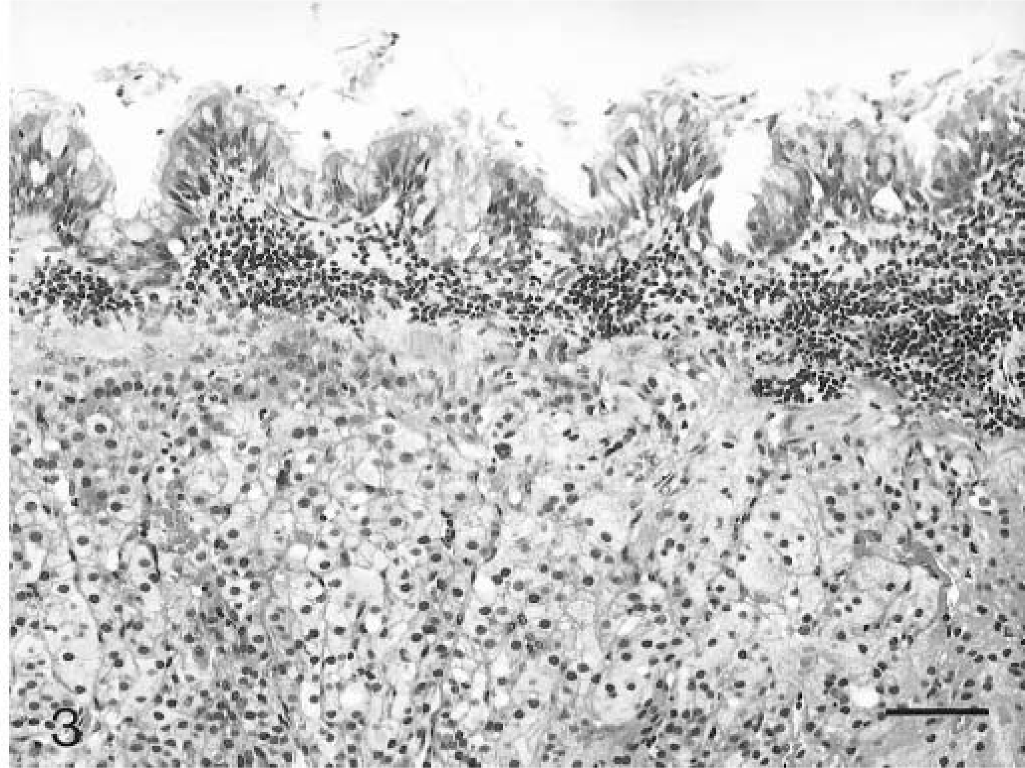
Adrenal teratoma from ferret No. 4. Mature endodermal element of well-differentiated ciliated respiratory epithelium overlying morphologically normal adrenocortical cells. HE. Bar = 37.5 µm.
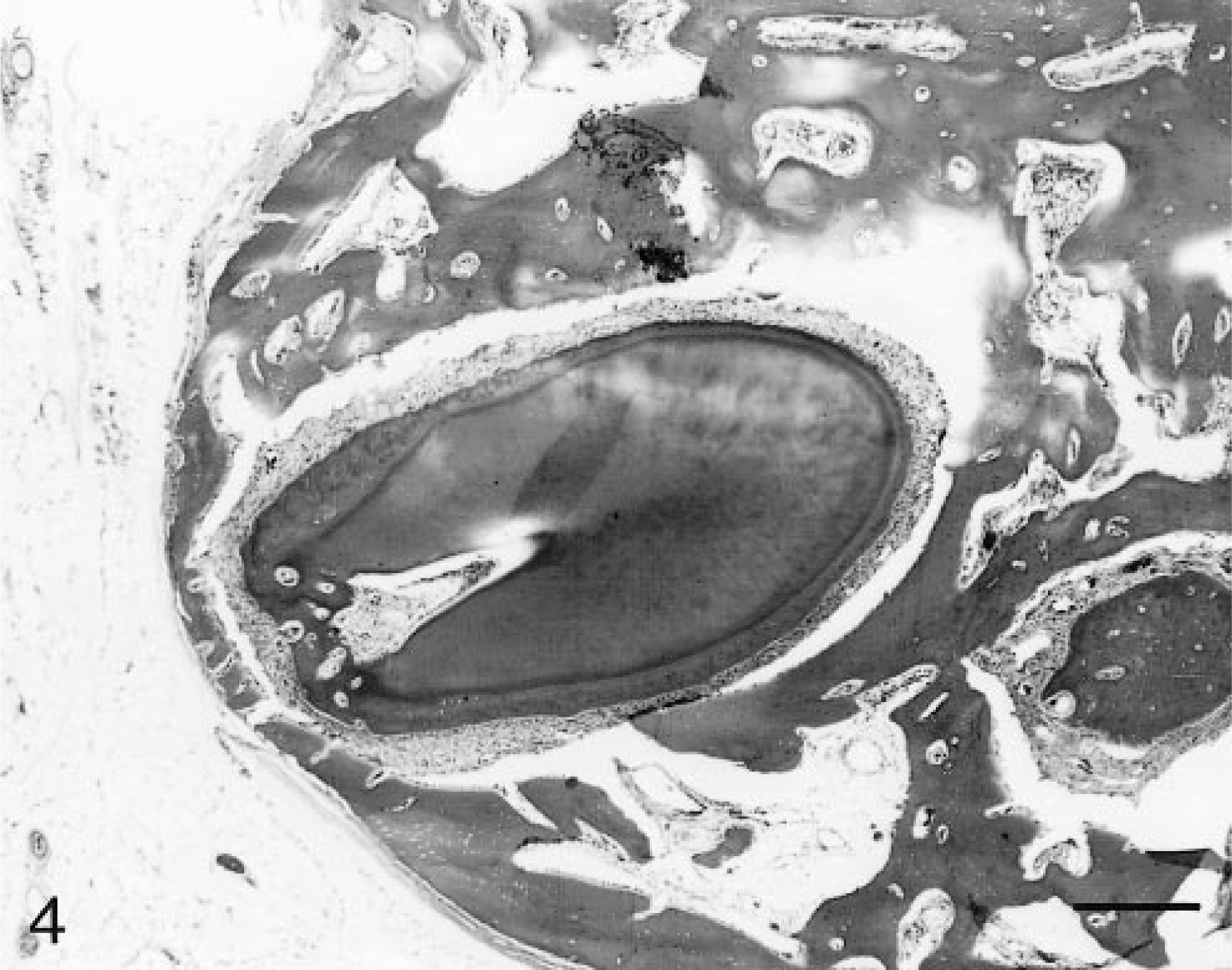
Adrenal teratoma from ferret No. 4. Mature mesodermal elements of a well-differentiated denticle surrounded by trabeculae of woven bone. HE on undecalcified section. Bar = 250 µm.
Teratomas are uncommon neoplasms of humans and animals and consist of tissues from multiple germ cell lines:ectoderm (epidermal structures of skin, nervous tissue, and the linings of the oral and nasal cavities), mesoderm (connective tissue, muscle, cartilage, bone, and structures of the cardiovascular, urinary, and genital systems), and endoderm (respiratory and gastrointestinal epithelium, including glands). These neoplasms have been reported in a wide variety of animals, including the dog, 22,23,25 cat, 6,23 horse, 19,23,24 laboratory rodents, 7,18,21,26,29,31 rabbits, 5 sheep, 1,23 ox, 13,15,23 poultry, 9,12,14 primates, 2,3,8,16,20,28,34 ferrets, 4,10,17 swine, 23 and a blue heron. 30
Due to the complex histological nature of these neoplasms as well as the varied and often conflicting theories of their development, an assortment of terms have been applied to them in the medical literature. The term “immature teratoma” has been used to describe a neoplasm that not only contains elements derived from the three germ cell layers but also contains rests of unrecognizable, poorly differentiated tissue, which is generally considered to be of neural origin. 28 If the teratoma is presumed to have arisen in the gonad from premeiotic diploid germ cells, postmeiotic haploid germ cells, or cells derived from extraembryonic components such as the yolk sac, the term “germ cell tumor” may be applied. 28,32
Teratomas may be benign (as is commonly the case in domestic species) or malignant, with metastasis of one or more components to distant sites (seen with a higher incidence in laboratory rodents and humans).
There is currently great debate over the histogenesis of teratomas as well as an increasing body of evidence that different origins exist for gonadal versus extragonadal teratomas. 28,32 Gonadal teratomas are presumed to develop from parthenogenetic development of sequestered haploid postmeiotic germ cells or diploid premeiotic germ cells in the gonad. The exact precursor cell is still unknown. 32 Extragonadal teratomas are usually congenital and are often present in the midline. While the pathogenesis of teratomas is still unclear, extragonadal teratomas likely originate from diploid pluripotent progenitor cells that escape embryonal organizers during migration. 32
In dogs and humans, teratomas account for less than 1% of all neoplasms but up to 25% of all ovarian neoplasms. 22 In inbred mouse strains 129 and LT, gonadal teratomas may be seen in up to 50% of females and 30% of males. 7
The incidence of extragonadal teratomas is very low in animals and humans. The most common form of extragonadal teratoma is the so-called dentigerous cyst of horses, 19 which usually occurs at the base of the ear. Extragonadal teratomas have also been documented in humans, 3,8,16,20,28,32,34 ducks, 14 a blue heron, 30 rabbits, 5 rats, 18,29 mice, 26 an ox, 15 a kitten, 6 a sheep, 1 and a calf. 13 Adrenal teratomas have been previously identified in humans, 16,20 an ox, 15 and a rat. 29
Adrenocortical neoplasms have been reported as the second most common neoplasm in the ferret. 17 These tumors are responsible for the liberation of excessive estrogen and its precursors and result in a syndrome of cutaneous, behavioral, and reproductive signs referred to as adrenal-associated endocrinopathy (AAE). Adrenocortical adenomas and carcinomas, as well as hyperplastic nodules, are hormonally active, resulting in characteristic clinical signs of AAE. 33 Spindle cell neoplasms of smooth muscle origin have also been reported in the adrenal gland of ferrets; 11 while these neoplasms may attain a large size, systemic hormonal effects have not been attributed to them.
Adrenal teratoma should be considered in the differential diagnosis for adrenal neoplasms in the ferret. As all four neoplasms in this report contained bone, this neoplasm may be most strongly suspected in animals with presumed adrenal masses, radiopaque densities in the area of the adrenal glands on survey radiographs, and an absence of clinical signs of hyperestrogenism.
Footnotes
Acknowledgements
We would like to thank Ms. Robin-Anne Veronica Ferris, MFS, and Mr. Douglas Landry for excellent photographic support and Drs. Charles A. Montgomery and James G. Fox for technical assistance in the production of this article.